Ashley Davidoff MD Barry Sacks MD
Copyright 2010
Types of Lesions
The macrofollicular component of a thyroid nodule is a normal variant while microfollicular component raises concern and warrants surgical excision.
|

Encapsulated Follicular Variant of Papillary Carcinoma |
|
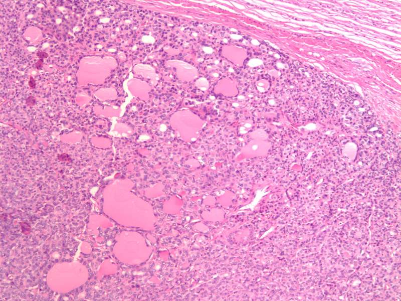
The histological section at 10X magnification using H&E stain shows a encapsulated follicular variant of papillary carcinoma (FVPC) with microfollicular architecture. There are a few MAcrofollicles scattered among the microfollicles
Image Courtesy Ashraf Khan MD. Department of Pathology, University of Massachusetts Medical School. 99397.8
|
|

Encapsulated Follicular Variant of Papillary Carcinoma
40X H&E |
|
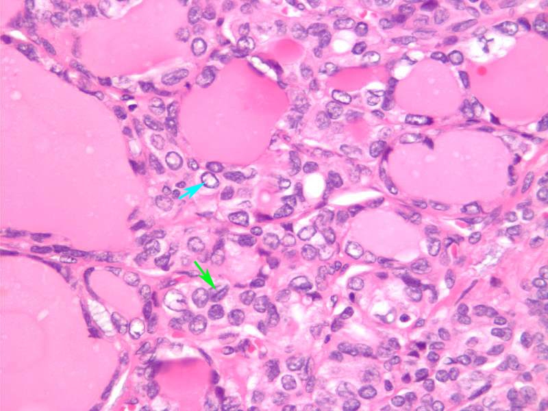
The histological section at 40X magnification using H&E stain shows a encapsulated follicular variant of papillary carcinoma (FVPC) with microfollicular architecture. This high power view shows papillary cancer nuclei with Orphan Annie nuclei and grooves. Orphan Annie nuclei a ?cleared-out? or empty appearance, similar to Little Orphan Annie?s eyes (light blue arrow). Nuclear grooves characteristic lines that run across the nuclei (green arrow).
Image Courtesy Ashraf Khan MD. Department of Pathology, University of Massachusetts Medical School. 99398.81
|

Mixed Microfollicular and MAcrofollicular Lesions |
|
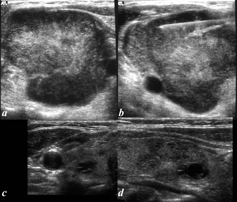
The ultrasound from two patients (a,b patient 1 and c,d patient 2) both show heterogeneous lesions with hypoechoic, isoechoic and hyperechoic texture (a,b, c,d)
Obvious calcifications nor haloes are present and the vascularity is not shown.
A biopsy (b, for first patient and c for second) supported the diagnosis of mixed micro-macrofollicular lesions in both cases. In the presence of microfollicles ? surgical resection is indicated.
Courtesy Barry Sacks MD 97202e.8L
|
DOMElement Object
(
    [schemaTypeInfo] =>
    [tagName] => table
    [firstElementChild] => (object value omitted)
    [lastElementChild] => (object value omitted)
    [childElementCount] => 1
    [previousElementSibling] => (object value omitted)
    [nextElementSibling] =>
    [nodeName] => table
    [nodeValue] =>


Mixed Microfollicular and MAcrofollicular Lesions


The ultrasound from two patients (a,b patient 1 and c,d patient 2) both  show heterogeneous lesions with hypoechoic, isoechoic and hyperechoic texture (a,b, c,d)
Obvious calcifications nor haloes are present and the vascularity is not shown.
A biopsy (b, for first patient and c for second) supported the diagnosis of mixed micro-macrofollicular lesions in both cases. In the presence of microfollicles ? surgical resection is indicated.
Courtesy Barry Sacks MD 97202e.8L


    [nodeType] => 1
    [parentNode] => (object value omitted)
    [childNodes] => (object value omitted)
    [firstChild] => (object value omitted)
    [lastChild] => (object value omitted)
    [previousSibling] => (object value omitted)
    [nextSibling] => (object value omitted)
    [attributes] => (object value omitted)
    [ownerDocument] => (object value omitted)
    [namespaceURI] =>
    [prefix] =>
    [localName] => table
    [baseURI] =>
    [textContent] =>


Mixed Microfollicular and MAcrofollicular Lesions


The ultrasound from two patients (a,b patient 1 and c,d patient 2) both  show heterogeneous lesions with hypoechoic, isoechoic and hyperechoic texture (a,b, c,d)
Obvious calcifications nor haloes are present and the vascularity is not shown.
A biopsy (b, for first patient and c for second) supported the diagnosis of mixed micro-macrofollicular lesions in both cases. In the presence of microfollicles ? surgical resection is indicated.
Courtesy Barry Sacks MD 97202e.8L


)
DOMElement Object
(
    [schemaTypeInfo] =>
    [tagName] => td
    [firstElementChild] => (object value omitted)
    [lastElementChild] => (object value omitted)
    [childElementCount] => 4
    [previousElementSibling] =>
    [nextElementSibling] =>
    [nodeName] => td
    [nodeValue] =>
The ultrasound from two patients (a,b patient 1 and c,d patient 2) both  show heterogeneous lesions with hypoechoic, isoechoic and hyperechoic texture (a,b, c,d)
Obvious calcifications nor haloes are present and the vascularity is not shown.
A biopsy (b, for first patient and c for second) supported the diagnosis of mixed micro-macrofollicular lesions in both cases. In the presence of microfollicles ? surgical resection is indicated.
Courtesy Barry Sacks MD 97202e.8L

    [nodeType] => 1
    [parentNode] => (object value omitted)
    [childNodes] => (object value omitted)
    [firstChild] => (object value omitted)
    [lastChild] => (object value omitted)
    [previousSibling] => (object value omitted)
    [nextSibling] => (object value omitted)
    [attributes] => (object value omitted)
    [ownerDocument] => (object value omitted)
    [namespaceURI] =>
    [prefix] =>
    [localName] => td
    [baseURI] =>
    [textContent] =>
The ultrasound from two patients (a,b patient 1 and c,d patient 2) both  show heterogeneous lesions with hypoechoic, isoechoic and hyperechoic texture (a,b, c,d)
Obvious calcifications nor haloes are present and the vascularity is not shown.
A biopsy (b, for first patient and c for second) supported the diagnosis of mixed micro-macrofollicular lesions in both cases. In the presence of microfollicles ? surgical resection is indicated.
Courtesy Barry Sacks MD 97202e.8L

)
DOMElement Object
(
    [schemaTypeInfo] =>
    [tagName] => td
    [firstElementChild] => (object value omitted)
    [lastElementChild] => (object value omitted)
    [childElementCount] => 2
    [previousElementSibling] =>
    [nextElementSibling] =>
    [nodeName] => td
    [nodeValue] =>
Mixed Microfollicular and MAcrofollicular Lesions
    [nodeType] => 1
    [parentNode] => (object value omitted)
    [childNodes] => (object value omitted)
    [firstChild] => (object value omitted)
    [lastChild] => (object value omitted)
    [previousSibling] => (object value omitted)
    [nextSibling] => (object value omitted)
    [attributes] => (object value omitted)
    [ownerDocument] => (object value omitted)
    [namespaceURI] =>
    [prefix] =>
    [localName] => td
    [baseURI] =>
    [textContent] =>
Mixed Microfollicular and MAcrofollicular Lesions
)
DOMElement Object
(
    [schemaTypeInfo] =>
    [tagName] => td
    [firstElementChild] => (object value omitted)
    [lastElementChild] => (object value omitted)
    [childElementCount] => 2
    [previousElementSibling] =>
    [nextElementSibling] =>
    [nodeName] => td
    [nodeValue] =>

Encapsulated Follicular Variant of Papillary Carcinoma
    [nodeType] => 1
    [parentNode] => (object value omitted)
    [childNodes] => (object value omitted)
    [firstChild] => (object value omitted)
    [lastChild] => (object value omitted)
    [previousSibling] => (object value omitted)
    [nextSibling] => (object value omitted)
    [attributes] => (object value omitted)
    [ownerDocument] => (object value omitted)
    [namespaceURI] =>
    [prefix] =>
    [localName] => td
    [baseURI] =>
    [textContent] =>

Encapsulated Follicular Variant of Papillary Carcinoma
)


