Ari Sacks MD
The Common Vein Copyright 2010
Definition
Multinodular Goiter is a benign neoplastic growth consisting of one or several hypoactive thyroid nodules that do not produce thyroid hormone. This condition is characterized by follicular epithelial cell hyperplasia. Causes of non-toxic goiters are either sporadic or endemic goiters from iodine deficiency. In the iodine-sufficient areas of the world, including United States, while prevalence of palpable nodules is about 5-6%, the prevalence of nodules visible upon ultrasound is 80-90% at the age of 60 years old.
Structural changes of the thyroid gland include enlargement and nodularity. There are typically no functional changes to thyroid hormone production or release.
Clinically patients will present with enlargement or feeling of a ?mass? in the anterior neck. Patients usually do not have any symptoms of either hypo or hyperthyroidism.
Imaging of the thyroid can be done with ultrasound and/or thyroid scintigraphy. Ultrasound can show the number, size, and complexity of the nodule(s). Sonographic appearance of the nodule can be used as a tool to predict risk of malignancy and guide biopsy. Thyroid scintigraphy of a non-toxic or ?cold? nodule would show little to no radioactive iodine uptake. Diagnosis should is best made with laboratory studies showing the patient to be euthyroid, physical exam and/or imaging revealing a non-toxic, benign nodule. Thyroid biopsy can give a definitive diagnosis of a non-toxic thyroid nodule. Treatment typically consists of observation. Occasionally if the goiter becomes symptomatic due to size, surgical removal or thyroidectomy can be performed.
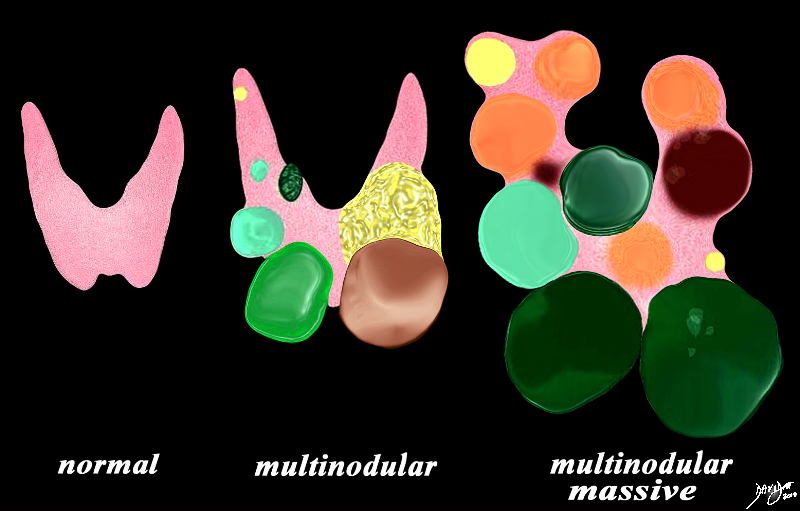
Normal and Multidular Goiter |
|
The diagram illustrates non toxic multinodular goiter together with a normal thyroid gland on the left. A moderately enlarged gland (middle) containing multiple variably sized non toxic nodules with different characteristics is shown together with a massively enlarged non toxic gland (right) When the gland becomes massively enlarged it may extend into the chest, compress the airway and neck veins as well as displace other structures such as the esophagus.
Courtesy Ashley Davidoff MD Copyright 2010 93852e07h01a03.8s
|
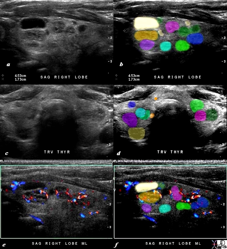
Multinodular Non Toxic Thyroid Gland
Not truly a Goiter Since It is Not Enlarged |
|
The 80 year old male presents with an asymptomatic multinodular goiter, consisting many nodules of varying size and echogenocity, as seen by sagittal ultrasound (a,b) and transverse imaging images (c,d), and Doppler imaging (e,f). The multiple nodules that are not border forming are within the confines of the parenchyma and do not alter the shape of the gland. The right lobe of the thyroid measures about 4.5 cms in sagittal 1.5cms.in A-P dimension, and 1.5cms in the transverse plane. The gland is therefore not enlarged. In addition the gland has a normal appearance in the transverse projection and the borders are not rounded to suggest enlargement.
The Doppler study shows no internal vascularity in any of the nodules visualized.
These findings are consistent with a non toxic multinodular thyroid gland, and not truly a goiter since the gland is not enlarged.
Courtesy Ashley Davidoff MD Copyright 2010 94780c04b02.8s
|
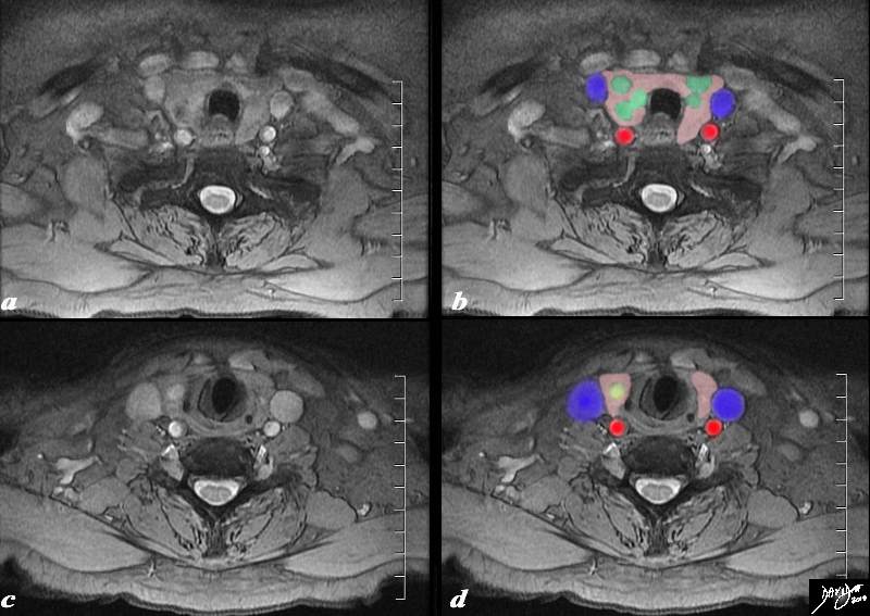
MRI of the Above Patient STIR Technique |
|
The MRI is from an 80 year old male presents with an asymptomatic multinodular goiter, consisting many nodules of varying size as identified by previous ultrasound.. The MRI using a STIR sequence shows a normal size and shape to the gland but multiple non border forming nodules (green nodules) are identified within the matrix of the gland (pink).One of the nodules (c,d) overlaid in yellow is cystic in nature. It is bright on the STIR sequence (c). The internal jugular veins (blue) form a lateral neighbor and deform the relatively soft gland. The common carotid arteries (red) form a posterolateral relation to the gland. The trachea (black) is surrounded anteriorly and laterally by the gland. These findings are consistent with a non toxic multinodular thyroid gland, – not truly a goiter since the gland is not enlarged.
Courtesy Ashley Davidoff MD Copyright 2010 94790c03.8s
|
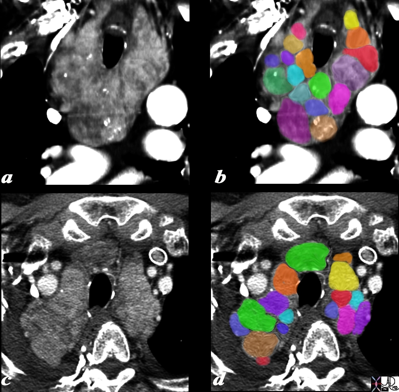
Multinodular Massive Retrosternal Goiter – CT scan |
|
The 75 year old male presents with an asymptomatic multinodular goiter, consisting many nodules of varying size and density, some with macrocalcifications as seen by coronal CT reconstruction (a,b) and axial images (c,d). The windows of the CTscan have been narrowed (a,c), to enhance the morphological characteristics of the components of retrosternal goiter. Each lobe of the thyroid measures about 12 cms in sagittal 6cms. in transverse and 12cms in A-P dimension. These findings are consistent with a non toxic multinodular goiter.
Courtesy Ashley Davidoff MD Copyright 2010 94712c01L01.8s
|
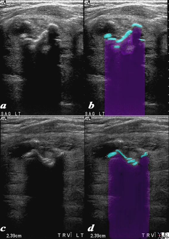
Heavy Calcification in a Multinodular Goiter Preclude Visualization of the Gland |
|
This 74year old female presents with a multinodular goiter. Images are taken in sagittal (a,b) and transverse (c,d). Multiple macrocalcifications (teal blue in b and d) preclude visualization of the gland because of extensive calcifications resulting in shadowing (black in a and c – purple in b and d) of the remaining thyroid parenchyma. These findings are consistent with a benign non toxic multinodular thyroid gland. The macrocalcifications are characteristic of benign disease.
Courtesy Ashley Davidoff MD Copyright 2010 95706c.8s
|
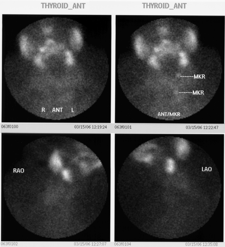
Enlarged Gland Minimal Uptake
Same Patient as Above |
|
This 74year old female presents with a multinodular goiter. Thyroid scan is shown. Markers were placed on the sternal notch. She was injected with 8.3mci of 99mTc pertechnitate. Images in the A-P (upper panel) and in right anterior oblique (lower panel right) and left anterior oblique (lower panel left). The images show minimal uptake in the by the enlarged gland. There is normal uptake in the salivary glands These findings are consistent with a benign non toxic multinodular thyroid gland.
Courtesy Alan Ashare MD Copyright 2010 95696bb02
|

Multinodular Goiter |
|
Enlarged thyroid gland Several large nodules of varying size are scattered throughout the gland. This basic appearance is characteristic of a multinodular goiter.
Courtesy Ashley Davidoff MD copyright 2010 all rights reserved 94460b16b.8s
|
DOMElement Object
(
    [schemaTypeInfo] =>
    [tagName] => table
    [firstElementChild] => (object value omitted)
    [lastElementChild] => (object value omitted)
    [childElementCount] => 1
    [previousElementSibling] => (object value omitted)
    [nextElementSibling] =>
    [nodeName] => table
    [nodeValue] =>


Multinodular Goiter


Enlarged thyroid gland Several large nodules of varying size are scattered throughout the gland. This basic appearance is characteristic of a multinodular goiter.
Courtesy Ashley Davidoff MD copyright 2010 all rights reserved 94460b16b.8s


    [nodeType] => 1
    [parentNode] => (object value omitted)
    [childNodes] => (object value omitted)
    [firstChild] => (object value omitted)
    [lastChild] => (object value omitted)
    [previousSibling] => (object value omitted)
    [nextSibling] => (object value omitted)
    [attributes] => (object value omitted)
    [ownerDocument] => (object value omitted)
    [namespaceURI] =>
    [prefix] =>
    [localName] => table
    [baseURI] =>
    [textContent] =>


Multinodular Goiter


Enlarged thyroid gland Several large nodules of varying size are scattered throughout the gland. This basic appearance is characteristic of a multinodular goiter.
Courtesy Ashley Davidoff MD copyright 2010 all rights reserved 94460b16b.8s


)
DOMElement Object
(
    [schemaTypeInfo] =>
    [tagName] => td
    [firstElementChild] => (object value omitted)
    [lastElementChild] => (object value omitted)
    [childElementCount] => 2
    [previousElementSibling] =>
    [nextElementSibling] =>
    [nodeName] => td
    [nodeValue] =>
Enlarged thyroid gland Several large nodules of varying size are scattered throughout the gland. This basic appearance is characteristic of a multinodular goiter.
Courtesy Ashley Davidoff MD copyright 2010 all rights reserved 94460b16b.8s

    [nodeType] => 1
    [parentNode] => (object value omitted)
    [childNodes] => (object value omitted)
    [firstChild] => (object value omitted)
    [lastChild] => (object value omitted)
    [previousSibling] => (object value omitted)
    [nextSibling] => (object value omitted)
    [attributes] => (object value omitted)
    [ownerDocument] => (object value omitted)
    [namespaceURI] =>
    [prefix] =>
    [localName] => td
    [baseURI] =>
    [textContent] =>
Enlarged thyroid gland Several large nodules of varying size are scattered throughout the gland. This basic appearance is characteristic of a multinodular goiter.
Courtesy Ashley Davidoff MD copyright 2010 all rights reserved 94460b16b.8s

)
DOMElement Object
(
    [schemaTypeInfo] =>
    [tagName] => td
    [firstElementChild] => (object value omitted)
    [lastElementChild] => (object value omitted)
    [childElementCount] => 2
    [previousElementSibling] =>
    [nextElementSibling] =>
    [nodeName] => td
    [nodeValue] =>
Multinodular Goiter
    [nodeType] => 1
    [parentNode] => (object value omitted)
    [childNodes] => (object value omitted)
    [firstChild] => (object value omitted)
    [lastChild] => (object value omitted)
    [previousSibling] => (object value omitted)
    [nextSibling] => (object value omitted)
    [attributes] => (object value omitted)
    [ownerDocument] => (object value omitted)
    [namespaceURI] =>
    [prefix] =>
    [localName] => td
    [baseURI] =>
    [textContent] =>
Multinodular Goiter
)
DOMElement Object
(
    [schemaTypeInfo] =>
    [tagName] => table
    [firstElementChild] => (object value omitted)
    [lastElementChild] => (object value omitted)
    [childElementCount] => 1
    [previousElementSibling] => (object value omitted)
    [nextElementSibling] => (object value omitted)
    [nodeName] => table
    [nodeValue] =>


Enlarged Gland Minimal Uptake
Same Patient as Above


This 74year old female presents with a multinodular goiter. Thyroid scan is shown. Markers were placed on the sternal notch. She was injected with 8.3mci of 99mTc pertechnitate. Images in the A-P (upper panel) and in right anterior oblique (lower panel right) and left anterior oblique (lower panel left). The images show minimal uptake in the by the enlarged gland. There is normal uptake in the salivary glands These findings are consistent with a benign non toxic multinodular thyroid gland.
Courtesy Alan Ashare MD Copyright 2010 95696bb02


    [nodeType] => 1
    [parentNode] => (object value omitted)
    [childNodes] => (object value omitted)
    [firstChild] => (object value omitted)
    [lastChild] => (object value omitted)
    [previousSibling] => (object value omitted)
    [nextSibling] => (object value omitted)
    [attributes] => (object value omitted)
    [ownerDocument] => (object value omitted)
    [namespaceURI] =>
    [prefix] =>
    [localName] => table
    [baseURI] =>
    [textContent] =>


Enlarged Gland Minimal Uptake
Same Patient as Above


This 74year old female presents with a multinodular goiter. Thyroid scan is shown. Markers were placed on the sternal notch. She was injected with 8.3mci of 99mTc pertechnitate. Images in the A-P (upper panel) and in right anterior oblique (lower panel right) and left anterior oblique (lower panel left). The images show minimal uptake in the by the enlarged gland. There is normal uptake in the salivary glands These findings are consistent with a benign non toxic multinodular thyroid gland.
Courtesy Alan Ashare MD Copyright 2010 95696bb02


)
DOMElement Object
(
    [schemaTypeInfo] =>
    [tagName] => td
    [firstElementChild] => (object value omitted)
    [lastElementChild] => (object value omitted)
    [childElementCount] => 2
    [previousElementSibling] =>
    [nextElementSibling] =>
    [nodeName] => td
    [nodeValue] =>
This 74year old female presents with a multinodular goiter. Thyroid scan is shown. Markers were placed on the sternal notch. She was injected with 8.3mci of 99mTc pertechnitate. Images in the A-P (upper panel) and in right anterior oblique (lower panel right) and left anterior oblique (lower panel left). The images show minimal uptake in the by the enlarged gland. There is normal uptake in the salivary glands These findings are consistent with a benign non toxic multinodular thyroid gland.
Courtesy Alan Ashare MD Copyright 2010 95696bb02

    [nodeType] => 1
    [parentNode] => (object value omitted)
    [childNodes] => (object value omitted)
    [firstChild] => (object value omitted)
    [lastChild] => (object value omitted)
    [previousSibling] => (object value omitted)
    [nextSibling] => (object value omitted)
    [attributes] => (object value omitted)
    [ownerDocument] => (object value omitted)
    [namespaceURI] =>
    [prefix] =>
    [localName] => td
    [baseURI] =>
    [textContent] =>
This 74year old female presents with a multinodular goiter. Thyroid scan is shown. Markers were placed on the sternal notch. She was injected with 8.3mci of 99mTc pertechnitate. Images in the A-P (upper panel) and in right anterior oblique (lower panel right) and left anterior oblique (lower panel left). The images show minimal uptake in the by the enlarged gland. There is normal uptake in the salivary glands These findings are consistent with a benign non toxic multinodular thyroid gland.
Courtesy Alan Ashare MD Copyright 2010 95696bb02

)
DOMElement Object
(
    [schemaTypeInfo] =>
    [tagName] => td
    [firstElementChild] => (object value omitted)
    [lastElementChild] => (object value omitted)
    [childElementCount] => 3
    [previousElementSibling] =>
    [nextElementSibling] =>
    [nodeName] => td
    [nodeValue] =>
Enlarged Gland Minimal Uptake
Same Patient as Above
    [nodeType] => 1
    [parentNode] => (object value omitted)
    [childNodes] => (object value omitted)
    [firstChild] => (object value omitted)
    [lastChild] => (object value omitted)
    [previousSibling] => (object value omitted)
    [nextSibling] => (object value omitted)
    [attributes] => (object value omitted)
    [ownerDocument] => (object value omitted)
    [namespaceURI] =>
    [prefix] =>
    [localName] => td
    [baseURI] =>
    [textContent] =>
Enlarged Gland Minimal Uptake
Same Patient as Above
)
DOMElement Object
(
    [schemaTypeInfo] =>
    [tagName] => table
    [firstElementChild] => (object value omitted)
    [lastElementChild] => (object value omitted)
    [childElementCount] => 1
    [previousElementSibling] => (object value omitted)
    [nextElementSibling] => (object value omitted)
    [nodeName] => table
    [nodeValue] =>


Heavy Calcification in a Multinodular Goiter Preclude Visualization of the Gland


This 74year old female presents with a multinodular goiter. Images are taken in sagittal (a,b) and transverse (c,d). Multiple macrocalcifications (teal blue in b and d) preclude visualization of the gland because of extensive calcifications resulting in shadowing (black in a and c – purple in b and d) of the remaining thyroid parenchyma. These findings are consistent with a benign non toxic multinodular thyroid gland. The macrocalcifications are characteristic of benign disease.
Courtesy Ashley Davidoff MD Copyright 2010 95706c.8s


    [nodeType] => 1
    [parentNode] => (object value omitted)
    [childNodes] => (object value omitted)
    [firstChild] => (object value omitted)
    [lastChild] => (object value omitted)
    [previousSibling] => (object value omitted)
    [nextSibling] => (object value omitted)
    [attributes] => (object value omitted)
    [ownerDocument] => (object value omitted)
    [namespaceURI] =>
    [prefix] =>
    [localName] => table
    [baseURI] =>
    [textContent] =>


Heavy Calcification in a Multinodular Goiter Preclude Visualization of the Gland


This 74year old female presents with a multinodular goiter. Images are taken in sagittal (a,b) and transverse (c,d). Multiple macrocalcifications (teal blue in b and d) preclude visualization of the gland because of extensive calcifications resulting in shadowing (black in a and c – purple in b and d) of the remaining thyroid parenchyma. These findings are consistent with a benign non toxic multinodular thyroid gland. The macrocalcifications are characteristic of benign disease.
Courtesy Ashley Davidoff MD Copyright 2010 95706c.8s


)
DOMElement Object
(
    [schemaTypeInfo] =>
    [tagName] => td
    [firstElementChild] => (object value omitted)
    [lastElementChild] => (object value omitted)
    [childElementCount] => 2
    [previousElementSibling] =>
    [nextElementSibling] =>
    [nodeName] => td
    [nodeValue] =>
This 74year old female presents with a multinodular goiter. Images are taken in sagittal (a,b) and transverse (c,d). Multiple macrocalcifications (teal blue in b and d) preclude visualization of the gland because of extensive calcifications resulting in shadowing (black in a and c – purple in b and d) of the remaining thyroid parenchyma. These findings are consistent with a benign non toxic multinodular thyroid gland. The macrocalcifications are characteristic of benign disease.
Courtesy Ashley Davidoff MD Copyright 2010 95706c.8s

    [nodeType] => 1
    [parentNode] => (object value omitted)
    [childNodes] => (object value omitted)
    [firstChild] => (object value omitted)
    [lastChild] => (object value omitted)
    [previousSibling] => (object value omitted)
    [nextSibling] => (object value omitted)
    [attributes] => (object value omitted)
    [ownerDocument] => (object value omitted)
    [namespaceURI] =>
    [prefix] =>
    [localName] => td
    [baseURI] =>
    [textContent] =>
This 74year old female presents with a multinodular goiter. Images are taken in sagittal (a,b) and transverse (c,d). Multiple macrocalcifications (teal blue in b and d) preclude visualization of the gland because of extensive calcifications resulting in shadowing (black in a and c – purple in b and d) of the remaining thyroid parenchyma. These findings are consistent with a benign non toxic multinodular thyroid gland. The macrocalcifications are characteristic of benign disease.
Courtesy Ashley Davidoff MD Copyright 2010 95706c.8s

)
DOMElement Object
(
    [schemaTypeInfo] =>
    [tagName] => td
    [firstElementChild] => (object value omitted)
    [lastElementChild] => (object value omitted)
    [childElementCount] => 2
    [previousElementSibling] =>
    [nextElementSibling] =>
    [nodeName] => td
    [nodeValue] =>
Heavy Calcification in a Multinodular Goiter Preclude Visualization of the Gland
    [nodeType] => 1
    [parentNode] => (object value omitted)
    [childNodes] => (object value omitted)
    [firstChild] => (object value omitted)
    [lastChild] => (object value omitted)
    [previousSibling] => (object value omitted)
    [nextSibling] => (object value omitted)
    [attributes] => (object value omitted)
    [ownerDocument] => (object value omitted)
    [namespaceURI] =>
    [prefix] =>
    [localName] => td
    [baseURI] =>
    [textContent] =>
Heavy Calcification in a Multinodular Goiter Preclude Visualization of the Gland
)
DOMElement Object
(
    [schemaTypeInfo] =>
    [tagName] => table
    [firstElementChild] => (object value omitted)
    [lastElementChild] => (object value omitted)
    [childElementCount] => 1
    [previousElementSibling] => (object value omitted)
    [nextElementSibling] => (object value omitted)
    [nodeName] => table
    [nodeValue] =>


Multinodular Massive Retrosternal Goiter – CT scan


The 75 year old male presents with an asymptomatic multinodular goiter, consisting many nodules of varying size and density, some with macrocalcifications as seen by coronal CT reconstruction (a,b) and axial images (c,d). The windows of the CTscan have been narrowed (a,c), to enhance the morphological characteristics of the components of retrosternal goiter. Each lobe of the thyroid measures about 12 cms in sagittal 6cms. in transverse and 12cms in A-P dimension. These findings are consistent with a non toxic multinodular goiter.
Courtesy Ashley Davidoff MD Copyright 2010 94712c01L01.8s


    [nodeType] => 1
    [parentNode] => (object value omitted)
    [childNodes] => (object value omitted)
    [firstChild] => (object value omitted)
    [lastChild] => (object value omitted)
    [previousSibling] => (object value omitted)
    [nextSibling] => (object value omitted)
    [attributes] => (object value omitted)
    [ownerDocument] => (object value omitted)
    [namespaceURI] =>
    [prefix] =>
    [localName] => table
    [baseURI] =>
    [textContent] =>


Multinodular Massive Retrosternal Goiter – CT scan


The 75 year old male presents with an asymptomatic multinodular goiter, consisting many nodules of varying size and density, some with macrocalcifications as seen by coronal CT reconstruction (a,b) and axial images (c,d). The windows of the CTscan have been narrowed (a,c), to enhance the morphological characteristics of the components of retrosternal goiter. Each lobe of the thyroid measures about 12 cms in sagittal 6cms. in transverse and 12cms in A-P dimension. These findings are consistent with a non toxic multinodular goiter.
Courtesy Ashley Davidoff MD Copyright 2010 94712c01L01.8s


)
DOMElement Object
(
    [schemaTypeInfo] =>
    [tagName] => td
    [firstElementChild] => (object value omitted)
    [lastElementChild] => (object value omitted)
    [childElementCount] => 2
    [previousElementSibling] =>
    [nextElementSibling] =>
    [nodeName] => td
    [nodeValue] =>
The 75 year old male presents with an asymptomatic multinodular goiter, consisting many nodules of varying size and density, some with macrocalcifications as seen by coronal CT reconstruction (a,b) and axial images (c,d). The windows of the CTscan have been narrowed (a,c), to enhance the morphological characteristics of the components of retrosternal goiter. Each lobe of the thyroid measures about 12 cms in sagittal 6cms. in transverse and 12cms in A-P dimension. These findings are consistent with a non toxic multinodular goiter.
Courtesy Ashley Davidoff MD Copyright 2010 94712c01L01.8s

    [nodeType] => 1
    [parentNode] => (object value omitted)
    [childNodes] => (object value omitted)
    [firstChild] => (object value omitted)
    [lastChild] => (object value omitted)
    [previousSibling] => (object value omitted)
    [nextSibling] => (object value omitted)
    [attributes] => (object value omitted)
    [ownerDocument] => (object value omitted)
    [namespaceURI] =>
    [prefix] =>
    [localName] => td
    [baseURI] =>
    [textContent] =>
The 75 year old male presents with an asymptomatic multinodular goiter, consisting many nodules of varying size and density, some with macrocalcifications as seen by coronal CT reconstruction (a,b) and axial images (c,d). The windows of the CTscan have been narrowed (a,c), to enhance the morphological characteristics of the components of retrosternal goiter. Each lobe of the thyroid measures about 12 cms in sagittal 6cms. in transverse and 12cms in A-P dimension. These findings are consistent with a non toxic multinodular goiter.
Courtesy Ashley Davidoff MD Copyright 2010 94712c01L01.8s

)
DOMElement Object
(
    [schemaTypeInfo] =>
    [tagName] => td
    [firstElementChild] => (object value omitted)
    [lastElementChild] => (object value omitted)
    [childElementCount] => 2
    [previousElementSibling] =>
    [nextElementSibling] =>
    [nodeName] => td
    [nodeValue] =>
Multinodular Massive Retrosternal Goiter – CT scan
    [nodeType] => 1
    [parentNode] => (object value omitted)
    [childNodes] => (object value omitted)
    [firstChild] => (object value omitted)
    [lastChild] => (object value omitted)
    [previousSibling] => (object value omitted)
    [nextSibling] => (object value omitted)
    [attributes] => (object value omitted)
    [ownerDocument] => (object value omitted)
    [namespaceURI] =>
    [prefix] =>
    [localName] => td
    [baseURI] =>
    [textContent] =>
Multinodular Massive Retrosternal Goiter – CT scan
)
DOMElement Object
(
    [schemaTypeInfo] =>
    [tagName] => table
    [firstElementChild] => (object value omitted)
    [lastElementChild] => (object value omitted)
    [childElementCount] => 1
    [previousElementSibling] => (object value omitted)
    [nextElementSibling] => (object value omitted)
    [nodeName] => table
    [nodeValue] =>


MRI of the Above Patient STIR Technique


The MRI is from an 80 year old male presents with an asymptomatic multinodular goiter, consisting many nodules of varying size as identified by previous ultrasound.. The MRI using a STIR sequence shows a normal size and shape to the gland but multiple non border forming nodules (green nodules) are identified within the matrix of the gland (pink).One of the nodules (c,d) overlaid in yellow is cystic in nature. It is bright on the STIR sequence (c). The internal jugular veins (blue) form a lateral neighbor and deform the relatively soft gland. The common carotid arteries (red) form a posterolateral relation to the gland. The trachea (black) is surrounded anteriorly and laterally by the gland. These findings are consistent with a non toxic multinodular thyroid gland, – not truly a goiter since the gland is not enlarged.
Courtesy Ashley Davidoff MD Copyright 2010 94790c03.8s


    [nodeType] => 1
    [parentNode] => (object value omitted)
    [childNodes] => (object value omitted)
    [firstChild] => (object value omitted)
    [lastChild] => (object value omitted)
    [previousSibling] => (object value omitted)
    [nextSibling] => (object value omitted)
    [attributes] => (object value omitted)
    [ownerDocument] => (object value omitted)
    [namespaceURI] =>
    [prefix] =>
    [localName] => table
    [baseURI] =>
    [textContent] =>


MRI of the Above Patient STIR Technique


The MRI is from an 80 year old male presents with an asymptomatic multinodular goiter, consisting many nodules of varying size as identified by previous ultrasound.. The MRI using a STIR sequence shows a normal size and shape to the gland but multiple non border forming nodules (green nodules) are identified within the matrix of the gland (pink).One of the nodules (c,d) overlaid in yellow is cystic in nature. It is bright on the STIR sequence (c). The internal jugular veins (blue) form a lateral neighbor and deform the relatively soft gland. The common carotid arteries (red) form a posterolateral relation to the gland. The trachea (black) is surrounded anteriorly and laterally by the gland. These findings are consistent with a non toxic multinodular thyroid gland, – not truly a goiter since the gland is not enlarged.
Courtesy Ashley Davidoff MD Copyright 2010 94790c03.8s


)
DOMElement Object
(
    [schemaTypeInfo] =>
    [tagName] => td
    [firstElementChild] => (object value omitted)
    [lastElementChild] => (object value omitted)
    [childElementCount] => 2
    [previousElementSibling] =>
    [nextElementSibling] =>
    [nodeName] => td
    [nodeValue] =>
The MRI is from an 80 year old male presents with an asymptomatic multinodular goiter, consisting many nodules of varying size as identified by previous ultrasound.. The MRI using a STIR sequence shows a normal size and shape to the gland but multiple non border forming nodules (green nodules) are identified within the matrix of the gland (pink).One of the nodules (c,d) overlaid in yellow is cystic in nature. It is bright on the STIR sequence (c). The internal jugular veins (blue) form a lateral neighbor and deform the relatively soft gland. The common carotid arteries (red) form a posterolateral relation to the gland. The trachea (black) is surrounded anteriorly and laterally by the gland. These findings are consistent with a non toxic multinodular thyroid gland, – not truly a goiter since the gland is not enlarged.
Courtesy Ashley Davidoff MD Copyright 2010 94790c03.8s

    [nodeType] => 1
    [parentNode] => (object value omitted)
    [childNodes] => (object value omitted)
    [firstChild] => (object value omitted)
    [lastChild] => (object value omitted)
    [previousSibling] => (object value omitted)
    [nextSibling] => (object value omitted)
    [attributes] => (object value omitted)
    [ownerDocument] => (object value omitted)
    [namespaceURI] =>
    [prefix] =>
    [localName] => td
    [baseURI] =>
    [textContent] =>
The MRI is from an 80 year old male presents with an asymptomatic multinodular goiter, consisting many nodules of varying size as identified by previous ultrasound.. The MRI using a STIR sequence shows a normal size and shape to the gland but multiple non border forming nodules (green nodules) are identified within the matrix of the gland (pink).One of the nodules (c,d) overlaid in yellow is cystic in nature. It is bright on the STIR sequence (c). The internal jugular veins (blue) form a lateral neighbor and deform the relatively soft gland. The common carotid arteries (red) form a posterolateral relation to the gland. The trachea (black) is surrounded anteriorly and laterally by the gland. These findings are consistent with a non toxic multinodular thyroid gland, – not truly a goiter since the gland is not enlarged.
Courtesy Ashley Davidoff MD Copyright 2010 94790c03.8s

)
DOMElement Object
(
    [schemaTypeInfo] =>
    [tagName] => td
    [firstElementChild] => (object value omitted)
    [lastElementChild] => (object value omitted)
    [childElementCount] => 2
    [previousElementSibling] =>
    [nextElementSibling] =>
    [nodeName] => td
    [nodeValue] =>
MRI of the Above Patient STIR Technique
    [nodeType] => 1
    [parentNode] => (object value omitted)
    [childNodes] => (object value omitted)
    [firstChild] => (object value omitted)
    [lastChild] => (object value omitted)
    [previousSibling] => (object value omitted)
    [nextSibling] => (object value omitted)
    [attributes] => (object value omitted)
    [ownerDocument] => (object value omitted)
    [namespaceURI] =>
    [prefix] =>
    [localName] => td
    [baseURI] =>
    [textContent] =>
MRI of the Above Patient STIR Technique
)
DOMElement Object
(
    [schemaTypeInfo] =>
    [tagName] => table
    [firstElementChild] => (object value omitted)
    [lastElementChild] => (object value omitted)
    [childElementCount] => 1
    [previousElementSibling] => (object value omitted)
    [nextElementSibling] => (object value omitted)
    [nodeName] => table
    [nodeValue] =>


Multinodular Non Toxic Thyroid Gland
Not truly a Goiter Since It is Not Enlarged


 The 80 year old male presents with an asymptomatic multinodular goiter, consisting many nodules of varying size and echogenocity, as seen by sagittal ultrasound (a,b) and transverse imaging images (c,d), and Doppler imaging (e,f). The multiple nodules that are not border forming are within the confines of the parenchyma and do not alter the shape of the gland. The right lobe of the thyroid measures about 4.5 cms in sagittal 1.5cms.in A-P dimension, and 1.5cms in the transverse plane. The gland is therefore not enlarged. In addition the gland has a normal appearance in the transverse projection and the borders are not rounded to suggest enlargement.
The Doppler study shows no internal vascularity in any of the nodules visualized.
These findings are consistent with a non toxic multinodular thyroid gland, and not truly a goiter since the gland is not enlarged.
Courtesy Ashley Davidoff MD Copyright 2010 94780c04b02.8s


    [nodeType] => 1
    [parentNode] => (object value omitted)
    [childNodes] => (object value omitted)
    [firstChild] => (object value omitted)
    [lastChild] => (object value omitted)
    [previousSibling] => (object value omitted)
    [nextSibling] => (object value omitted)
    [attributes] => (object value omitted)
    [ownerDocument] => (object value omitted)
    [namespaceURI] =>
    [prefix] =>
    [localName] => table
    [baseURI] =>
    [textContent] =>


Multinodular Non Toxic Thyroid Gland
Not truly a Goiter Since It is Not Enlarged


 The 80 year old male presents with an asymptomatic multinodular goiter, consisting many nodules of varying size and echogenocity, as seen by sagittal ultrasound (a,b) and transverse imaging images (c,d), and Doppler imaging (e,f). The multiple nodules that are not border forming are within the confines of the parenchyma and do not alter the shape of the gland. The right lobe of the thyroid measures about 4.5 cms in sagittal 1.5cms.in A-P dimension, and 1.5cms in the transverse plane. The gland is therefore not enlarged. In addition the gland has a normal appearance in the transverse projection and the borders are not rounded to suggest enlargement.
The Doppler study shows no internal vascularity in any of the nodules visualized.
These findings are consistent with a non toxic multinodular thyroid gland, and not truly a goiter since the gland is not enlarged.
Courtesy Ashley Davidoff MD Copyright 2010 94780c04b02.8s


)
DOMElement Object
(
    [schemaTypeInfo] =>
    [tagName] => td
    [firstElementChild] => (object value omitted)
    [lastElementChild] => (object value omitted)
    [childElementCount] => 4
    [previousElementSibling] =>
    [nextElementSibling] =>
    [nodeName] => td
    [nodeValue] =>
 The 80 year old male presents with an asymptomatic multinodular goiter, consisting many nodules of varying size and echogenocity, as seen by sagittal ultrasound (a,b) and transverse imaging images (c,d), and Doppler imaging (e,f). The multiple nodules that are not border forming are within the confines of the parenchyma and do not alter the shape of the gland. The right lobe of the thyroid measures about 4.5 cms in sagittal 1.5cms.in A-P dimension, and 1.5cms in the transverse plane. The gland is therefore not enlarged. In addition the gland has a normal appearance in the transverse projection and the borders are not rounded to suggest enlargement.
The Doppler study shows no internal vascularity in any of the nodules visualized.
These findings are consistent with a non toxic multinodular thyroid gland, and not truly a goiter since the gland is not enlarged.
Courtesy Ashley Davidoff MD Copyright 2010 94780c04b02.8s

    [nodeType] => 1
    [parentNode] => (object value omitted)
    [childNodes] => (object value omitted)
    [firstChild] => (object value omitted)
    [lastChild] => (object value omitted)
    [previousSibling] => (object value omitted)
    [nextSibling] => (object value omitted)
    [attributes] => (object value omitted)
    [ownerDocument] => (object value omitted)
    [namespaceURI] =>
    [prefix] =>
    [localName] => td
    [baseURI] =>
    [textContent] =>
 The 80 year old male presents with an asymptomatic multinodular goiter, consisting many nodules of varying size and echogenocity, as seen by sagittal ultrasound (a,b) and transverse imaging images (c,d), and Doppler imaging (e,f). The multiple nodules that are not border forming are within the confines of the parenchyma and do not alter the shape of the gland. The right lobe of the thyroid measures about 4.5 cms in sagittal 1.5cms.in A-P dimension, and 1.5cms in the transverse plane. The gland is therefore not enlarged. In addition the gland has a normal appearance in the transverse projection and the borders are not rounded to suggest enlargement.
The Doppler study shows no internal vascularity in any of the nodules visualized.
These findings are consistent with a non toxic multinodular thyroid gland, and not truly a goiter since the gland is not enlarged.
Courtesy Ashley Davidoff MD Copyright 2010 94780c04b02.8s

)
DOMElement Object
(
    [schemaTypeInfo] =>
    [tagName] => td
    [firstElementChild] => (object value omitted)
    [lastElementChild] => (object value omitted)
    [childElementCount] => 3
    [previousElementSibling] =>
    [nextElementSibling] =>
    [nodeName] => td
    [nodeValue] =>
Multinodular Non Toxic Thyroid Gland
Not truly a Goiter Since It is Not Enlarged
    [nodeType] => 1
    [parentNode] => (object value omitted)
    [childNodes] => (object value omitted)
    [firstChild] => (object value omitted)
    [lastChild] => (object value omitted)
    [previousSibling] => (object value omitted)
    [nextSibling] => (object value omitted)
    [attributes] => (object value omitted)
    [ownerDocument] => (object value omitted)
    [namespaceURI] =>
    [prefix] =>
    [localName] => td
    [baseURI] =>
    [textContent] =>
Multinodular Non Toxic Thyroid Gland
Not truly a Goiter Since It is Not Enlarged
)
DOMElement Object
(
    [schemaTypeInfo] =>
    [tagName] => table
    [firstElementChild] => (object value omitted)
    [lastElementChild] => (object value omitted)
    [childElementCount] => 1
    [previousElementSibling] => (object value omitted)
    [nextElementSibling] => (object value omitted)
    [nodeName] => table
    [nodeValue] =>


Normal and Multidular Goiter


The diagram illustrates non toxic multinodular goiter together with a normal thyroid gland on the left. A moderately enlarged gland (middle) containing multiple variably sized non toxic nodules with different characteristics is shown together with a massively enlarged non toxic gland (right) When the gland becomes massively enlarged it may extend into the chest, compress the airway and neck veins as well as displace other structures such as the esophagus.
Courtesy Ashley Davidoff MD Copyright 2010 93852e07h01a03.8s


    [nodeType] => 1
    [parentNode] => (object value omitted)
    [childNodes] => (object value omitted)
    [firstChild] => (object value omitted)
    [lastChild] => (object value omitted)
    [previousSibling] => (object value omitted)
    [nextSibling] => (object value omitted)
    [attributes] => (object value omitted)
    [ownerDocument] => (object value omitted)
    [namespaceURI] =>
    [prefix] =>
    [localName] => table
    [baseURI] =>
    [textContent] =>


Normal and Multidular Goiter


The diagram illustrates non toxic multinodular goiter together with a normal thyroid gland on the left. A moderately enlarged gland (middle) containing multiple variably sized non toxic nodules with different characteristics is shown together with a massively enlarged non toxic gland (right) When the gland becomes massively enlarged it may extend into the chest, compress the airway and neck veins as well as displace other structures such as the esophagus.
Courtesy Ashley Davidoff MD Copyright 2010 93852e07h01a03.8s


)
DOMElement Object
(
    [schemaTypeInfo] =>
    [tagName] => td
    [firstElementChild] => (object value omitted)
    [lastElementChild] => (object value omitted)
    [childElementCount] => 2
    [previousElementSibling] =>
    [nextElementSibling] =>
    [nodeName] => td
    [nodeValue] =>
The diagram illustrates non toxic multinodular goiter together with a normal thyroid gland on the left. A moderately enlarged gland (middle) containing multiple variably sized non toxic nodules with different characteristics is shown together with a massively enlarged non toxic gland (right) When the gland becomes massively enlarged it may extend into the chest, compress the airway and neck veins as well as displace other structures such as the esophagus.
Courtesy Ashley Davidoff MD Copyright 2010 93852e07h01a03.8s

    [nodeType] => 1
    [parentNode] => (object value omitted)
    [childNodes] => (object value omitted)
    [firstChild] => (object value omitted)
    [lastChild] => (object value omitted)
    [previousSibling] => (object value omitted)
    [nextSibling] => (object value omitted)
    [attributes] => (object value omitted)
    [ownerDocument] => (object value omitted)
    [namespaceURI] =>
    [prefix] =>
    [localName] => td
    [baseURI] =>
    [textContent] =>
The diagram illustrates non toxic multinodular goiter together with a normal thyroid gland on the left. A moderately enlarged gland (middle) containing multiple variably sized non toxic nodules with different characteristics is shown together with a massively enlarged non toxic gland (right) When the gland becomes massively enlarged it may extend into the chest, compress the airway and neck veins as well as displace other structures such as the esophagus.
Courtesy Ashley Davidoff MD Copyright 2010 93852e07h01a03.8s

)
DOMElement Object
(
    [schemaTypeInfo] =>
    [tagName] => td
    [firstElementChild] => (object value omitted)
    [lastElementChild] => (object value omitted)
    [childElementCount] => 2
    [previousElementSibling] =>
    [nextElementSibling] =>
    [nodeName] => td
    [nodeValue] =>
Normal and Multidular Goiter
    [nodeType] => 1
    [parentNode] => (object value omitted)
    [childNodes] => (object value omitted)
    [firstChild] => (object value omitted)
    [lastChild] => (object value omitted)
    [previousSibling] => (object value omitted)
    [nextSibling] => (object value omitted)
    [attributes] => (object value omitted)
    [ownerDocument] => (object value omitted)
    [namespaceURI] =>
    [prefix] =>
    [localName] => td
    [baseURI] =>
    [textContent] =>
Normal and Multidular Goiter
)


