Ashley Davidoff MD
The Common Vein Copyright 2010
Introduction
A complex cyst is a fluid filled structure in the thyroid that shows complexity to its morphology including mural thickening, complex septations coarse calcifications and usually demonstartes intrinsic blood flow. They are usually as a result of a hemorrhagic nodule or due to necrosis within a tumor.

Complex Lesion Dominantly Solid with Cystic Components
Medullary Cell Carcinoma |
|
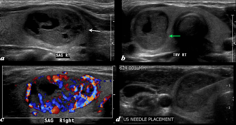
A large complex mass occupies more than half the right lobe of the thyroid gland is seen by ultrasound. The mass measures 2.6cms in sagittal and in A-P dimension measures 1.7cms(a). In transverse it measure 1.7cms. It is characterized by a mildly hypoechoic matrix with cystic type serpiginous components. (a) The margins are irregular (white arrow, a) There is an incomplete halo which shows regions of irregularity (green arrow) The lesion is extremely vascular both peripherally and centrally (c) A biopsy was performed and showed medullary carcinoma.
Courtesy Barry Sacks MD Copyright 2010 97339cL01.8
|

Hurthle Cell Carcinoma |
|
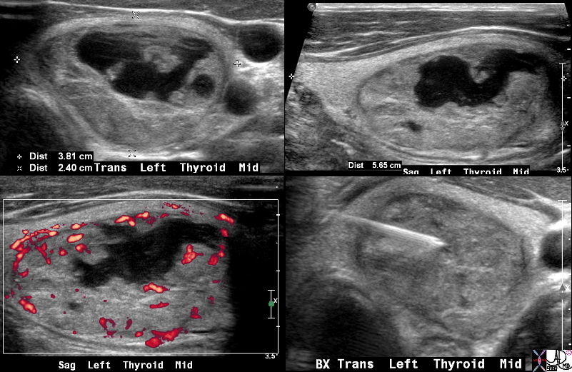
This is a thyroid scan from a 31 year old female with a clinical history of a thyroid nodule. The scan shows a heterogeneous and complex mass involving almost the entire left lobe that is well defined both on the transverse view (a) as well as the sagittal view (b). The left lobe measures 5.7X 3.8 X 2. 4 cms, resulting in a grossly enlarged left lobe as a result of the mass. An serpiginous cystic appearing component is seen in images a,b,and c and contains complex fluid and nodular excrescences. Blood flow is mostly on the periphery with the cystic component being avascular. A biopsy was performed (d), which revealed a Hurthle cell carcinoma
Courtesy Barry Sacks MD Copyright 2010 99394c.8s
|
DOMElement Object
(
    [schemaTypeInfo] =>
    [tagName] => table
    [firstElementChild] => (object value omitted)
    [lastElementChild] => (object value omitted)
    [childElementCount] => 1
    [previousElementSibling] => (object value omitted)
    [nextElementSibling] =>
    [nodeName] => table
    [nodeValue] =>


Hurthle Cell Carcinoma


This is a thyroid scan from a 31 year old female with a clinical history of a thyroid nodule. The scan shows a heterogeneous and complex mass involving almost the entire left lobe that is well defined both on the transverse view (a) as well as the sagittal view (b). The left lobe measures 5.7X 3.8 X 2. 4 cms, resulting in a grossly enlarged left lobe as a result of the mass. An serpiginous cystic appearing component is seen in images a,b,and c and contains complex fluid and nodular excrescences. Blood flow is mostly on the periphery with the cystic component being avascular. A biopsy was performed (d), which revealed a Hurthle cell carcinoma
Courtesy Barry Sacks MD Copyright 2010 99394c.8s


    [nodeType] => 1
    [parentNode] => (object value omitted)
    [childNodes] => (object value omitted)
    [firstChild] => (object value omitted)
    [lastChild] => (object value omitted)
    [previousSibling] => (object value omitted)
    [nextSibling] => (object value omitted)
    [attributes] => (object value omitted)
    [ownerDocument] => (object value omitted)
    [namespaceURI] =>
    [prefix] =>
    [localName] => table
    [baseURI] =>
    [textContent] =>


Hurthle Cell Carcinoma


This is a thyroid scan from a 31 year old female with a clinical history of a thyroid nodule. The scan shows a heterogeneous and complex mass involving almost the entire left lobe that is well defined both on the transverse view (a) as well as the sagittal view (b). The left lobe measures 5.7X 3.8 X 2. 4 cms, resulting in a grossly enlarged left lobe as a result of the mass. An serpiginous cystic appearing component is seen in images a,b,and c and contains complex fluid and nodular excrescences. Blood flow is mostly on the periphery with the cystic component being avascular. A biopsy was performed (d), which revealed a Hurthle cell carcinoma
Courtesy Barry Sacks MD Copyright 2010 99394c.8s


)
DOMElement Object
(
    [schemaTypeInfo] =>
    [tagName] => td
    [firstElementChild] => (object value omitted)
    [lastElementChild] => (object value omitted)
    [childElementCount] => 2
    [previousElementSibling] =>
    [nextElementSibling] =>
    [nodeName] => td
    [nodeValue] =>
This is a thyroid scan from a 31 year old female with a clinical history of a thyroid nodule. The scan shows a heterogeneous and complex mass involving almost the entire left lobe that is well defined both on the transverse view (a) as well as the sagittal view (b). The left lobe measures 5.7X 3.8 X 2. 4 cms, resulting in a grossly enlarged left lobe as a result of the mass. An serpiginous cystic appearing component is seen in images a,b,and c and contains complex fluid and nodular excrescences. Blood flow is mostly on the periphery with the cystic component being avascular. A biopsy was performed (d), which revealed a Hurthle cell carcinoma
Courtesy Barry Sacks MD Copyright 2010 99394c.8s

    [nodeType] => 1
    [parentNode] => (object value omitted)
    [childNodes] => (object value omitted)
    [firstChild] => (object value omitted)
    [lastChild] => (object value omitted)
    [previousSibling] => (object value omitted)
    [nextSibling] => (object value omitted)
    [attributes] => (object value omitted)
    [ownerDocument] => (object value omitted)
    [namespaceURI] =>
    [prefix] =>
    [localName] => td
    [baseURI] =>
    [textContent] =>
This is a thyroid scan from a 31 year old female with a clinical history of a thyroid nodule. The scan shows a heterogeneous and complex mass involving almost the entire left lobe that is well defined both on the transverse view (a) as well as the sagittal view (b). The left lobe measures 5.7X 3.8 X 2. 4 cms, resulting in a grossly enlarged left lobe as a result of the mass. An serpiginous cystic appearing component is seen in images a,b,and c and contains complex fluid and nodular excrescences. Blood flow is mostly on the periphery with the cystic component being avascular. A biopsy was performed (d), which revealed a Hurthle cell carcinoma
Courtesy Barry Sacks MD Copyright 2010 99394c.8s

)
DOMElement Object
(
    [schemaTypeInfo] =>
    [tagName] => td
    [firstElementChild] => (object value omitted)
    [lastElementChild] => (object value omitted)
    [childElementCount] => 2
    [previousElementSibling] =>
    [nextElementSibling] =>
    [nodeName] => td
    [nodeValue] =>
Hurthle Cell Carcinoma
    [nodeType] => 1
    [parentNode] => (object value omitted)
    [childNodes] => (object value omitted)
    [firstChild] => (object value omitted)
    [lastChild] => (object value omitted)
    [previousSibling] => (object value omitted)
    [nextSibling] => (object value omitted)
    [attributes] => (object value omitted)
    [ownerDocument] => (object value omitted)
    [namespaceURI] =>
    [prefix] =>
    [localName] => td
    [baseURI] =>
    [textContent] =>
Hurthle Cell Carcinoma
)
DOMElement Object
(
    [schemaTypeInfo] =>
    [tagName] => table
    [firstElementChild] => (object value omitted)
    [lastElementChild] => (object value omitted)
    [childElementCount] => 1
    [previousElementSibling] => (object value omitted)
    [nextElementSibling] => (object value omitted)
    [nodeName] => table
    [nodeValue] =>


Complex Lesion Dominantly Solid with Cystic Components
Medullary Cell Carcinoma


A large complex mass occupies more than half the right lobe of the thyroid gland is seen by ultrasound. The mass measures 2.6cms in sagittal and in A-P dimension measures 1.7cms(a). In transverse it measure 1.7cms. It is characterized by a mildly hypoechoic matrix with cystic type serpiginous components. (a) The margins are irregular (white arrow, a) There is an incomplete halo which shows regions of irregularity (green arrow) The lesion is extremely vascular both peripherally and centrally (c) A biopsy was performed and showed medullary carcinoma.
Courtesy Barry Sacks MD Copyright 2010 97339cL01.8


    [nodeType] => 1
    [parentNode] => (object value omitted)
    [childNodes] => (object value omitted)
    [firstChild] => (object value omitted)
    [lastChild] => (object value omitted)
    [previousSibling] => (object value omitted)
    [nextSibling] => (object value omitted)
    [attributes] => (object value omitted)
    [ownerDocument] => (object value omitted)
    [namespaceURI] =>
    [prefix] =>
    [localName] => table
    [baseURI] =>
    [textContent] =>


Complex Lesion Dominantly Solid with Cystic Components
Medullary Cell Carcinoma


A large complex mass occupies more than half the right lobe of the thyroid gland is seen by ultrasound. The mass measures 2.6cms in sagittal and in A-P dimension measures 1.7cms(a). In transverse it measure 1.7cms. It is characterized by a mildly hypoechoic matrix with cystic type serpiginous components. (a) The margins are irregular (white arrow, a) There is an incomplete halo which shows regions of irregularity (green arrow) The lesion is extremely vascular both peripherally and centrally (c) A biopsy was performed and showed medullary carcinoma.
Courtesy Barry Sacks MD Copyright 2010 97339cL01.8


)
DOMElement Object
(
    [schemaTypeInfo] =>
    [tagName] => td
    [firstElementChild] => (object value omitted)
    [lastElementChild] => (object value omitted)
    [childElementCount] => 2
    [previousElementSibling] =>
    [nextElementSibling] =>
    [nodeName] => td
    [nodeValue] =>
A large complex mass occupies more than half the right lobe of the thyroid gland is seen by ultrasound. The mass measures 2.6cms in sagittal and in A-P dimension measures 1.7cms(a). In transverse it measure 1.7cms. It is characterized by a mildly hypoechoic matrix with cystic type serpiginous components. (a) The margins are irregular (white arrow, a) There is an incomplete halo which shows regions of irregularity (green arrow) The lesion is extremely vascular both peripherally and centrally (c) A biopsy was performed and showed medullary carcinoma.
Courtesy Barry Sacks MD Copyright 2010 97339cL01.8

    [nodeType] => 1
    [parentNode] => (object value omitted)
    [childNodes] => (object value omitted)
    [firstChild] => (object value omitted)
    [lastChild] => (object value omitted)
    [previousSibling] => (object value omitted)
    [nextSibling] => (object value omitted)
    [attributes] => (object value omitted)
    [ownerDocument] => (object value omitted)
    [namespaceURI] =>
    [prefix] =>
    [localName] => td
    [baseURI] =>
    [textContent] =>
A large complex mass occupies more than half the right lobe of the thyroid gland is seen by ultrasound. The mass measures 2.6cms in sagittal and in A-P dimension measures 1.7cms(a). In transverse it measure 1.7cms. It is characterized by a mildly hypoechoic matrix with cystic type serpiginous components. (a) The margins are irregular (white arrow, a) There is an incomplete halo which shows regions of irregularity (green arrow) The lesion is extremely vascular both peripherally and centrally (c) A biopsy was performed and showed medullary carcinoma.
Courtesy Barry Sacks MD Copyright 2010 97339cL01.8

)
DOMElement Object
(
    [schemaTypeInfo] =>
    [tagName] => td
    [firstElementChild] => (object value omitted)
    [lastElementChild] => (object value omitted)
    [childElementCount] => 3
    [previousElementSibling] =>
    [nextElementSibling] =>
    [nodeName] => td
    [nodeValue] =>
Complex Lesion Dominantly Solid with Cystic Components
Medullary Cell Carcinoma
    [nodeType] => 1
    [parentNode] => (object value omitted)
    [childNodes] => (object value omitted)
    [firstChild] => (object value omitted)
    [lastChild] => (object value omitted)
    [previousSibling] => (object value omitted)
    [nextSibling] => (object value omitted)
    [attributes] => (object value omitted)
    [ownerDocument] => (object value omitted)
    [namespaceURI] =>
    [prefix] =>
    [localName] => td
    [baseURI] =>
    [textContent] =>
Complex Lesion Dominantly Solid with Cystic Components
Medullary Cell Carcinoma
)


