Histology
The Common Vein copyright 2009
Histology is the science of the microscopic structure of body tissues.
Parts
Cellular Makeup
Stromal and Connective Tissues
Extracellular Space
Blood Supply
Venous Drainage
Lymphatics
Nerves

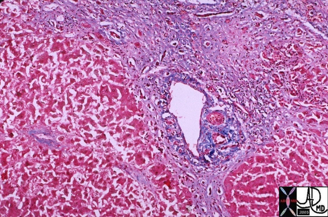
Portal vein |
| 00376 liver hepatic cirrhosis fx fibrosis fx nodules fx bridging between the triads portal triad hepatic artery portal vein bile duct histopathology Courtesy Barbara Banner |
Within the cell, the receiving, processing, and exporting functions occur between spatially oriented and connected organelles.
DOMElement Object
(
    [schemaTypeInfo] =>
    [tagName] => table
    [firstElementChild] => (object value omitted)
    [lastElementChild] => (object value omitted)
    [childElementCount] => 1
    [previousElementSibling] => (object value omitted)
    [nextElementSibling] =>
    [nodeName] => table
    [nodeValue] =>


Receiving Processing Exporting
Optimal Spatial Relationships Necessary


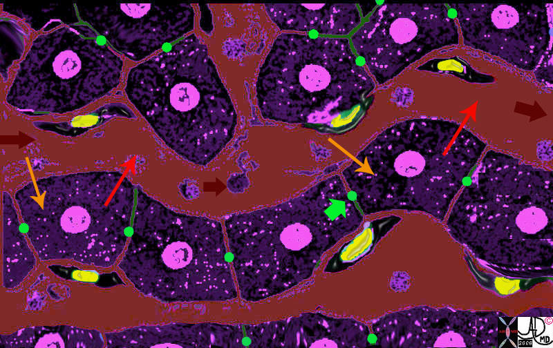
When the metabolic products enter the cell (orange arrow) they also have to enter a factory line that has a different arrangement than liver cord/sinusoid arrangement of the liver lobule.   The organelles  in the cell have to be spatially positioned and connected for the metabolic and biochemical processes to take place.  The exact mechanisms and organelles responsible for the processwill be discused in detail in future modules and are represented here as organelles with cogs situated within the cell cytoplasm.  The metabolic substrates enter the cell (light orange arrow) and are processed by the dark blue organelle, then exported to the light green organelle and so on, until the final step where a by product called bile (lime green arrow) is transported to the bile canaliculus ( green sphere between two cells) and the primary product is exported  (red arrow) into the portal circulation (maroon).
 13062b.4k08.8 Davidoff MD D avidoff art


    [nodeType] => 1
    [parentNode] => (object value omitted)
    [childNodes] => (object value omitted)
    [firstChild] => (object value omitted)
    [lastChild] => (object value omitted)
    [previousSibling] => (object value omitted)
    [nextSibling] => (object value omitted)
    [attributes] => (object value omitted)
    [ownerDocument] => (object value omitted)
    [namespaceURI] =>
    [prefix] =>
    [localName] => table
    [baseURI] =>
    [textContent] =>


Receiving Processing Exporting
Optimal Spatial Relationships Necessary


When the metabolic products enter the cell (orange arrow) they also have to enter a factory line that has a different arrangement than liver cord/sinusoid arrangement of the liver lobule.   The organelles  in the cell have to be spatially positioned and connected for the metabolic and biochemical processes to take place.  The exact mechanisms and organelles responsible for the processwill be discused in detail in future modules and are represented here as organelles with cogs situated within the cell cytoplasm.  The metabolic substrates enter the cell (light orange arrow) and are processed by the dark blue organelle, then exported to the light green organelle and so on, until the final step where a by product called bile (lime green arrow) is transported to the bile canaliculus ( green sphere between two cells) and the primary product is exported  (red arrow) into the portal circulation (maroon).
 13062b.4k08.8 Davidoff MD D avidoff art


)
DOMElement Object
(
    [schemaTypeInfo] =>
    [tagName] => td
    [firstElementChild] => (object value omitted)
    [lastElementChild] => (object value omitted)
    [childElementCount] => 2
    [previousElementSibling] =>
    [nextElementSibling] =>
    [nodeName] => td
    [nodeValue] => When the metabolic products enter the cell (orange arrow) they also have to enter a factory line that has a different arrangement than liver cord/sinusoid arrangement of the liver lobule.   The organelles  in the cell have to be spatially positioned and connected for the metabolic and biochemical processes to take place.  The exact mechanisms and organelles responsible for the processwill be discused in detail in future modules and are represented here as organelles with cogs situated within the cell cytoplasm.  The metabolic substrates enter the cell (light orange arrow) and are processed by the dark blue organelle, then exported to the light green organelle and so on, until the final step where a by product called bile (lime green arrow) is transported to the bile canaliculus ( green sphere between two cells) and the primary product is exported  (red arrow) into the portal circulation (maroon).
 13062b.4k08.8 Davidoff MD D avidoff art
    [nodeType] => 1
    [parentNode] => (object value omitted)
    [childNodes] => (object value omitted)
    [firstChild] => (object value omitted)
    [lastChild] => (object value omitted)
    [previousSibling] => (object value omitted)
    [nextSibling] => (object value omitted)
    [attributes] => (object value omitted)
    [ownerDocument] => (object value omitted)
    [namespaceURI] =>
    [prefix] =>
    [localName] => td
    [baseURI] =>
    [textContent] => When the metabolic products enter the cell (orange arrow) they also have to enter a factory line that has a different arrangement than liver cord/sinusoid arrangement of the liver lobule.   The organelles  in the cell have to be spatially positioned and connected for the metabolic and biochemical processes to take place.  The exact mechanisms and organelles responsible for the processwill be discused in detail in future modules and are represented here as organelles with cogs situated within the cell cytoplasm.  The metabolic substrates enter the cell (light orange arrow) and are processed by the dark blue organelle, then exported to the light green organelle and so on, until the final step where a by product called bile (lime green arrow) is transported to the bile canaliculus ( green sphere between two cells) and the primary product is exported  (red arrow) into the portal circulation (maroon).
 13062b.4k08.8 Davidoff MD D avidoff art
)
DOMElement Object
(
    [schemaTypeInfo] =>
    [tagName] => td
    [firstElementChild] => (object value omitted)
    [lastElementChild] => (object value omitted)
    [childElementCount] => 3
    [previousElementSibling] =>
    [nextElementSibling] =>
    [nodeName] => td
    [nodeValue] =>
Receiving Processing Exporting
Optimal Spatial Relationships Necessary

    [nodeType] => 1
    [parentNode] => (object value omitted)
    [childNodes] => (object value omitted)
    [firstChild] => (object value omitted)
    [lastChild] => (object value omitted)
    [previousSibling] => (object value omitted)
    [nextSibling] => (object value omitted)
    [attributes] => (object value omitted)
    [ownerDocument] => (object value omitted)
    [namespaceURI] =>
    [prefix] =>
    [localName] => td
    [baseURI] =>
    [textContent] =>
Receiving Processing Exporting
Optimal Spatial Relationships Necessary

)
DOMElement Object
(
    [schemaTypeInfo] =>
    [tagName] => table
    [firstElementChild] => (object value omitted)
    [lastElementChild] => (object value omitted)
    [childElementCount] => 1
    [previousElementSibling] => (object value omitted)
    [nextElementSibling] => (object value omitted)
    [nodeName] => table
    [nodeValue] =>


Positioning of the Liver Cell Around the Sinusoids


The liver cells (purple with central pink nucleus) are arranged in cords along the sinusoid (maroon) which is the smallest blood vessel of the liver.  The portal vein brings metabolic products from the gastrointestinal tract and combines with the hepatic artery which brings oxygen to the liver cell, to form the sinusoid.  The liver cell imports the raw products and oxygen (light orange arrow) from the sinusoid, processes them  and exports the product (red arrow) back into the sinusoid.  The sinusoids eventually deliver the metabolic products into the central vein which connects with inferior vena cava and the heart.
13062b05b04.8 liver hepatocyte sinusoid Kupffer cell space of Disse bile canaliculus import export excrete transport portal blood hepatic arterial blood Davidoff MD Davidoff art


    [nodeType] => 1
    [parentNode] => (object value omitted)
    [childNodes] => (object value omitted)
    [firstChild] => (object value omitted)
    [lastChild] => (object value omitted)
    [previousSibling] => (object value omitted)
    [nextSibling] => (object value omitted)
    [attributes] => (object value omitted)
    [ownerDocument] => (object value omitted)
    [namespaceURI] =>
    [prefix] =>
    [localName] => table
    [baseURI] =>
    [textContent] =>


Positioning of the Liver Cell Around the Sinusoids


The liver cells (purple with central pink nucleus) are arranged in cords along the sinusoid (maroon) which is the smallest blood vessel of the liver.  The portal vein brings metabolic products from the gastrointestinal tract and combines with the hepatic artery which brings oxygen to the liver cell, to form the sinusoid.  The liver cell imports the raw products and oxygen (light orange arrow) from the sinusoid, processes them  and exports the product (red arrow) back into the sinusoid.  The sinusoids eventually deliver the metabolic products into the central vein which connects with inferior vena cava and the heart.
13062b05b04.8 liver hepatocyte sinusoid Kupffer cell space of Disse bile canaliculus import export excrete transport portal blood hepatic arterial blood Davidoff MD Davidoff art


)
DOMElement Object
(
    [schemaTypeInfo] =>
    [tagName] => td
    [firstElementChild] => (object value omitted)
    [lastElementChild] => (object value omitted)
    [childElementCount] => 2
    [previousElementSibling] =>
    [nextElementSibling] =>
    [nodeName] => td
    [nodeValue] =>
The liver cells (purple with central pink nucleus) are arranged in cords along the sinusoid (maroon) which is the smallest blood vessel of the liver.  The portal vein brings metabolic products from the gastrointestinal tract and combines with the hepatic artery which brings oxygen to the liver cell, to form the sinusoid.  The liver cell imports the raw products and oxygen (light orange arrow) from the sinusoid, processes them  and exports the product (red arrow) back into the sinusoid.  The sinusoids eventually deliver the metabolic products into the central vein which connects with inferior vena cava and the heart.
13062b05b04.8 liver hepatocyte sinusoid Kupffer cell space of Disse bile canaliculus import export excrete transport portal blood hepatic arterial blood Davidoff MD Davidoff art
    [nodeType] => 1
    [parentNode] => (object value omitted)
    [childNodes] => (object value omitted)
    [firstChild] => (object value omitted)
    [lastChild] => (object value omitted)
    [previousSibling] => (object value omitted)
    [nextSibling] => (object value omitted)
    [attributes] => (object value omitted)
    [ownerDocument] => (object value omitted)
    [namespaceURI] =>
    [prefix] =>
    [localName] => td
    [baseURI] =>
    [textContent] =>
The liver cells (purple with central pink nucleus) are arranged in cords along the sinusoid (maroon) which is the smallest blood vessel of the liver.  The portal vein brings metabolic products from the gastrointestinal tract and combines with the hepatic artery which brings oxygen to the liver cell, to form the sinusoid.  The liver cell imports the raw products and oxygen (light orange arrow) from the sinusoid, processes them  and exports the product (red arrow) back into the sinusoid.  The sinusoids eventually deliver the metabolic products into the central vein which connects with inferior vena cava and the heart.
13062b05b04.8 liver hepatocyte sinusoid Kupffer cell space of Disse bile canaliculus import export excrete transport portal blood hepatic arterial blood Davidoff MD Davidoff art
)
DOMElement Object
(
    [schemaTypeInfo] =>
    [tagName] => td
    [firstElementChild] => (object value omitted)
    [lastElementChild] => (object value omitted)
    [childElementCount] => 2
    [previousElementSibling] =>
    [nextElementSibling] =>
    [nodeName] => td
    [nodeValue] =>
Positioning of the Liver Cell Around the Sinusoids

    [nodeType] => 1
    [parentNode] => (object value omitted)
    [childNodes] => (object value omitted)
    [firstChild] => (object value omitted)
    [lastChild] => (object value omitted)
    [previousSibling] => (object value omitted)
    [nextSibling] => (object value omitted)
    [attributes] => (object value omitted)
    [ownerDocument] => (object value omitted)
    [namespaceURI] =>
    [prefix] =>
    [localName] => td
    [baseURI] =>
    [textContent] =>
Positioning of the Liver Cell Around the Sinusoids

)
DOMElement Object
(
    [schemaTypeInfo] =>
    [tagName] => table
    [firstElementChild] => (object value omitted)
    [lastElementChild] => (object value omitted)
    [childElementCount] => 1
    [previousElementSibling] => (object value omitted)
    [nextElementSibling] => (object value omitted)
    [nodeName] => table
    [nodeValue] =>


Portal vein


00376 liver hepatic cirrhosis fx fibrosis fx nodules fx bridging between the triads portal triad hepatic artery portal vein bile duct histopathology Courtesy Barbara Banner


    [nodeType] => 1
    [parentNode] => (object value omitted)
    [childNodes] => (object value omitted)
    [firstChild] => (object value omitted)
    [lastChild] => (object value omitted)
    [previousSibling] => (object value omitted)
    [nextSibling] => (object value omitted)
    [attributes] => (object value omitted)
    [ownerDocument] => (object value omitted)
    [namespaceURI] =>
    [prefix] =>
    [localName] => table
    [baseURI] =>
    [textContent] =>


Portal vein


00376 liver hepatic cirrhosis fx fibrosis fx nodules fx bridging between the triads portal triad hepatic artery portal vein bile duct histopathology Courtesy Barbara Banner


)
DOMElement Object
(
    [schemaTypeInfo] =>
    [tagName] => td
    [firstElementChild] => (object value omitted)
    [lastElementChild] => (object value omitted)
    [childElementCount] => 1
    [previousElementSibling] =>
    [nextElementSibling] =>
    [nodeName] => td
    [nodeValue] => 00376 liver hepatic cirrhosis fx fibrosis fx nodules fx bridging between the triads portal triad hepatic artery portal vein bile duct histopathology Courtesy Barbara Banner
    [nodeType] => 1
    [parentNode] => (object value omitted)
    [childNodes] => (object value omitted)
    [firstChild] => (object value omitted)
    [lastChild] => (object value omitted)
    [previousSibling] => (object value omitted)
    [nextSibling] => (object value omitted)
    [attributes] => (object value omitted)
    [ownerDocument] => (object value omitted)
    [namespaceURI] =>
    [prefix] =>
    [localName] => td
    [baseURI] =>
    [textContent] => 00376 liver hepatic cirrhosis fx fibrosis fx nodules fx bridging between the triads portal triad hepatic artery portal vein bile duct histopathology Courtesy Barbara Banner
)
DOMElement Object
(
    [schemaTypeInfo] =>
    [tagName] => td
    [firstElementChild] => (object value omitted)
    [lastElementChild] => (object value omitted)
    [childElementCount] => 2
    [previousElementSibling] =>
    [nextElementSibling] =>
    [nodeName] => td
    [nodeValue] =>
Portal vein

    [nodeType] => 1
    [parentNode] => (object value omitted)
    [childNodes] => (object value omitted)
    [firstChild] => (object value omitted)
    [lastChild] => (object value omitted)
    [previousSibling] => (object value omitted)
    [nextSibling] => (object value omitted)
    [attributes] => (object value omitted)
    [ownerDocument] => (object value omitted)
    [namespaceURI] =>
    [prefix] =>
    [localName] => td
    [baseURI] =>
    [textContent] =>
Portal vein

)


