The Common Vein
Copyright 2011
Introduction
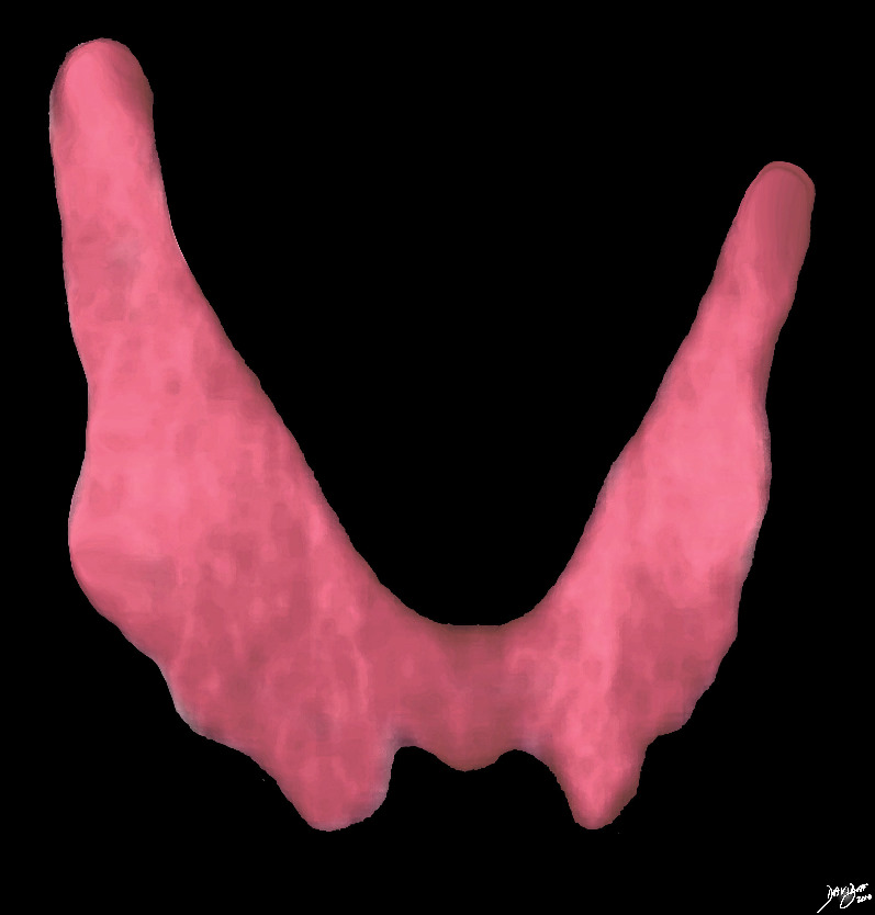
The thyroid is a butterfly-shaped gland with two lobes each an inverted cone. Together their shape varies from an H to a U shape in the coronal plane. It is also U-shaped or horseshoe shaped in the axial projection.
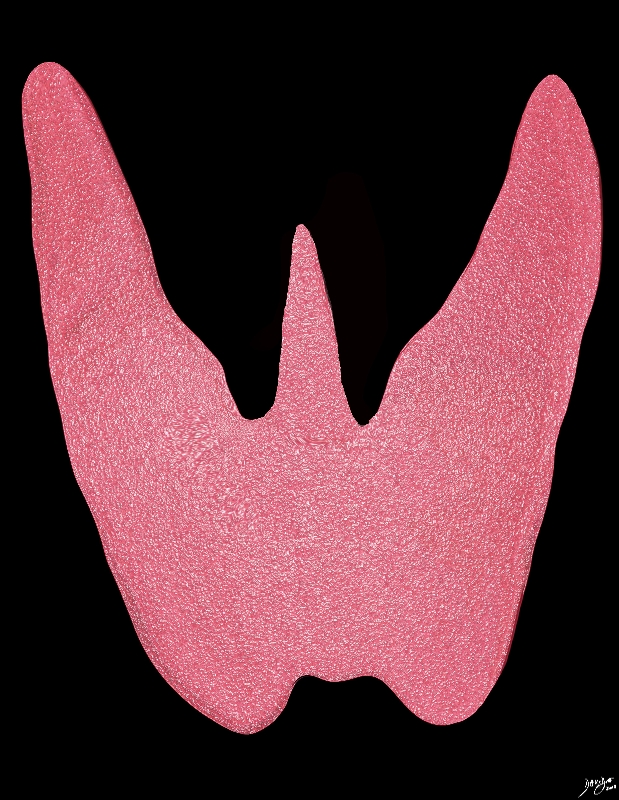
The thyroid gland gets its name from the Greek word for shield given its shape and the shape of the thyroid cartilage on which it rests. In some patients a pyramidal lobe extends cranially from the isthmus representing residual thyroglossal duct tissue that assumes thyroid character.
The shape of the thyroid cannot be observed through clinical examination; however, the ?thyroid neck check,? which requires a patient to observe his/her neck for asymmetrical bulges while swallowing, may reveal irregularities in its shape.
|
The Butterfly Shaped Organ |
|
The butterfly shaped form of the thyroid gland is appreciated in this image. Courtesy Ashley Davidoff MD copyright 2010 all rights reserved 93816d02.2kd07b10b.8s |
|
“H” or “V”or ?U? shaped Gland |
|
The H V or U shaped form of the thyroid gland is appreciated in this image. The isthmus may be broad in its craniocaudad dimension or may be relatively narrow. In the anteroposterior dimension it is only about 3-5mm thick Courtesy Ashley Davidoff MD copyright 2010 all rights reserved 93852..4kd06.8s |
|
The Pyramidal Lobe |
|
A variation in this shape of the thyroid gland is created by the presence of a pyramidal lobe which is an embryological remnant of the thyroglossal duct. It is characterized by a third small cone that lies between the lobes and sometimes originates off one of the lobes more commonly the left. Courtesy Ashley Davidoff MD Copyright 2010 93852.4kd02b02g.8s |
In the Transverse or Axial Projection
|
Thyroid Gland in Axial Projection – CT scan |
|
The axial CTscan of the neck shows the thyroid gland with the right and left lobes (pink) The gland is horseshoe shaped, and while on the horse theme the gland in this projection could also be viewed as a saddle with saddle bags n either side. The thyroid surrounds the similarly shaped trachea (black). Note in this case the right lobe is slightly larger than the left. Courtesy Ashley Davidoff MD copyright 2010 all rights reserved 30491c01d01b.8s |
|
Axial Image – Ultrasound Scan |
|
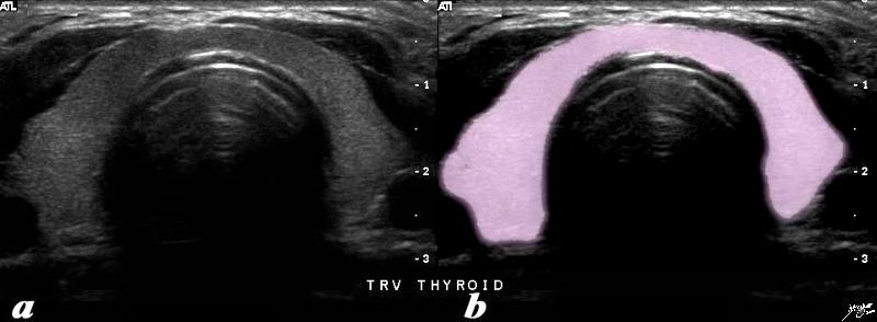
The transverse ultrasound of the thyroid gland shows the asymmetric symmetry of the lobes. In this cut the right lobe is slightly larger than the left and the isthmus consists of a narrow band that is only about 5mms thick. Courtesy Ashley Davidoff MD copyright 2010 all rights reserved 77510c02.81s |
|
Multinodular Thyroid The Distinction between Normal Displaced Thyroid Tissues and Nodule is Sometimes Difficult to Discern |
|
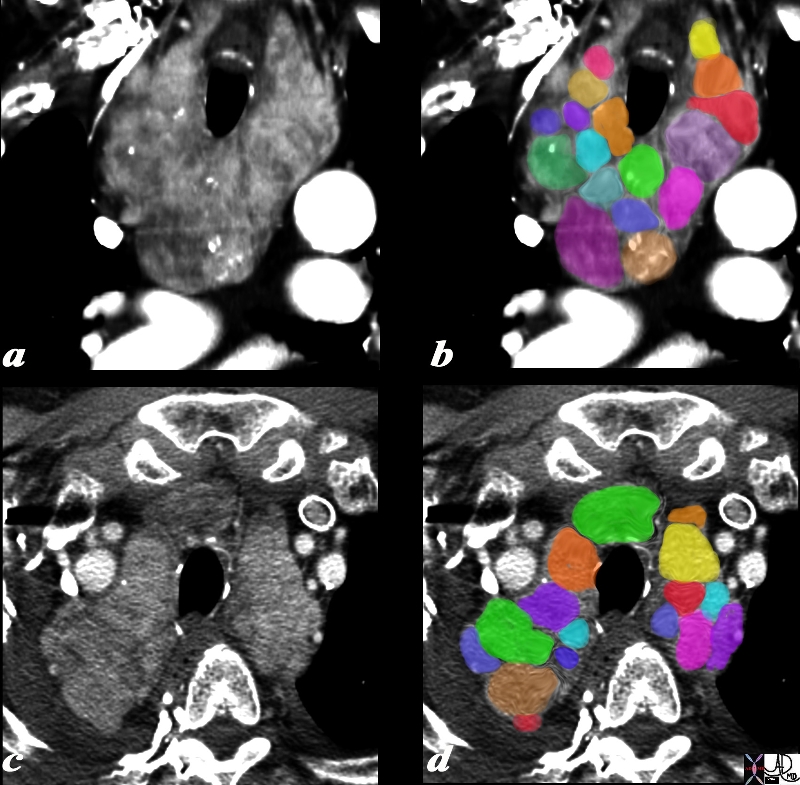
The 75 year old male presents with an asymptomatic multinodular goiter, consisting many nodules of varying size and density, some with macrocalcifications as seen by coronal CT reconstruction (a,b) and axial images (c,d). The windows of the CTscan have been narrowed (a,c), to enhance the morphological characteristics of the components of retrosternal goiter. Each lobe of the thyroid measures about 12 cms in sagittal 6cms. in transverse and 12cms in A-P dimension. These findings are consistent with a non toxic multinodular goiter. Courtesy Ashley Davidoff MD Copyright 2010 94712c01L01.8s |
|
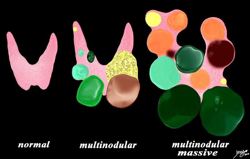
Types of Goiter That Change the Shape of the Gland |
|
The diagram illustrates non toxic multinodular goiter together with a normal thyroid gland on the left. A moderately enlarged gland (middle) containing multiple variably sized non toxic nodules with different characteristics is shown together with a massively enlarged non toxic gland (right) When the gland becomes massively enlarged it may extend into the chest, compress the airway and neck veins as well as displace other structures such as the esophagus. Courtesy Ashley Davidoff MD Copyright 2010 93852e07h01a03.8s |
DOMElement Object
(
    [schemaTypeInfo] =>
    [tagName] => table
    [firstElementChild] => (object value omitted)
    [lastElementChild] => (object value omitted)
    [childElementCount] => 1
    [previousElementSibling] => (object value omitted)
    [nextElementSibling] =>
    [nodeName] => table
    [nodeValue] =>


Types of Goiter That Change the Shape of the Gland


The diagram illustrates non toxic multinodular goiter together with a normal thyroid gland on the left. A moderately enlarged gland (middle) containing multiple variably sized non toxic nodules with different characteristics is shown together with a massively enlarged non toxic gland (right) When the gland becomes massively enlarged it may extend into the chest, compress the airway and neck veins as well as displace other structures such as the esophagus.
Courtesy Ashley Davidoff MD Copyright 2010 93852e07h01a03.8s


    [nodeType] => 1
    [parentNode] => (object value omitted)
    [childNodes] => (object value omitted)
    [firstChild] => (object value omitted)
    [lastChild] => (object value omitted)
    [previousSibling] => (object value omitted)
    [nextSibling] => (object value omitted)
    [attributes] => (object value omitted)
    [ownerDocument] => (object value omitted)
    [namespaceURI] =>
    [prefix] =>
    [localName] => table
    [baseURI] =>
    [textContent] =>


Types of Goiter That Change the Shape of the Gland


The diagram illustrates non toxic multinodular goiter together with a normal thyroid gland on the left. A moderately enlarged gland (middle) containing multiple variably sized non toxic nodules with different characteristics is shown together with a massively enlarged non toxic gland (right) When the gland becomes massively enlarged it may extend into the chest, compress the airway and neck veins as well as displace other structures such as the esophagus.
Courtesy Ashley Davidoff MD Copyright 2010 93852e07h01a03.8s


)
DOMElement Object
(
    [schemaTypeInfo] =>
    [tagName] => td
    [firstElementChild] => (object value omitted)
    [lastElementChild] => (object value omitted)
    [childElementCount] => 2
    [previousElementSibling] =>
    [nextElementSibling] =>
    [nodeName] => td
    [nodeValue] =>
The diagram illustrates non toxic multinodular goiter together with a normal thyroid gland on the left. A moderately enlarged gland (middle) containing multiple variably sized non toxic nodules with different characteristics is shown together with a massively enlarged non toxic gland (right) When the gland becomes massively enlarged it may extend into the chest, compress the airway and neck veins as well as displace other structures such as the esophagus.
Courtesy Ashley Davidoff MD Copyright 2010 93852e07h01a03.8s

    [nodeType] => 1
    [parentNode] => (object value omitted)
    [childNodes] => (object value omitted)
    [firstChild] => (object value omitted)
    [lastChild] => (object value omitted)
    [previousSibling] => (object value omitted)
    [nextSibling] => (object value omitted)
    [attributes] => (object value omitted)
    [ownerDocument] => (object value omitted)
    [namespaceURI] =>
    [prefix] =>
    [localName] => td
    [baseURI] =>
    [textContent] =>
The diagram illustrates non toxic multinodular goiter together with a normal thyroid gland on the left. A moderately enlarged gland (middle) containing multiple variably sized non toxic nodules with different characteristics is shown together with a massively enlarged non toxic gland (right) When the gland becomes massively enlarged it may extend into the chest, compress the airway and neck veins as well as displace other structures such as the esophagus.
Courtesy Ashley Davidoff MD Copyright 2010 93852e07h01a03.8s

)
DOMElement Object
(
    [schemaTypeInfo] =>
    [tagName] => td
    [firstElementChild] => (object value omitted)
    [lastElementChild] => (object value omitted)
    [childElementCount] => 2
    [previousElementSibling] =>
    [nextElementSibling] =>
    [nodeName] => td
    [nodeValue] =>

Types of Goiter That Change the Shape of the Gland

    [nodeType] => 1
    [parentNode] => (object value omitted)
    [childNodes] => (object value omitted)
    [firstChild] => (object value omitted)
    [lastChild] => (object value omitted)
    [previousSibling] => (object value omitted)
    [nextSibling] => (object value omitted)
    [attributes] => (object value omitted)
    [ownerDocument] => (object value omitted)
    [namespaceURI] =>
    [prefix] =>
    [localName] => td
    [baseURI] =>
    [textContent] =>

Types of Goiter That Change the Shape of the Gland

)
DOMElement Object
(
    [schemaTypeInfo] =>
    [tagName] => table
    [firstElementChild] => (object value omitted)
    [lastElementChild] => (object value omitted)
    [childElementCount] => 1
    [previousElementSibling] => (object value omitted)
    [nextElementSibling] => (object value omitted)
    [nodeName] => table
    [nodeValue] =>


Multinodular Thyroid
The Distinction between Normal Displaced Thyroid Tissues and Nodule is Sometimes Difficult to Discern


The 75 year old male presents with an asymptomatic multinodular goiter, consisting many nodules of varying size and density, some with macrocalcifications as seen by coronal CT reconstruction (a,b) and axial images (c,d). The windows of the CTscan have been narrowed (a,c), to enhance the morphological characteristics of the components of retrosternal goiter. Each lobe of the thyroid measures about 12 cms in sagittal 6cms. in transverse and 12cms in A-P dimension. These findings are consistent with a non toxic multinodular goiter.
Courtesy Ashley Davidoff MD Copyright 2010 94712c01L01.8s


    [nodeType] => 1
    [parentNode] => (object value omitted)
    [childNodes] => (object value omitted)
    [firstChild] => (object value omitted)
    [lastChild] => (object value omitted)
    [previousSibling] => (object value omitted)
    [nextSibling] => (object value omitted)
    [attributes] => (object value omitted)
    [ownerDocument] => (object value omitted)
    [namespaceURI] =>
    [prefix] =>
    [localName] => table
    [baseURI] =>
    [textContent] =>


Multinodular Thyroid
The Distinction between Normal Displaced Thyroid Tissues and Nodule is Sometimes Difficult to Discern


The 75 year old male presents with an asymptomatic multinodular goiter, consisting many nodules of varying size and density, some with macrocalcifications as seen by coronal CT reconstruction (a,b) and axial images (c,d). The windows of the CTscan have been narrowed (a,c), to enhance the morphological characteristics of the components of retrosternal goiter. Each lobe of the thyroid measures about 12 cms in sagittal 6cms. in transverse and 12cms in A-P dimension. These findings are consistent with a non toxic multinodular goiter.
Courtesy Ashley Davidoff MD Copyright 2010 94712c01L01.8s


)
DOMElement Object
(
    [schemaTypeInfo] =>
    [tagName] => td
    [firstElementChild] => (object value omitted)
    [lastElementChild] => (object value omitted)
    [childElementCount] => 2
    [previousElementSibling] =>
    [nextElementSibling] =>
    [nodeName] => td
    [nodeValue] =>
The 75 year old male presents with an asymptomatic multinodular goiter, consisting many nodules of varying size and density, some with macrocalcifications as seen by coronal CT reconstruction (a,b) and axial images (c,d). The windows of the CTscan have been narrowed (a,c), to enhance the morphological characteristics of the components of retrosternal goiter. Each lobe of the thyroid measures about 12 cms in sagittal 6cms. in transverse and 12cms in A-P dimension. These findings are consistent with a non toxic multinodular goiter.
Courtesy Ashley Davidoff MD Copyright 2010 94712c01L01.8s

    [nodeType] => 1
    [parentNode] => (object value omitted)
    [childNodes] => (object value omitted)
    [firstChild] => (object value omitted)
    [lastChild] => (object value omitted)
    [previousSibling] => (object value omitted)
    [nextSibling] => (object value omitted)
    [attributes] => (object value omitted)
    [ownerDocument] => (object value omitted)
    [namespaceURI] =>
    [prefix] =>
    [localName] => td
    [baseURI] =>
    [textContent] =>
The 75 year old male presents with an asymptomatic multinodular goiter, consisting many nodules of varying size and density, some with macrocalcifications as seen by coronal CT reconstruction (a,b) and axial images (c,d). The windows of the CTscan have been narrowed (a,c), to enhance the morphological characteristics of the components of retrosternal goiter. Each lobe of the thyroid measures about 12 cms in sagittal 6cms. in transverse and 12cms in A-P dimension. These findings are consistent with a non toxic multinodular goiter.
Courtesy Ashley Davidoff MD Copyright 2010 94712c01L01.8s

)
DOMElement Object
(
    [schemaTypeInfo] =>
    [tagName] => td
    [firstElementChild] => (object value omitted)
    [lastElementChild] => (object value omitted)
    [childElementCount] => 3
    [previousElementSibling] =>
    [nextElementSibling] =>
    [nodeName] => td
    [nodeValue] =>

Multinodular Thyroid
The Distinction between Normal Displaced Thyroid Tissues and Nodule is Sometimes Difficult to Discern

    [nodeType] => 1
    [parentNode] => (object value omitted)
    [childNodes] => (object value omitted)
    [firstChild] => (object value omitted)
    [lastChild] => (object value omitted)
    [previousSibling] => (object value omitted)
    [nextSibling] => (object value omitted)
    [attributes] => (object value omitted)
    [ownerDocument] => (object value omitted)
    [namespaceURI] =>
    [prefix] =>
    [localName] => td
    [baseURI] =>
    [textContent] =>

Multinodular Thyroid
The Distinction between Normal Displaced Thyroid Tissues and Nodule is Sometimes Difficult to Discern

)
DOMElement Object
(
    [schemaTypeInfo] =>
    [tagName] => table
    [firstElementChild] => (object value omitted)
    [lastElementChild] => (object value omitted)
    [childElementCount] => 1
    [previousElementSibling] => (object value omitted)
    [nextElementSibling] => (object value omitted)
    [nodeName] => table
    [nodeValue] =>


Axial Image – Ultrasound Scan


The transverse ultrasound of the thyroid gland shows the asymmetric symmetry of the lobes. In this cut the right lobe is slightly larger than the left and the isthmus consists of a narrow band that is only about 5mms thick.
Courtesy Ashley Davidoff MD copyright 2010 all rights reserved 77510c02.81s


    [nodeType] => 1
    [parentNode] => (object value omitted)
    [childNodes] => (object value omitted)
    [firstChild] => (object value omitted)
    [lastChild] => (object value omitted)
    [previousSibling] => (object value omitted)
    [nextSibling] => (object value omitted)
    [attributes] => (object value omitted)
    [ownerDocument] => (object value omitted)
    [namespaceURI] =>
    [prefix] =>
    [localName] => table
    [baseURI] =>
    [textContent] =>


Axial Image – Ultrasound Scan


The transverse ultrasound of the thyroid gland shows the asymmetric symmetry of the lobes. In this cut the right lobe is slightly larger than the left and the isthmus consists of a narrow band that is only about 5mms thick.
Courtesy Ashley Davidoff MD copyright 2010 all rights reserved 77510c02.81s


)
DOMElement Object
(
    [schemaTypeInfo] =>
    [tagName] => td
    [firstElementChild] => (object value omitted)
    [lastElementChild] => (object value omitted)
    [childElementCount] => 2
    [previousElementSibling] =>
    [nextElementSibling] =>
    [nodeName] => td
    [nodeValue] =>
The transverse ultrasound of the thyroid gland shows the asymmetric symmetry of the lobes. In this cut the right lobe is slightly larger than the left and the isthmus consists of a narrow band that is only about 5mms thick.
Courtesy Ashley Davidoff MD copyright 2010 all rights reserved 77510c02.81s

    [nodeType] => 1
    [parentNode] => (object value omitted)
    [childNodes] => (object value omitted)
    [firstChild] => (object value omitted)
    [lastChild] => (object value omitted)
    [previousSibling] => (object value omitted)
    [nextSibling] => (object value omitted)
    [attributes] => (object value omitted)
    [ownerDocument] => (object value omitted)
    [namespaceURI] =>
    [prefix] =>
    [localName] => td
    [baseURI] =>
    [textContent] =>
The transverse ultrasound of the thyroid gland shows the asymmetric symmetry of the lobes. In this cut the right lobe is slightly larger than the left and the isthmus consists of a narrow band that is only about 5mms thick.
Courtesy Ashley Davidoff MD copyright 2010 all rights reserved 77510c02.81s

)
DOMElement Object
(
    [schemaTypeInfo] =>
    [tagName] => td
    [firstElementChild] => (object value omitted)
    [lastElementChild] => (object value omitted)
    [childElementCount] => 2
    [previousElementSibling] =>
    [nextElementSibling] =>
    [nodeName] => td
    [nodeValue] =>

Axial Image – Ultrasound Scan

    [nodeType] => 1
    [parentNode] => (object value omitted)
    [childNodes] => (object value omitted)
    [firstChild] => (object value omitted)
    [lastChild] => (object value omitted)
    [previousSibling] => (object value omitted)
    [nextSibling] => (object value omitted)
    [attributes] => (object value omitted)
    [ownerDocument] => (object value omitted)
    [namespaceURI] =>
    [prefix] =>
    [localName] => td
    [baseURI] =>
    [textContent] =>

Axial Image – Ultrasound Scan

)
DOMElement Object
(
    [schemaTypeInfo] =>
    [tagName] => table
    [firstElementChild] => (object value omitted)
    [lastElementChild] => (object value omitted)
    [childElementCount] => 1
    [previousElementSibling] => (object value omitted)
    [nextElementSibling] => (object value omitted)
    [nodeName] => table
    [nodeValue] =>


Thyroid Gland in Axial Projection – CT scan


The axial CTscan of the neck shows the thyroid gland with the right and left lobes (pink) The gland is horseshoe shaped, and while on the horse theme the gland in this projection could also be viewed as a saddle with saddle bags n either side. The thyroid surrounds the similarly shaped trachea (black).  Note in this case the right lobe is slightly larger than the left.
Courtesy Ashley Davidoff MD copyright 2010 all rights reserved 30491c01d01b.8s


    [nodeType] => 1
    [parentNode] => (object value omitted)
    [childNodes] => (object value omitted)
    [firstChild] => (object value omitted)
    [lastChild] => (object value omitted)
    [previousSibling] => (object value omitted)
    [nextSibling] => (object value omitted)
    [attributes] => (object value omitted)
    [ownerDocument] => (object value omitted)
    [namespaceURI] =>
    [prefix] =>
    [localName] => table
    [baseURI] =>
    [textContent] =>


Thyroid Gland in Axial Projection – CT scan


The axial CTscan of the neck shows the thyroid gland with the right and left lobes (pink) The gland is horseshoe shaped, and while on the horse theme the gland in this projection could also be viewed as a saddle with saddle bags n either side. The thyroid surrounds the similarly shaped trachea (black).  Note in this case the right lobe is slightly larger than the left.
Courtesy Ashley Davidoff MD copyright 2010 all rights reserved 30491c01d01b.8s


)
DOMElement Object
(
    [schemaTypeInfo] =>
    [tagName] => td
    [firstElementChild] => (object value omitted)
    [lastElementChild] => (object value omitted)
    [childElementCount] => 2
    [previousElementSibling] =>
    [nextElementSibling] =>
    [nodeName] => td
    [nodeValue] =>
The axial CTscan of the neck shows the thyroid gland with the right and left lobes (pink) The gland is horseshoe shaped, and while on the horse theme the gland in this projection could also be viewed as a saddle with saddle bags n either side. The thyroid surrounds the similarly shaped trachea (black).  Note in this case the right lobe is slightly larger than the left.
Courtesy Ashley Davidoff MD copyright 2010 all rights reserved 30491c01d01b.8s

    [nodeType] => 1
    [parentNode] => (object value omitted)
    [childNodes] => (object value omitted)
    [firstChild] => (object value omitted)
    [lastChild] => (object value omitted)
    [previousSibling] => (object value omitted)
    [nextSibling] => (object value omitted)
    [attributes] => (object value omitted)
    [ownerDocument] => (object value omitted)
    [namespaceURI] =>
    [prefix] =>
    [localName] => td
    [baseURI] =>
    [textContent] =>
The axial CTscan of the neck shows the thyroid gland with the right and left lobes (pink) The gland is horseshoe shaped, and while on the horse theme the gland in this projection could also be viewed as a saddle with saddle bags n either side. The thyroid surrounds the similarly shaped trachea (black).  Note in this case the right lobe is slightly larger than the left.
Courtesy Ashley Davidoff MD copyright 2010 all rights reserved 30491c01d01b.8s

)
DOMElement Object
(
    [schemaTypeInfo] =>
    [tagName] => td
    [firstElementChild] => (object value omitted)
    [lastElementChild] => (object value omitted)
    [childElementCount] => 2
    [previousElementSibling] =>
    [nextElementSibling] =>
    [nodeName] => td
    [nodeValue] =>

Thyroid Gland in Axial Projection – CT scan

    [nodeType] => 1
    [parentNode] => (object value omitted)
    [childNodes] => (object value omitted)
    [firstChild] => (object value omitted)
    [lastChild] => (object value omitted)
    [previousSibling] => (object value omitted)
    [nextSibling] => (object value omitted)
    [attributes] => (object value omitted)
    [ownerDocument] => (object value omitted)
    [namespaceURI] =>
    [prefix] =>
    [localName] => td
    [baseURI] =>
    [textContent] =>

Thyroid Gland in Axial Projection – CT scan

)
DOMElement Object
(
    [schemaTypeInfo] =>
    [tagName] => table
    [firstElementChild] => (object value omitted)
    [lastElementChild] => (object value omitted)
    [childElementCount] => 1
    [previousElementSibling] => (object value omitted)
    [nextElementSibling] => (object value omitted)
    [nodeName] => table
    [nodeValue] =>


The Pyramidal Lobe


A variation in this shape of the thyroid gland is created by the presence of a pyramidal lobe which is an embryological remnant of the thyroglossal duct.  It is characterized by a third small cone that lies between the lobes and sometimes originates off one of the lobes more commonly the left.
Courtesy Ashley Davidoff MD Copyright 2010 93852.4kd02b02g.8s


    [nodeType] => 1
    [parentNode] => (object value omitted)
    [childNodes] => (object value omitted)
    [firstChild] => (object value omitted)
    [lastChild] => (object value omitted)
    [previousSibling] => (object value omitted)
    [nextSibling] => (object value omitted)
    [attributes] => (object value omitted)
    [ownerDocument] => (object value omitted)
    [namespaceURI] =>
    [prefix] =>
    [localName] => table
    [baseURI] =>
    [textContent] =>


The Pyramidal Lobe


A variation in this shape of the thyroid gland is created by the presence of a pyramidal lobe which is an embryological remnant of the thyroglossal duct.  It is characterized by a third small cone that lies between the lobes and sometimes originates off one of the lobes more commonly the left.
Courtesy Ashley Davidoff MD Copyright 2010 93852.4kd02b02g.8s


)
DOMElement Object
(
    [schemaTypeInfo] =>
    [tagName] => td
    [firstElementChild] => (object value omitted)
    [lastElementChild] => (object value omitted)
    [childElementCount] => 2
    [previousElementSibling] =>
    [nextElementSibling] =>
    [nodeName] => td
    [nodeValue] =>
A variation in this shape of the thyroid gland is created by the presence of a pyramidal lobe which is an embryological remnant of the thyroglossal duct.  It is characterized by a third small cone that lies between the lobes and sometimes originates off one of the lobes more commonly the left.
Courtesy Ashley Davidoff MD Copyright 2010 93852.4kd02b02g.8s

    [nodeType] => 1
    [parentNode] => (object value omitted)
    [childNodes] => (object value omitted)
    [firstChild] => (object value omitted)
    [lastChild] => (object value omitted)
    [previousSibling] => (object value omitted)
    [nextSibling] => (object value omitted)
    [attributes] => (object value omitted)
    [ownerDocument] => (object value omitted)
    [namespaceURI] =>
    [prefix] =>
    [localName] => td
    [baseURI] =>
    [textContent] =>
A variation in this shape of the thyroid gland is created by the presence of a pyramidal lobe which is an embryological remnant of the thyroglossal duct.  It is characterized by a third small cone that lies between the lobes and sometimes originates off one of the lobes more commonly the left.
Courtesy Ashley Davidoff MD Copyright 2010 93852.4kd02b02g.8s

)
DOMElement Object
(
    [schemaTypeInfo] =>
    [tagName] => td
    [firstElementChild] => (object value omitted)
    [lastElementChild] => (object value omitted)
    [childElementCount] => 2
    [previousElementSibling] =>
    [nextElementSibling] =>
    [nodeName] => td
    [nodeValue] =>

The Pyramidal Lobe

    [nodeType] => 1
    [parentNode] => (object value omitted)
    [childNodes] => (object value omitted)
    [firstChild] => (object value omitted)
    [lastChild] => (object value omitted)
    [previousSibling] => (object value omitted)
    [nextSibling] => (object value omitted)
    [attributes] => (object value omitted)
    [ownerDocument] => (object value omitted)
    [namespaceURI] =>
    [prefix] =>
    [localName] => td
    [baseURI] =>
    [textContent] =>

The Pyramidal Lobe

)
DOMElement Object
(
    [schemaTypeInfo] =>
    [tagName] => table
    [firstElementChild] => (object value omitted)
    [lastElementChild] => (object value omitted)
    [childElementCount] => 1
    [previousElementSibling] => (object value omitted)
    [nextElementSibling] => (object value omitted)
    [nodeName] => table
    [nodeValue] =>


Variation in the Normal Shape of the Thyroid


The shape of the thyroid is well appreciated with the two lobes connected by the isthmus.  The size and shapes of the two lobes may vary.
Courtesy Ashley Davidoff MD  93852.8kb15.3kb09b01.8s


    [nodeType] => 1
    [parentNode] => (object value omitted)
    [childNodes] => (object value omitted)
    [firstChild] => (object value omitted)
    [lastChild] => (object value omitted)
    [previousSibling] => (object value omitted)
    [nextSibling] => (object value omitted)
    [attributes] => (object value omitted)
    [ownerDocument] => (object value omitted)
    [namespaceURI] =>
    [prefix] =>
    [localName] => table
    [baseURI] =>
    [textContent] =>


Variation in the Normal Shape of the Thyroid


The shape of the thyroid is well appreciated with the two lobes connected by the isthmus.  The size and shapes of the two lobes may vary.
Courtesy Ashley Davidoff MD  93852.8kb15.3kb09b01.8s


)
DOMElement Object
(
    [schemaTypeInfo] =>
    [tagName] => td
    [firstElementChild] => (object value omitted)
    [lastElementChild] => (object value omitted)
    [childElementCount] => 2
    [previousElementSibling] =>
    [nextElementSibling] =>
    [nodeName] => td
    [nodeValue] =>
The shape of the thyroid is well appreciated with the two lobes connected by the isthmus.  The size and shapes of the two lobes may vary.
Courtesy Ashley Davidoff MD  93852.8kb15.3kb09b01.8s

    [nodeType] => 1
    [parentNode] => (object value omitted)
    [childNodes] => (object value omitted)
    [firstChild] => (object value omitted)
    [lastChild] => (object value omitted)
    [previousSibling] => (object value omitted)
    [nextSibling] => (object value omitted)
    [attributes] => (object value omitted)
    [ownerDocument] => (object value omitted)
    [namespaceURI] =>
    [prefix] =>
    [localName] => td
    [baseURI] =>
    [textContent] =>
The shape of the thyroid is well appreciated with the two lobes connected by the isthmus.  The size and shapes of the two lobes may vary.
Courtesy Ashley Davidoff MD  93852.8kb15.3kb09b01.8s

)
DOMElement Object
(
    [schemaTypeInfo] =>
    [tagName] => td
    [firstElementChild] => (object value omitted)
    [lastElementChild] => (object value omitted)
    [childElementCount] => 2
    [previousElementSibling] =>
    [nextElementSibling] =>
    [nodeName] => td
    [nodeValue] =>

Variation in the Normal Shape of the Thyroid

    [nodeType] => 1
    [parentNode] => (object value omitted)
    [childNodes] => (object value omitted)
    [firstChild] => (object value omitted)
    [lastChild] => (object value omitted)
    [previousSibling] => (object value omitted)
    [nextSibling] => (object value omitted)
    [attributes] => (object value omitted)
    [ownerDocument] => (object value omitted)
    [namespaceURI] =>
    [prefix] =>
    [localName] => td
    [baseURI] =>
    [textContent] =>

Variation in the Normal Shape of the Thyroid

)
DOMElement Object
(
    [schemaTypeInfo] =>
    [tagName] => table
    [firstElementChild] => (object value omitted)
    [lastElementChild] => (object value omitted)
    [childElementCount] => 1
    [previousElementSibling] => (object value omitted)
    [nextElementSibling] => (object value omitted)
    [nodeName] => table
    [nodeValue] =>


“H” or “V”or ?U? shaped Gland


The H V or U  shaped form of the thyroid gland is appreciated in this image. The isthmus may be broad in its craniocaudad dimension or may be relatively narrow. In the anteroposterior dimension it is only about 3-5mm thick
Courtesy Ashley Davidoff MD copyright 2010 all rights reserved 93852..4kd06.8s


    [nodeType] => 1
    [parentNode] => (object value omitted)
    [childNodes] => (object value omitted)
    [firstChild] => (object value omitted)
    [lastChild] => (object value omitted)
    [previousSibling] => (object value omitted)
    [nextSibling] => (object value omitted)
    [attributes] => (object value omitted)
    [ownerDocument] => (object value omitted)
    [namespaceURI] =>
    [prefix] =>
    [localName] => table
    [baseURI] =>
    [textContent] =>


“H” or “V”or ?U? shaped Gland


The H V or U  shaped form of the thyroid gland is appreciated in this image. The isthmus may be broad in its craniocaudad dimension or may be relatively narrow. In the anteroposterior dimension it is only about 3-5mm thick
Courtesy Ashley Davidoff MD copyright 2010 all rights reserved 93852..4kd06.8s


)
DOMElement Object
(
    [schemaTypeInfo] =>
    [tagName] => td
    [firstElementChild] => (object value omitted)
    [lastElementChild] => (object value omitted)
    [childElementCount] => 2
    [previousElementSibling] =>
    [nextElementSibling] =>
    [nodeName] => td
    [nodeValue] =>
The H V or U  shaped form of the thyroid gland is appreciated in this image. The isthmus may be broad in its craniocaudad dimension or may be relatively narrow. In the anteroposterior dimension it is only about 3-5mm thick
Courtesy Ashley Davidoff MD copyright 2010 all rights reserved 93852..4kd06.8s

    [nodeType] => 1
    [parentNode] => (object value omitted)
    [childNodes] => (object value omitted)
    [firstChild] => (object value omitted)
    [lastChild] => (object value omitted)
    [previousSibling] => (object value omitted)
    [nextSibling] => (object value omitted)
    [attributes] => (object value omitted)
    [ownerDocument] => (object value omitted)
    [namespaceURI] =>
    [prefix] =>
    [localName] => td
    [baseURI] =>
    [textContent] =>
The H V or U  shaped form of the thyroid gland is appreciated in this image. The isthmus may be broad in its craniocaudad dimension or may be relatively narrow. In the anteroposterior dimension it is only about 3-5mm thick
Courtesy Ashley Davidoff MD copyright 2010 all rights reserved 93852..4kd06.8s

)
DOMElement Object
(
    [schemaTypeInfo] =>
    [tagName] => td
    [firstElementChild] => (object value omitted)
    [lastElementChild] => (object value omitted)
    [childElementCount] => 2
    [previousElementSibling] =>
    [nextElementSibling] =>
    [nodeName] => td
    [nodeValue] =>

“H” or “V”or ?U? shaped Gland

    [nodeType] => 1
    [parentNode] => (object value omitted)
    [childNodes] => (object value omitted)
    [firstChild] => (object value omitted)
    [lastChild] => (object value omitted)
    [previousSibling] => (object value omitted)
    [nextSibling] => (object value omitted)
    [attributes] => (object value omitted)
    [ownerDocument] => (object value omitted)
    [namespaceURI] =>
    [prefix] =>
    [localName] => td
    [baseURI] =>
    [textContent] =>

“H” or “V”or ?U? shaped Gland

)
DOMElement Object
(
    [schemaTypeInfo] =>
    [tagName] => table
    [firstElementChild] => (object value omitted)
    [lastElementChild] => (object value omitted)
    [childElementCount] => 1
    [previousElementSibling] => (object value omitted)
    [nextElementSibling] => (object value omitted)
    [nodeName] => table
    [nodeValue] =>


The Butterfly Shaped Organ


The butterfly shaped form of the thyroid gland is appreciated in this image.
Courtesy Ashley Davidoff MD copyright 2010 all rights reserved 93816d02.2kd07b10b.8s


    [nodeType] => 1
    [parentNode] => (object value omitted)
    [childNodes] => (object value omitted)
    [firstChild] => (object value omitted)
    [lastChild] => (object value omitted)
    [previousSibling] => (object value omitted)
    [nextSibling] => (object value omitted)
    [attributes] => (object value omitted)
    [ownerDocument] => (object value omitted)
    [namespaceURI] =>
    [prefix] =>
    [localName] => table
    [baseURI] =>
    [textContent] =>


The Butterfly Shaped Organ


The butterfly shaped form of the thyroid gland is appreciated in this image.
Courtesy Ashley Davidoff MD copyright 2010 all rights reserved 93816d02.2kd07b10b.8s


)
DOMElement Object
(
    [schemaTypeInfo] =>
    [tagName] => td
    [firstElementChild] => (object value omitted)
    [lastElementChild] => (object value omitted)
    [childElementCount] => 2
    [previousElementSibling] =>
    [nextElementSibling] =>
    [nodeName] => td
    [nodeValue] =>
The butterfly shaped form of the thyroid gland is appreciated in this image.
Courtesy Ashley Davidoff MD copyright 2010 all rights reserved 93816d02.2kd07b10b.8s

    [nodeType] => 1
    [parentNode] => (object value omitted)
    [childNodes] => (object value omitted)
    [firstChild] => (object value omitted)
    [lastChild] => (object value omitted)
    [previousSibling] => (object value omitted)
    [nextSibling] => (object value omitted)
    [attributes] => (object value omitted)
    [ownerDocument] => (object value omitted)
    [namespaceURI] =>
    [prefix] =>
    [localName] => td
    [baseURI] =>
    [textContent] =>
The butterfly shaped form of the thyroid gland is appreciated in this image.
Courtesy Ashley Davidoff MD copyright 2010 all rights reserved 93816d02.2kd07b10b.8s

)
DOMElement Object
(
    [schemaTypeInfo] =>
    [tagName] => td
    [firstElementChild] => (object value omitted)
    [lastElementChild] => (object value omitted)
    [childElementCount] => 2
    [previousElementSibling] =>
    [nextElementSibling] =>
    [nodeName] => td
    [nodeValue] =>

The Butterfly Shaped Organ

    [nodeType] => 1
    [parentNode] => (object value omitted)
    [childNodes] => (object value omitted)
    [firstChild] => (object value omitted)
    [lastChild] => (object value omitted)
    [previousSibling] => (object value omitted)
    [nextSibling] => (object value omitted)
    [attributes] => (object value omitted)
    [ownerDocument] => (object value omitted)
    [namespaceURI] =>
    [prefix] =>
    [localName] => td
    [baseURI] =>
    [textContent] =>

The Butterfly Shaped Organ

)


