49671c01
Ashley Davidoff MD
TheCommonVein.net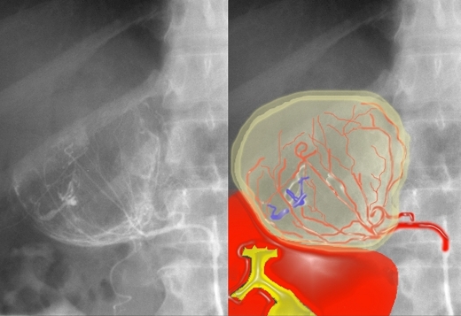
In this injection of the middle adrenal artery, a mass is apparent in the right adrenal gland. The branches of the artery are distorted and, rather than a triangular shape as seen on previous image, we see a rounded mass. There is evidence of early venous filling (blue overlay) reflecting an arteriovenous shunt, characteristic of a hypervascular tumor.
39538
Ashley Davidoff MD
TheCommonVein.net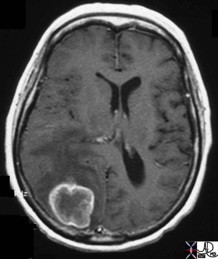
Metastasis from Endometrial Carcinoma
21694
Ashley Davidoff MD
TheCommonVein.net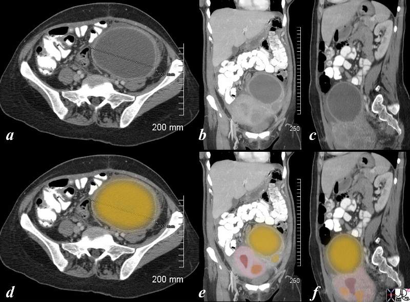
The CT is from a 36 year old female recently post c section who developed pain fever and rigors in the left lower quadrant. She has a known fibroid uterus but a new tender mass was found in her left adnexa. The CTscan shows a large cystic collection in the left adnexa with an enhancing rind. This was aspirated under ultrasound guidance , pus was aspirated and a diagnosis of an ovarian abscess was made. She was treated on antibiotics and she subsequently her pain and fever resolved pain
83287c03.8s
Ashley Davidoff MD
TheCommonVein.net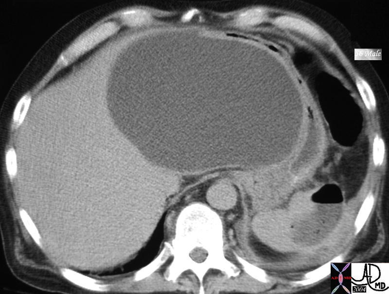
40116
Ashley Davidoff MD
TheCommonVein.net
42262 42262 b01 42269 42269b01 42271b 42271b01
Ashley Davidoff MD
TheCommonVein.net
42262 42262 b01 42269 42269b01 42271b 42271b01
Ashley Davidoff MD
TheCommonVein.net
42262 42262 b01 42269 42269b01 42271b 42271b01
Ashley Davidoff MD
TheCommonVein.net
42262 42262 b01 42269 42269b01 42271b 42271b01
Ashley Davidoff MD
TheCommonVein.net
42262 42262 b01 42269 42269b01 42271b 42271b01
Ashley Davidoff MD
TheCommonVein.net
42262 42262 b01 42269 42269b01 42271b 42271b01
Ashley Davidoff MD
TheCommonVein.net
42244c01
Ashley Davidoff MD
TheCommonVein.net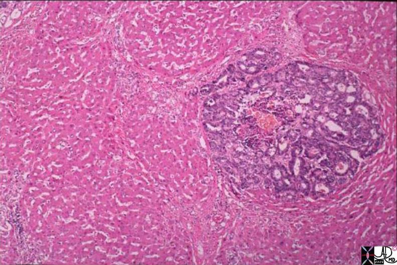
The histopathological specimen obtained from autopsy biopsy is from a liver and shows discrete metastatic deposits of glandular type metastases separated by spaces within the lesion of mucus consistent with metastatic adenocarcinoma. The distinct blueness and “badness of the malignant tissue is reflective of the hyperchromicity of the nuclii and their dominance in the cell. The malignant unit occupies space and displaces normal liver tissue. Note also the roundness in shape that is characteristic of malignant tissue
02938b01.8s
Ashley Davidoff MD
TheCommonVein.net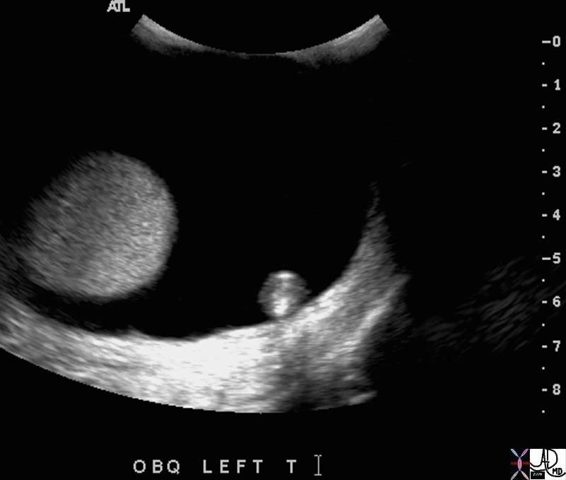
Elderly man with left testicular pain
47719
Ashley Davidoff MD
TheCommonVein.net
Elderly man with left testicular pain testis hydrocele under pressure
47735
Ashley Davidoff MD
TheCommonVein.net
The CTscan of the abdominal aorta reveals an acute rupture characterized by a high density crescent shaped density of acute blood within the chronic thrombus of the aortic lumen. The extraluminal component of the rupture is seen as high density acute blood in the retroperitoneum.
27175
Ashley Davidoff MD
TheCommonVein.net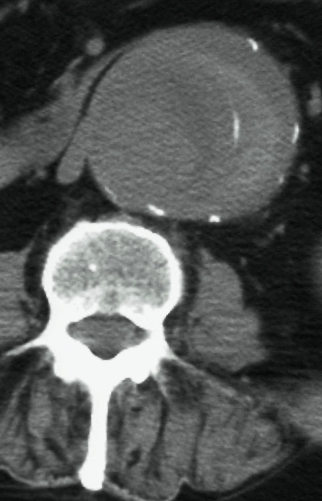
25254b
Ashley Davidoff MD
TheCommonVein.net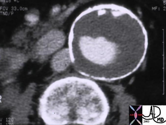
This magnified view of the infra-renal aorta using CT, shows an 6.5cms aneurysm, with a narrowed lumen and a wall thickened by thrombus. The vertebral body is 5.5cms in transverse dimension and it can be used as a reference structure to assess the size of the aorta. ie if the aorta reaches the size the vertebra, it is time to repair it.
10208
Ashley Davidoff MD
TheCommonVein.net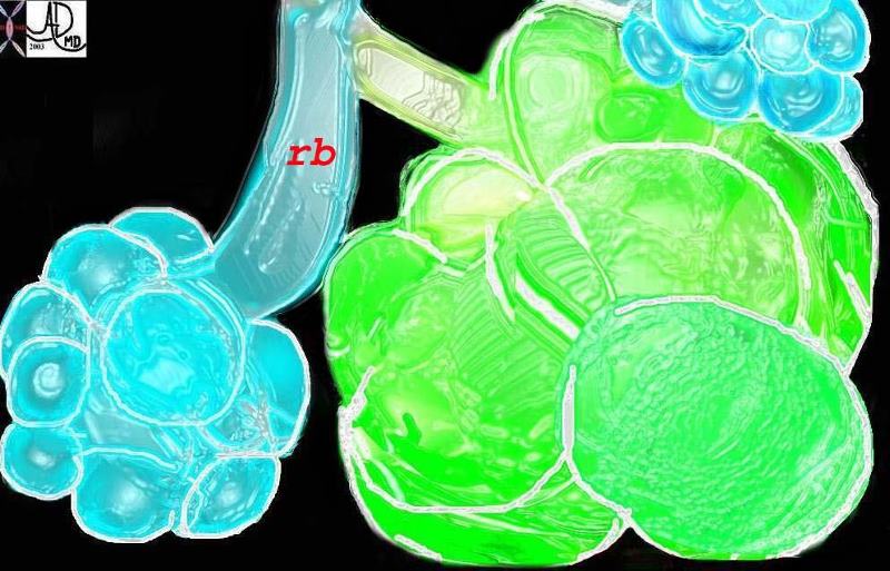
This diagram shows alveoli and respiratory bronchioles that are too large due to loss of elasticity, so that air cannot be moved efficiently through them This is a diagram of emphysema causing hyperinflated lungs
keywords lung Davidoff tree branching emphysema size enlarged alveoli enlarged airways respiratory bronchiole
32645b01.800
Ashley Davidoff MD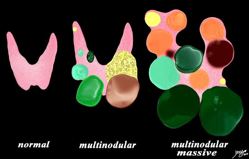
The diagram illustrates non toxic multinodular goiter together with a normal thyroid gland on the left. A moderately enlarged gland (middle) containing multiple variably sized non toxic nodules with different characteristics is shown together with a massively enlarged non toxic gland (right) When the gland becomes massively enlarged it may extend into the chest, compress the airway and neck veins as well as displace other structures such as the esophagus.
93852e07h01a03.8s
Ashley Davidoff MD
TheCommonVein.net
