Introduction
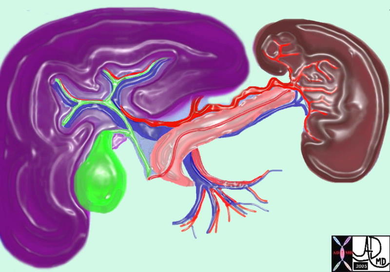
The portal vein is a medium sized to large vein structurally characterised by its portal nature providing connections between liver, spleen, and the gastrointestinal tract and its position in the porta hepatis as it branches to segments of the right lobe of the liver functionally characterised by being the conduit for metabolically rich mesenteric blood and splenic blood arising from the confluens of the splenic, mesenteric and gastric venous circulations branching into right and left portal veins common diseases include portal hypertension portal vein thrombosis congestion clinical presentation portal hypertension hematemesis splenomegaly ascites diagnostic studies include US, CT, MRI, angiography treatment is commonly by interventional radiology endoscopic ablation of varices surgery
The portal system includes all the veins, which drain the blood from the abdominal part of the digestive tube, including the pancreas, spleen, and gall-bladder. The blood and other products of the digestive process are conveyed to the liver by the portal vein. The blood enters the sinusoids, which are the unique capillary system of the liver. The end point of the sinusoid is the central venule. Blood is transferred into the hepatic veins, and eventually to the inferior vena cava (IVC). The portal vein is responsible for 75-80% of the blood flow to the liver. The hepatic artery transports oxygenated blood, while the portal vein transports metabolic products to the liver.
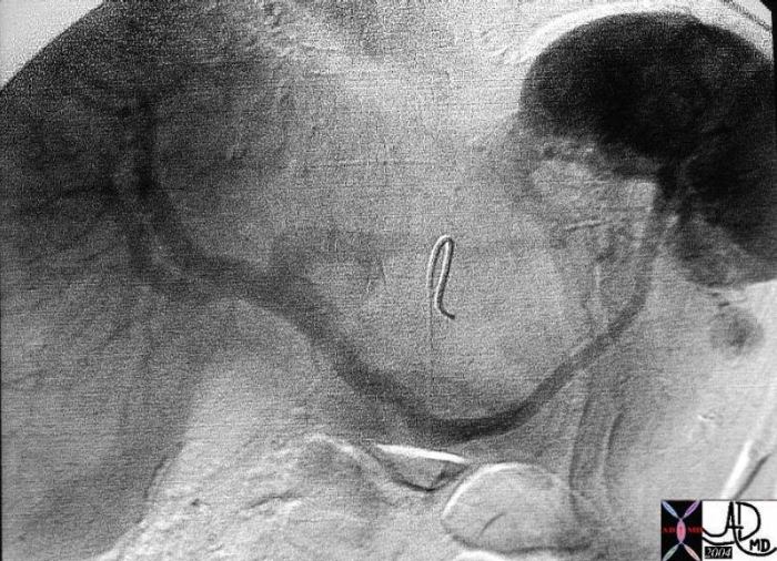 
Splenic Vein and Portal Vein |
|
26625 liver + vein + portal + fx normal + anatomy angiography Copyright 2012 Courtesy Ashley Davidoff MD 26625 |
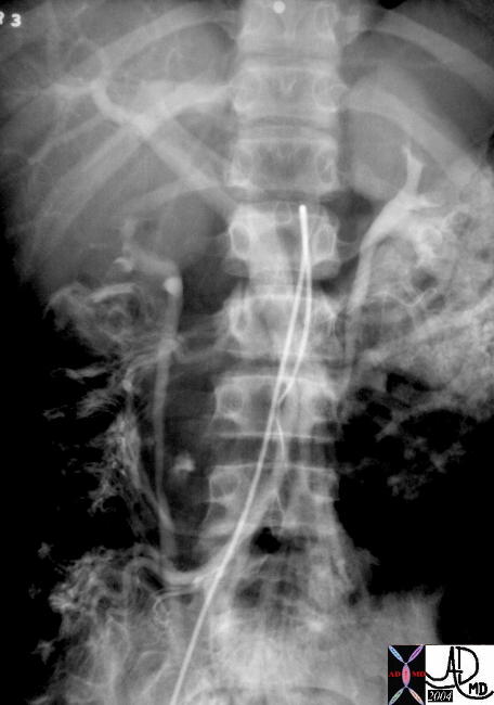 
SMV and Portal Vein |
|
small bowel liver colon vein venous drainage SMV portal vein fx normal angiography anatomy Copyright 2012 Courtesy Ashley Davidoff MD 26567 |
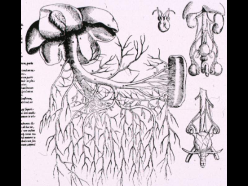
The Portal vein of Vesalius |
|
liver small bowel ? portal vein liver anatomy Vesalius historical drawing 13190 |
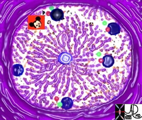
The portal triad is the group of connecting structures consisting of hepatic artery portal vein bile duct enclosed in a connective tissue bundle of Glisson’s capsule structurally characterised by position in the periphery of the lobule functionally characterised by contains the structural and functional connections of the liver part of liver capsule and hilum dividing into smaller triads common diseases include diseases of the peritoneum hepatic artery bile duct clinical presentation no specific clinical correlate diagnostic studies include US, CT, MRI treatment no specific treatment is directed to portal triad.
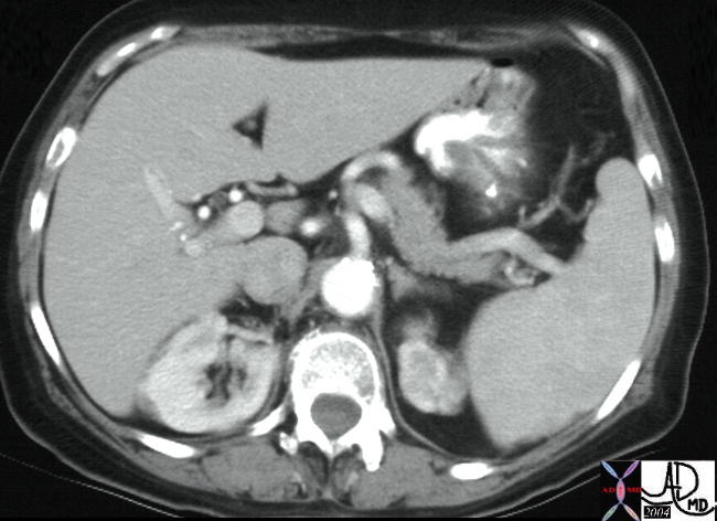 
24963 |
|
24963 liver artery celiac axis hepatic artery potra hepatic normal anatomy Copyright 2012 Courtesy Ashley Davidoff MD 24963 |
 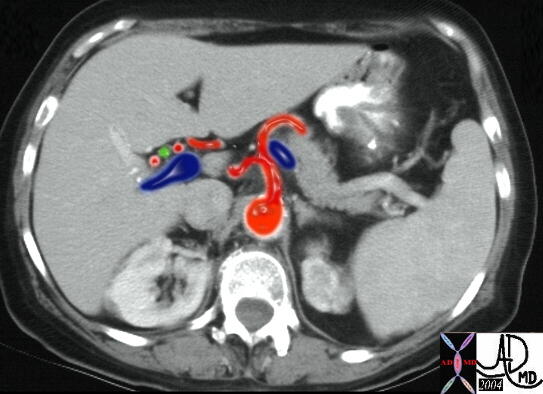
Celiac Axis to the Porta hepatis Meeting of the Triad at the Gates |
|
24963 W liver artery celiac axis hepatic artery potra hepatic normal anatomy Copyright 2012 Courtesy Ashley Davidoff MD 24963 |
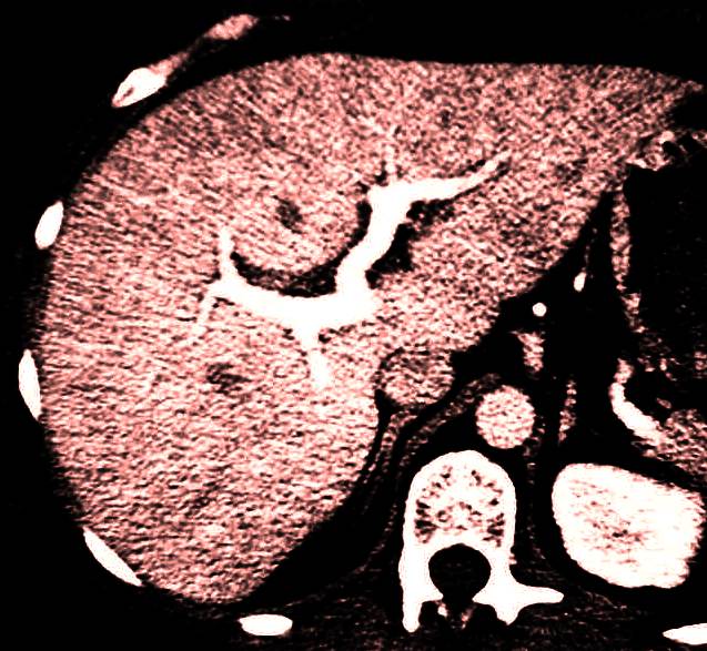
Intrahepatic Portal Vein Right and left Branches |
|
CODE liver portal vein blood supply normal anatomy CTscan Davidoff Art Copyright 2012 Coureesy Ashley Davidoff 27084b |
Right portal vein
The right portal vein is a medium sized vein structurally characterised by its portal nature providing connections between liver, spleen, and the gastrointestinal tract and its position in the porta hepatis as it branches to the segments of the right lobe of the liver functionally characterised by being the conduit for mesenteric blood and to lesser extent splenic blood arising from the main portal vein entering into the right segmental veins common diseases include portal hypertension portal vein thrombosis congestion clinical presentation part of portal hypertensive presentation diagnostic studies include US, CT, MRI, angiography treatment is commonly by interventional radiology surgery
Left portal vein
The left portal vein is a medium sized vein structurally characterised by its its portal nature providing connections between liver, spleen, and the gastrointestinal tract and its position in the porta hepatis as it branches to the segments of the right lobe of the liver functionally characterised by being the conduit for mesenteric blood and to lesser extent splenic blood arising from the main portal vein entering into the right segmental veins common diseases include portal hypertension portal vein thrombosis congestion clinical presentation part of portal hypertensive presentation diagnostic studies include US, CT, MRI, angiography treatment is commonly by interventional radiology surgery
Applied Anatomy
Acute portal vein thrombosis is an acute thrombotic circulatory disorder of the liver caused by thrombosis resulting in acute loss of portal blood supply characterised by partial or complete obstruction of the portal circulation maintenance of perfusion by hepatic artery pathogenesis disorder causes liver injury results in variable outcome resolution fibrosis acute or chronic liver failure structural disorder acute inflammatory infiltrate swelling hyperemia functional disorder usually of no functional significance until liver extensively involved clinical presentation local pain jaundice mild hepatomegaly systemic fever diagnostic studies include CT treatment is commonly by surgery if unstable
The radiographic feature of portal venous gas is gas that extends almost to the capsule of the liver. There are numerous causes for portal venous gas among the most common is mesenteric ischemia. Many intra-abdominal inflammatory conditions have been reported to cause portal venous gas, including inflammatory bowel disease and intra-abdominal abscesses. Among the rare causes are ingestion of corrosives, bronchopneumonia, and postperoxide enema.

Portal Venous Gas |
|
liver hx M52 vein portal fx air small bowel dilated dx ischemiv bowel imaging plain film KUB Copyright 2012 Courtesy Ashley Davidoff MD 02107 |
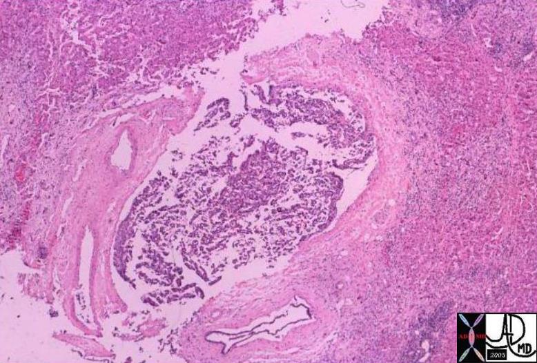
HCC Portal Vein Invasion |
|
liver fx mass portal vein fx invasion dx HCC hepatocellular carcinoma hepatoma histopathology Courtesy Barabara Banner MD 03463 |
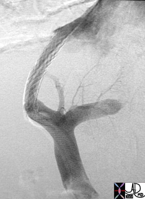 
TIPS procedure Porto Systemic Shunt |
|
liver + vein + portal + dx cirrhosis portal hypertension + TIPS + angiography + MITx Copyright 2012 Courtesy Ashley Davidoff MD 22367 |
Historical Perspectives
DOMElement Object
(
    [schemaTypeInfo] =>
    [tagName] => table
    [firstElementChild] => (object value omitted)
    [lastElementChild] => (object value omitted)
    [childElementCount] => 1
    [previousElementSibling] => (object value omitted)
    [nextElementSibling] => (object value omitted)
    [nodeName] => table
    [nodeValue] =>


Portal vein After Galen


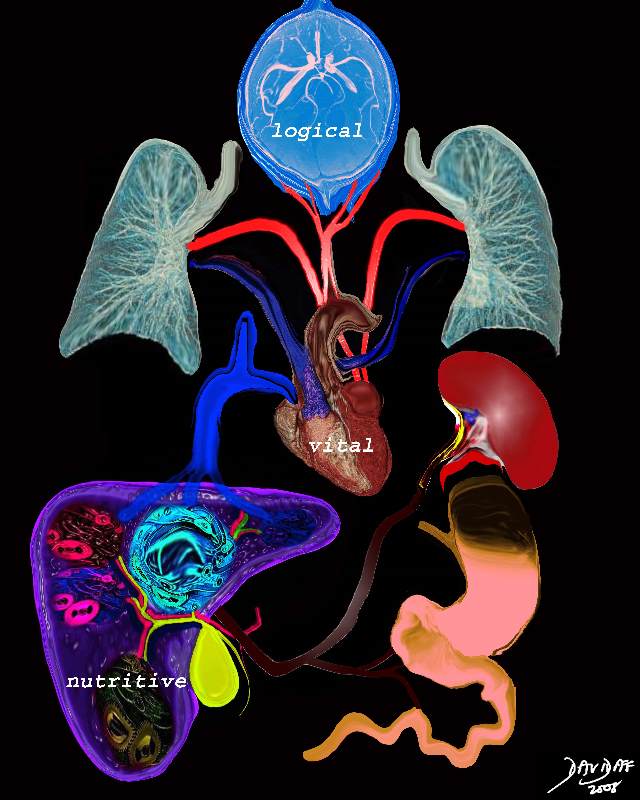
Heat plays a central role part in the theory of Galen. The three ?faculties? of the body are the nutritive, vital and logical faculties. The nutritive faculty is related to the stomach which “cooks” the food and converts it into chyle. The chyle is transported to the liver by the portal vein. In the liver further heat converts the food into blood and adds natural spirit. Some of the blood is transported via the veins to the heart where more heat is added to create vital spirit. The blood becomes thinner is distributed to the body by the arteries giving warmth and enables growth. The vital spirit is measured through the pulse. The brain adds psychic pneuma, which provides the rational and logical faculty in the form of thought will and choice. These are distributed to the body via the nerves. The logical faculty reigns supreme and is followed in orderof importance by the vital and nutrtive faculties. The transport systems of the body include the nerves which transmit the logical faculty, the arteries which transport the vital spirit, and the veins which transport the blood with nutritive faculty from the liver.
Copyright 2012 Courtesy Ashley Davidoff 13169c18b01.8s


    [nodeType] => 1
    [parentNode] => (object value omitted)
    [childNodes] => (object value omitted)
    [firstChild] => (object value omitted)
    [lastChild] => (object value omitted)
    [previousSibling] => (object value omitted)
    [nextSibling] => (object value omitted)
    [attributes] => (object value omitted)
    [ownerDocument] => (object value omitted)
    [namespaceURI] =>
    [prefix] =>
    [localName] => table
    [baseURI] =>
    [textContent] =>


Portal vein After Galen


Heat plays a central role part in the theory of Galen. The three ?faculties? of the body are the nutritive, vital and logical faculties. The nutritive faculty is related to the stomach which “cooks” the food and converts it into chyle. The chyle is transported to the liver by the portal vein. In the liver further heat converts the food into blood and adds natural spirit. Some of the blood is transported via the veins to the heart where more heat is added to create vital spirit. The blood becomes thinner is distributed to the body by the arteries giving warmth and enables growth. The vital spirit is measured through the pulse. The brain adds psychic pneuma, which provides the rational and logical faculty in the form of thought will and choice. These are distributed to the body via the nerves. The logical faculty reigns supreme and is followed in orderof importance by the vital and nutrtive faculties. The transport systems of the body include the nerves which transmit the logical faculty, the arteries which transport the vital spirit, and the veins which transport the blood with nutritive faculty from the liver.
Copyright 2012 Courtesy Ashley Davidoff 13169c18b01.8s


)
DOMElement Object
(
    [schemaTypeInfo] =>
    [tagName] => td
    [firstElementChild] => (object value omitted)
    [lastElementChild] => (object value omitted)
    [childElementCount] => 2
    [previousElementSibling] =>
    [nextElementSibling] =>
    [nodeName] => td
    [nodeValue] =>
Heat plays a central role part in the theory of Galen. The three ?faculties? of the body are the nutritive, vital and logical faculties. The nutritive faculty is related to the stomach which “cooks” the food and converts it into chyle. The chyle is transported to the liver by the portal vein. In the liver further heat converts the food into blood and adds natural spirit. Some of the blood is transported via the veins to the heart where more heat is added to create vital spirit. The blood becomes thinner is distributed to the body by the arteries giving warmth and enables growth. The vital spirit is measured through the pulse. The brain adds psychic pneuma, which provides the rational and logical faculty in the form of thought will and choice. These are distributed to the body via the nerves. The logical faculty reigns supreme and is followed in orderof importance by the vital and nutrtive faculties. The transport systems of the body include the nerves which transmit the logical faculty, the arteries which transport the vital spirit, and the veins which transport the blood with nutritive faculty from the liver.
Copyright 2012 Courtesy Ashley Davidoff 13169c18b01.8s

    [nodeType] => 1
    [parentNode] => (object value omitted)
    [childNodes] => (object value omitted)
    [firstChild] => (object value omitted)
    [lastChild] => (object value omitted)
    [previousSibling] => (object value omitted)
    [nextSibling] => (object value omitted)
    [attributes] => (object value omitted)
    [ownerDocument] => (object value omitted)
    [namespaceURI] =>
    [prefix] =>
    [localName] => td
    [baseURI] =>
    [textContent] =>
Heat plays a central role part in the theory of Galen. The three ?faculties? of the body are the nutritive, vital and logical faculties. The nutritive faculty is related to the stomach which “cooks” the food and converts it into chyle. The chyle is transported to the liver by the portal vein. In the liver further heat converts the food into blood and adds natural spirit. Some of the blood is transported via the veins to the heart where more heat is added to create vital spirit. The blood becomes thinner is distributed to the body by the arteries giving warmth and enables growth. The vital spirit is measured through the pulse. The brain adds psychic pneuma, which provides the rational and logical faculty in the form of thought will and choice. These are distributed to the body via the nerves. The logical faculty reigns supreme and is followed in orderof importance by the vital and nutrtive faculties. The transport systems of the body include the nerves which transmit the logical faculty, the arteries which transport the vital spirit, and the veins which transport the blood with nutritive faculty from the liver.
Copyright 2012 Courtesy Ashley Davidoff 13169c18b01.8s

)
DOMElement Object
(
    [schemaTypeInfo] =>
    [tagName] => td
    [firstElementChild] => (object value omitted)
    [lastElementChild] => (object value omitted)
    [childElementCount] => 2
    [previousElementSibling] =>
    [nextElementSibling] =>
    [nodeName] => td
    [nodeValue] =>
Portal vein After Galen

    [nodeType] => 1
    [parentNode] => (object value omitted)
    [childNodes] => (object value omitted)
    [firstChild] => (object value omitted)
    [lastChild] => (object value omitted)
    [previousSibling] => (object value omitted)
    [nextSibling] => (object value omitted)
    [attributes] => (object value omitted)
    [ownerDocument] => (object value omitted)
    [namespaceURI] =>
    [prefix] =>
    [localName] => td
    [baseURI] =>
    [textContent] =>
Portal vein After Galen

)
DOMElement Object
(
    [schemaTypeInfo] =>
    [tagName] => table
    [firstElementChild] => (object value omitted)
    [lastElementChild] => (object value omitted)
    [childElementCount] => 1
    [previousElementSibling] => (object value omitted)
    [nextElementSibling] => (object value omitted)
    [nodeName] => table
    [nodeValue] =>


TIPS procedure Porto Systemic Shunt


liver + vein + portal + dx cirrhosis portal hypertension + TIPS + angiography + MITx
Copyright 2012  Courtesy Ashley Davidoff MD 22367


    [nodeType] => 1
    [parentNode] => (object value omitted)
    [childNodes] => (object value omitted)
    [firstChild] => (object value omitted)
    [lastChild] => (object value omitted)
    [previousSibling] => (object value omitted)
    [nextSibling] => (object value omitted)
    [attributes] => (object value omitted)
    [ownerDocument] => (object value omitted)
    [namespaceURI] =>
    [prefix] =>
    [localName] => table
    [baseURI] =>
    [textContent] =>


TIPS procedure Porto Systemic Shunt


liver + vein + portal + dx cirrhosis portal hypertension + TIPS + angiography + MITx
Copyright 2012  Courtesy Ashley Davidoff MD 22367


)
DOMElement Object
(
    [schemaTypeInfo] =>
    [tagName] => td
    [firstElementChild] => (object value omitted)
    [lastElementChild] => (object value omitted)
    [childElementCount] => 2
    [previousElementSibling] =>
    [nextElementSibling] =>
    [nodeName] => td
    [nodeValue] =>
liver + vein + portal + dx cirrhosis portal hypertension + TIPS + angiography + MITx
Copyright 2012  Courtesy Ashley Davidoff MD 22367

    [nodeType] => 1
    [parentNode] => (object value omitted)
    [childNodes] => (object value omitted)
    [firstChild] => (object value omitted)
    [lastChild] => (object value omitted)
    [previousSibling] => (object value omitted)
    [nextSibling] => (object value omitted)
    [attributes] => (object value omitted)
    [ownerDocument] => (object value omitted)
    [namespaceURI] =>
    [prefix] =>
    [localName] => td
    [baseURI] =>
    [textContent] =>
liver + vein + portal + dx cirrhosis portal hypertension + TIPS + angiography + MITx
Copyright 2012  Courtesy Ashley Davidoff MD 22367

)
DOMElement Object
(
    [schemaTypeInfo] =>
    [tagName] => td
    [firstElementChild] => (object value omitted)
    [lastElementChild] => (object value omitted)
    [childElementCount] => 2
    [previousElementSibling] =>
    [nextElementSibling] =>
    [nodeName] => td
    [nodeValue] =>
TIPS procedure Porto Systemic Shunt
    [nodeType] => 1
    [parentNode] => (object value omitted)
    [childNodes] => (object value omitted)
    [firstChild] => (object value omitted)
    [lastChild] => (object value omitted)
    [previousSibling] => (object value omitted)
    [nextSibling] => (object value omitted)
    [attributes] => (object value omitted)
    [ownerDocument] => (object value omitted)
    [namespaceURI] =>
    [prefix] =>
    [localName] => td
    [baseURI] =>
    [textContent] =>
TIPS procedure Porto Systemic Shunt
)
DOMElement Object
(
    [schemaTypeInfo] =>
    [tagName] => table
    [firstElementChild] => (object value omitted)
    [lastElementChild] => (object value omitted)
    [childElementCount] => 1
    [previousElementSibling] => (object value omitted)
    [nextElementSibling] => (object value omitted)
    [nodeName] => table
    [nodeValue] =>


Cirrhossis
Bridging Between Portal Triads


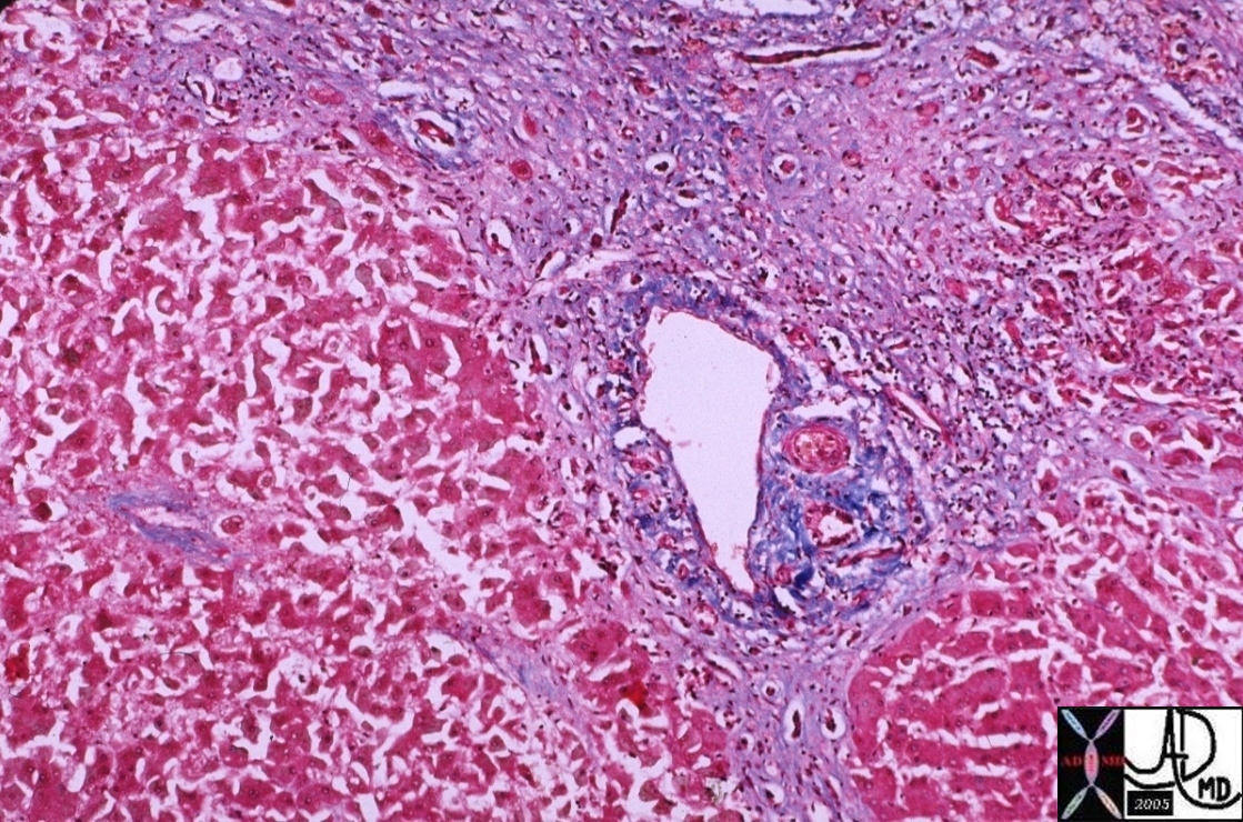
liver hepatic cirrhosis fx fibrosis fx nodules fx bridging between the triads portal triad hepatic artery portal vein bile duct histopathology
Courtesy Barbara Banner 00376


    [nodeType] => 1
    [parentNode] => (object value omitted)
    [childNodes] => (object value omitted)
    [firstChild] => (object value omitted)
    [lastChild] => (object value omitted)
    [previousSibling] => (object value omitted)
    [nextSibling] => (object value omitted)
    [attributes] => (object value omitted)
    [ownerDocument] => (object value omitted)
    [namespaceURI] =>
    [prefix] =>
    [localName] => table
    [baseURI] =>
    [textContent] =>


Cirrhossis
Bridging Between Portal Triads


liver hepatic cirrhosis fx fibrosis fx nodules fx bridging between the triads portal triad hepatic artery portal vein bile duct histopathology
Courtesy Barbara Banner 00376


)
DOMElement Object
(
    [schemaTypeInfo] =>
    [tagName] => td
    [firstElementChild] => (object value omitted)
    [lastElementChild] => (object value omitted)
    [childElementCount] => 2
    [previousElementSibling] =>
    [nextElementSibling] =>
    [nodeName] => td
    [nodeValue] =>
liver hepatic cirrhosis fx fibrosis fx nodules fx bridging between the triads portal triad hepatic artery portal vein bile duct histopathology
Courtesy Barbara Banner 00376
    [nodeType] => 1
    [parentNode] => (object value omitted)
    [childNodes] => (object value omitted)
    [firstChild] => (object value omitted)
    [lastChild] => (object value omitted)
    [previousSibling] => (object value omitted)
    [nextSibling] => (object value omitted)
    [attributes] => (object value omitted)
    [ownerDocument] => (object value omitted)
    [namespaceURI] =>
    [prefix] =>
    [localName] => td
    [baseURI] =>
    [textContent] =>
liver hepatic cirrhosis fx fibrosis fx nodules fx bridging between the triads portal triad hepatic artery portal vein bile duct histopathology
Courtesy Barbara Banner 00376
)
DOMElement Object
(
    [schemaTypeInfo] =>
    [tagName] => td
    [firstElementChild] => (object value omitted)
    [lastElementChild] => (object value omitted)
    [childElementCount] => 3
    [previousElementSibling] =>
    [nextElementSibling] =>
    [nodeName] => td
    [nodeValue] =>
Cirrhossis
Bridging Between Portal Triads
    [nodeType] => 1
    [parentNode] => (object value omitted)
    [childNodes] => (object value omitted)
    [firstChild] => (object value omitted)
    [lastChild] => (object value omitted)
    [previousSibling] => (object value omitted)
    [nextSibling] => (object value omitted)
    [attributes] => (object value omitted)
    [ownerDocument] => (object value omitted)
    [namespaceURI] =>
    [prefix] =>
    [localName] => td
    [baseURI] =>
    [textContent] =>
Cirrhossis
Bridging Between Portal Triads
)
DOMElement Object
(
    [schemaTypeInfo] =>
    [tagName] => table
    [firstElementChild] => (object value omitted)
    [lastElementChild] => (object value omitted)
    [childElementCount] => 1
    [previousElementSibling] => (object value omitted)
    [nextElementSibling] => (object value omitted)
    [nodeName] => table
    [nodeValue] =>


HCC Portal Vein Invasion


 liver fx mass portal vein fx invasion dx HCC hepatocellular carcinoma hepatoma histopathology
Courtesy Barabara Banner MD 03463


    [nodeType] => 1
    [parentNode] => (object value omitted)
    [childNodes] => (object value omitted)
    [firstChild] => (object value omitted)
    [lastChild] => (object value omitted)
    [previousSibling] => (object value omitted)
    [nextSibling] => (object value omitted)
    [attributes] => (object value omitted)
    [ownerDocument] => (object value omitted)
    [namespaceURI] =>
    [prefix] =>
    [localName] => table
    [baseURI] =>
    [textContent] =>


HCC Portal Vein Invasion


 liver fx mass portal vein fx invasion dx HCC hepatocellular carcinoma hepatoma histopathology
Courtesy Barabara Banner MD 03463


)
DOMElement Object
(
    [schemaTypeInfo] =>
    [tagName] => td
    [firstElementChild] => (object value omitted)
    [lastElementChild] => (object value omitted)
    [childElementCount] => 2
    [previousElementSibling] =>
    [nextElementSibling] =>
    [nodeName] => td
    [nodeValue] =>
 liver fx mass portal vein fx invasion dx HCC hepatocellular carcinoma hepatoma histopathology
Courtesy Barabara Banner MD 03463

    [nodeType] => 1
    [parentNode] => (object value omitted)
    [childNodes] => (object value omitted)
    [firstChild] => (object value omitted)
    [lastChild] => (object value omitted)
    [previousSibling] => (object value omitted)
    [nextSibling] => (object value omitted)
    [attributes] => (object value omitted)
    [ownerDocument] => (object value omitted)
    [namespaceURI] =>
    [prefix] =>
    [localName] => td
    [baseURI] =>
    [textContent] =>
 liver fx mass portal vein fx invasion dx HCC hepatocellular carcinoma hepatoma histopathology
Courtesy Barabara Banner MD 03463

)
DOMElement Object
(
    [schemaTypeInfo] =>
    [tagName] => td
    [firstElementChild] => (object value omitted)
    [lastElementChild] => (object value omitted)
    [childElementCount] => 2
    [previousElementSibling] =>
    [nextElementSibling] =>
    [nodeName] => td
    [nodeValue] =>
HCC Portal Vein Invasion

    [nodeType] => 1
    [parentNode] => (object value omitted)
    [childNodes] => (object value omitted)
    [firstChild] => (object value omitted)
    [lastChild] => (object value omitted)
    [previousSibling] => (object value omitted)
    [nextSibling] => (object value omitted)
    [attributes] => (object value omitted)
    [ownerDocument] => (object value omitted)
    [namespaceURI] =>
    [prefix] =>
    [localName] => td
    [baseURI] =>
    [textContent] =>
HCC Portal Vein Invasion

)
DOMElement Object
(
    [schemaTypeInfo] =>
    [tagName] => table
    [firstElementChild] => (object value omitted)
    [lastElementChild] => (object value omitted)
    [childElementCount] => 1
    [previousElementSibling] => (object value omitted)
    [nextElementSibling] => (object value omitted)
    [nodeName] => table
    [nodeValue] =>


Portal Venous Gas


liver hx M52 vein portal fx air small bowel dilated dx ischemiv bowel imaging plain film KUB
Copyright 2012 Courtesy Ashley Davidoff MD 02107


    [nodeType] => 1
    [parentNode] => (object value omitted)
    [childNodes] => (object value omitted)
    [firstChild] => (object value omitted)
    [lastChild] => (object value omitted)
    [previousSibling] => (object value omitted)
    [nextSibling] => (object value omitted)
    [attributes] => (object value omitted)
    [ownerDocument] => (object value omitted)
    [namespaceURI] =>
    [prefix] =>
    [localName] => table
    [baseURI] =>
    [textContent] =>


Portal Venous Gas


liver hx M52 vein portal fx air small bowel dilated dx ischemiv bowel imaging plain film KUB
Copyright 2012 Courtesy Ashley Davidoff MD 02107


)
DOMElement Object
(
    [schemaTypeInfo] =>
    [tagName] => td
    [firstElementChild] => (object value omitted)
    [lastElementChild] => (object value omitted)
    [childElementCount] => 2
    [previousElementSibling] =>
    [nextElementSibling] =>
    [nodeName] => td
    [nodeValue] =>
liver hx M52 vein portal fx air small bowel dilated dx ischemiv bowel imaging plain film KUB
Copyright 2012 Courtesy Ashley Davidoff MD 02107

    [nodeType] => 1
    [parentNode] => (object value omitted)
    [childNodes] => (object value omitted)
    [firstChild] => (object value omitted)
    [lastChild] => (object value omitted)
    [previousSibling] => (object value omitted)
    [nextSibling] => (object value omitted)
    [attributes] => (object value omitted)
    [ownerDocument] => (object value omitted)
    [namespaceURI] =>
    [prefix] =>
    [localName] => td
    [baseURI] =>
    [textContent] =>
liver hx M52 vein portal fx air small bowel dilated dx ischemiv bowel imaging plain film KUB
Copyright 2012 Courtesy Ashley Davidoff MD 02107

)
DOMElement Object
(
    [schemaTypeInfo] =>
    [tagName] => td
    [firstElementChild] => (object value omitted)
    [lastElementChild] => (object value omitted)
    [childElementCount] => 2
    [previousElementSibling] =>
    [nextElementSibling] =>
    [nodeName] => td
    [nodeValue] =>
Portal Venous Gas

    [nodeType] => 1
    [parentNode] => (object value omitted)
    [childNodes] => (object value omitted)
    [firstChild] => (object value omitted)
    [lastChild] => (object value omitted)
    [previousSibling] => (object value omitted)
    [nextSibling] => (object value omitted)
    [attributes] => (object value omitted)
    [ownerDocument] => (object value omitted)
    [namespaceURI] =>
    [prefix] =>
    [localName] => td
    [baseURI] =>
    [textContent] =>
Portal Venous Gas

)
DOMElement Object
(
    [schemaTypeInfo] =>
    [tagName] => table
    [firstElementChild] => (object value omitted)
    [lastElementChild] => (object value omitted)
    [childElementCount] => 1
    [previousElementSibling] => (object value omitted)
    [nextElementSibling] => (object value omitted)
    [nodeName] => table
    [nodeValue] =>


The Portal Triad and the Sinusoids


13009 W13 liver + portal triad lobule + anatomy histology portal vein bile duct hepatic artery central vein hepatic vein Davidoff art
Copyright 2012 Courtesy Ashley Davidoff MD  13009 W13


    [nodeType] => 1
    [parentNode] => (object value omitted)
    [childNodes] => (object value omitted)
    [firstChild] => (object value omitted)
    [lastChild] => (object value omitted)
    [previousSibling] => (object value omitted)
    [nextSibling] => (object value omitted)
    [attributes] => (object value omitted)
    [ownerDocument] => (object value omitted)
    [namespaceURI] =>
    [prefix] =>
    [localName] => table
    [baseURI] =>
    [textContent] =>


The Portal Triad and the Sinusoids


13009 W13 liver + portal triad lobule + anatomy histology portal vein bile duct hepatic artery central vein hepatic vein Davidoff art
Copyright 2012 Courtesy Ashley Davidoff MD  13009 W13


)
DOMElement Object
(
    [schemaTypeInfo] =>
    [tagName] => td
    [firstElementChild] => (object value omitted)
    [lastElementChild] => (object value omitted)
    [childElementCount] => 2
    [previousElementSibling] =>
    [nextElementSibling] =>
    [nodeName] => td
    [nodeValue] =>
13009 W13 liver + portal triad lobule + anatomy histology portal vein bile duct hepatic artery central vein hepatic vein Davidoff art
Copyright 2012 Courtesy Ashley Davidoff MD  13009 W13

    [nodeType] => 1
    [parentNode] => (object value omitted)
    [childNodes] => (object value omitted)
    [firstChild] => (object value omitted)
    [lastChild] => (object value omitted)
    [previousSibling] => (object value omitted)
    [nextSibling] => (object value omitted)
    [attributes] => (object value omitted)
    [ownerDocument] => (object value omitted)
    [namespaceURI] =>
    [prefix] =>
    [localName] => td
    [baseURI] =>
    [textContent] =>
13009 W13 liver + portal triad lobule + anatomy histology portal vein bile duct hepatic artery central vein hepatic vein Davidoff art
Copyright 2012 Courtesy Ashley Davidoff MD  13009 W13

)
DOMElement Object
(
    [schemaTypeInfo] =>
    [tagName] => td
    [firstElementChild] => (object value omitted)
    [lastElementChild] => (object value omitted)
    [childElementCount] => 2
    [previousElementSibling] =>
    [nextElementSibling] =>
    [nodeName] => td
    [nodeValue] =>
The Portal Triad and the Sinusoids

    [nodeType] => 1
    [parentNode] => (object value omitted)
    [childNodes] => (object value omitted)
    [firstChild] => (object value omitted)
    [lastChild] => (object value omitted)
    [previousSibling] => (object value omitted)
    [nextSibling] => (object value omitted)
    [attributes] => (object value omitted)
    [ownerDocument] => (object value omitted)
    [namespaceURI] =>
    [prefix] =>
    [localName] => td
    [baseURI] =>
    [textContent] =>
The Portal Triad and the Sinusoids

)
DOMElement Object
(
    [schemaTypeInfo] =>
    [tagName] => table
    [firstElementChild] => (object value omitted)
    [lastElementChild] => (object value omitted)
    [childElementCount] => 1
    [previousElementSibling] => (object value omitted)
    [nextElementSibling] => (object value omitted)
    [nodeName] => table
    [nodeValue] =>


Intrahepatic Portal Vein
Right and left Branches


CODE liver portal vein blood supply normal anatomy CTscan Davidoff Art
Copyright 2012 Coureesy Ashley Davidoff 27084b


    [nodeType] => 1
    [parentNode] => (object value omitted)
    [childNodes] => (object value omitted)
    [firstChild] => (object value omitted)
    [lastChild] => (object value omitted)
    [previousSibling] => (object value omitted)
    [nextSibling] => (object value omitted)
    [attributes] => (object value omitted)
    [ownerDocument] => (object value omitted)
    [namespaceURI] =>
    [prefix] =>
    [localName] => table
    [baseURI] =>
    [textContent] =>


Intrahepatic Portal Vein
Right and left Branches


CODE liver portal vein blood supply normal anatomy CTscan Davidoff Art
Copyright 2012 Coureesy Ashley Davidoff 27084b


)
DOMElement Object
(
    [schemaTypeInfo] =>
    [tagName] => td
    [firstElementChild] => (object value omitted)
    [lastElementChild] => (object value omitted)
    [childElementCount] => 2
    [previousElementSibling] =>
    [nextElementSibling] =>
    [nodeName] => td
    [nodeValue] =>
CODE liver portal vein blood supply normal anatomy CTscan Davidoff Art
Copyright 2012 Coureesy Ashley Davidoff 27084b

    [nodeType] => 1
    [parentNode] => (object value omitted)
    [childNodes] => (object value omitted)
    [firstChild] => (object value omitted)
    [lastChild] => (object value omitted)
    [previousSibling] => (object value omitted)
    [nextSibling] => (object value omitted)
    [attributes] => (object value omitted)
    [ownerDocument] => (object value omitted)
    [namespaceURI] =>
    [prefix] =>
    [localName] => td
    [baseURI] =>
    [textContent] =>
CODE liver portal vein blood supply normal anatomy CTscan Davidoff Art
Copyright 2012 Coureesy Ashley Davidoff 27084b

)
DOMElement Object
(
    [schemaTypeInfo] =>
    [tagName] => td
    [firstElementChild] => (object value omitted)
    [lastElementChild] => (object value omitted)
    [childElementCount] => 3
    [previousElementSibling] =>
    [nextElementSibling] =>
    [nodeName] => td
    [nodeValue] =>
Intrahepatic Portal Vein
Right and left Branches

    [nodeType] => 1
    [parentNode] => (object value omitted)
    [childNodes] => (object value omitted)
    [firstChild] => (object value omitted)
    [lastChild] => (object value omitted)
    [previousSibling] => (object value omitted)
    [nextSibling] => (object value omitted)
    [attributes] => (object value omitted)
    [ownerDocument] => (object value omitted)
    [namespaceURI] =>
    [prefix] =>
    [localName] => td
    [baseURI] =>
    [textContent] =>
Intrahepatic Portal Vein
Right and left Branches

)
DOMElement Object
(
    [schemaTypeInfo] =>
    [tagName] => table
    [firstElementChild] => (object value omitted)
    [lastElementChild] => (object value omitted)
    [childElementCount] => 1
    [previousElementSibling] => (object value omitted)
    [nextElementSibling] => (object value omitted)
    [nodeName] => table
    [nodeValue] =>


Mickey Mouse and the Portal Triad


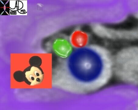
Here’s Mickey again! The main portal vein enters the porta hepatis with its companions, the hepatic artery and the common hepatic duct. Together they are known as the portal triad. The main portal vein lies posterior to the hepatic artery and bile duct at the porta hepatis.
Copyright 2012 Courtesy Ashley Davidoff MD 24964 R 3


    [nodeType] => 1
    [parentNode] => (object value omitted)
    [childNodes] => (object value omitted)
    [firstChild] => (object value omitted)
    [lastChild] => (object value omitted)
    [previousSibling] => (object value omitted)
    [nextSibling] => (object value omitted)
    [attributes] => (object value omitted)
    [ownerDocument] => (object value omitted)
    [namespaceURI] =>
    [prefix] =>
    [localName] => table
    [baseURI] =>
    [textContent] =>


Mickey Mouse and the Portal Triad


Here’s Mickey again! The main portal vein enters the porta hepatis with its companions, the hepatic artery and the common hepatic duct. Together they are known as the portal triad. The main portal vein lies posterior to the hepatic artery and bile duct at the porta hepatis.
Copyright 2012 Courtesy Ashley Davidoff MD 24964 R 3


)
DOMElement Object
(
    [schemaTypeInfo] =>
    [tagName] => td
    [firstElementChild] => (object value omitted)
    [lastElementChild] => (object value omitted)
    [childElementCount] => 3
    [previousElementSibling] =>
    [nextElementSibling] =>
    [nodeName] => td
    [nodeValue] =>
Here’s Mickey again! The main portal vein enters the porta hepatis with its companions, the hepatic artery and the common hepatic duct. Together they are known as the portal triad. The main portal vein lies posterior to the hepatic artery and bile duct at the porta hepatis.
Copyright 2012 Courtesy Ashley Davidoff MD 24964 R 3

    [nodeType] => 1
    [parentNode] => (object value omitted)
    [childNodes] => (object value omitted)
    [firstChild] => (object value omitted)
    [lastChild] => (object value omitted)
    [previousSibling] => (object value omitted)
    [nextSibling] => (object value omitted)
    [attributes] => (object value omitted)
    [ownerDocument] => (object value omitted)
    [namespaceURI] =>
    [prefix] =>
    [localName] => td
    [baseURI] =>
    [textContent] =>
Here’s Mickey again! The main portal vein enters the porta hepatis with its companions, the hepatic artery and the common hepatic duct. Together they are known as the portal triad. The main portal vein lies posterior to the hepatic artery and bile duct at the porta hepatis.
Copyright 2012 Courtesy Ashley Davidoff MD 24964 R 3

)
DOMElement Object
(
    [schemaTypeInfo] =>
    [tagName] => td
    [firstElementChild] => (object value omitted)
    [lastElementChild] => (object value omitted)
    [childElementCount] => 2
    [previousElementSibling] =>
    [nextElementSibling] =>
    [nodeName] => td
    [nodeValue] =>
Mickey Mouse and the Portal Triad

    [nodeType] => 1
    [parentNode] => (object value omitted)
    [childNodes] => (object value omitted)
    [firstChild] => (object value omitted)
    [lastChild] => (object value omitted)
    [previousSibling] => (object value omitted)
    [nextSibling] => (object value omitted)
    [attributes] => (object value omitted)
    [ownerDocument] => (object value omitted)
    [namespaceURI] =>
    [prefix] =>
    [localName] => td
    [baseURI] =>
    [textContent] =>
Mickey Mouse and the Portal Triad

)
DOMElement Object
(
    [schemaTypeInfo] =>
    [tagName] => table
    [firstElementChild] => (object value omitted)
    [lastElementChild] => (object value omitted)
    [childElementCount] => 1
    [previousElementSibling] => (object value omitted)
    [nextElementSibling] => (object value omitted)
    [nodeName] => table
    [nodeValue] =>


Celiac Axis to the Porta hepatis
Meeting of the Triad at the Gates


24963 W liver artery celiac axis hepatic artery potra hepatic normal anatomy
Copyright 2012 Courtesy Ashley Davidoff MD 24963


    [nodeType] => 1
    [parentNode] => (object value omitted)
    [childNodes] => (object value omitted)
    [firstChild] => (object value omitted)
    [lastChild] => (object value omitted)
    [previousSibling] => (object value omitted)
    [nextSibling] => (object value omitted)
    [attributes] => (object value omitted)
    [ownerDocument] => (object value omitted)
    [namespaceURI] =>
    [prefix] =>
    [localName] => table
    [baseURI] =>
    [textContent] =>


Celiac Axis to the Porta hepatis
Meeting of the Triad at the Gates


24963 W liver artery celiac axis hepatic artery potra hepatic normal anatomy
Copyright 2012 Courtesy Ashley Davidoff MD 24963


)
DOMElement Object
(
    [schemaTypeInfo] =>
    [tagName] => td
    [firstElementChild] => (object value omitted)
    [lastElementChild] => (object value omitted)
    [childElementCount] => 2
    [previousElementSibling] =>
    [nextElementSibling] =>
    [nodeName] => td
    [nodeValue] =>
24963 W liver artery celiac axis hepatic artery potra hepatic normal anatomy
Copyright 2012 Courtesy Ashley Davidoff MD 24963

    [nodeType] => 1
    [parentNode] => (object value omitted)
    [childNodes] => (object value omitted)
    [firstChild] => (object value omitted)
    [lastChild] => (object value omitted)
    [previousSibling] => (object value omitted)
    [nextSibling] => (object value omitted)
    [attributes] => (object value omitted)
    [ownerDocument] => (object value omitted)
    [namespaceURI] =>
    [prefix] =>
    [localName] => td
    [baseURI] =>
    [textContent] =>
24963 W liver artery celiac axis hepatic artery potra hepatic normal anatomy
Copyright 2012 Courtesy Ashley Davidoff MD 24963

)
DOMElement Object
(
    [schemaTypeInfo] =>
    [tagName] => td
    [firstElementChild] => (object value omitted)
    [lastElementChild] => (object value omitted)
    [childElementCount] => 3
    [previousElementSibling] =>
    [nextElementSibling] =>
    [nodeName] => td
    [nodeValue] =>
Celiac Axis to the Porta hepatis
Meeting of the Triad at the Gates

    [nodeType] => 1
    [parentNode] => (object value omitted)
    [childNodes] => (object value omitted)
    [firstChild] => (object value omitted)
    [lastChild] => (object value omitted)
    [previousSibling] => (object value omitted)
    [nextSibling] => (object value omitted)
    [attributes] => (object value omitted)
    [ownerDocument] => (object value omitted)
    [namespaceURI] =>
    [prefix] =>
    [localName] => td
    [baseURI] =>
    [textContent] =>
Celiac Axis to the Porta hepatis
Meeting of the Triad at the Gates

)
DOMElement Object
(
    [schemaTypeInfo] =>
    [tagName] => table
    [firstElementChild] => (object value omitted)
    [lastElementChild] => (object value omitted)
    [childElementCount] => 1
    [previousElementSibling] => (object value omitted)
    [nextElementSibling] => (object value omitted)
    [nodeName] => table
    [nodeValue] =>


24963


24963 liver artery celiac axis hepatic artery potra hepatic normal anatomy
Copyright 2012 Courtesy Ashley Davidoff MD 24963


    [nodeType] => 1
    [parentNode] => (object value omitted)
    [childNodes] => (object value omitted)
    [firstChild] => (object value omitted)
    [lastChild] => (object value omitted)
    [previousSibling] => (object value omitted)
    [nextSibling] => (object value omitted)
    [attributes] => (object value omitted)
    [ownerDocument] => (object value omitted)
    [namespaceURI] =>
    [prefix] =>
    [localName] => table
    [baseURI] =>
    [textContent] =>


24963


24963 liver artery celiac axis hepatic artery potra hepatic normal anatomy
Copyright 2012 Courtesy Ashley Davidoff MD 24963


)
DOMElement Object
(
    [schemaTypeInfo] =>
    [tagName] => td
    [firstElementChild] => (object value omitted)
    [lastElementChild] => (object value omitted)
    [childElementCount] => 2
    [previousElementSibling] =>
    [nextElementSibling] =>
    [nodeName] => td
    [nodeValue] =>
24963 liver artery celiac axis hepatic artery potra hepatic normal anatomy
Copyright 2012 Courtesy Ashley Davidoff MD 24963

    [nodeType] => 1
    [parentNode] => (object value omitted)
    [childNodes] => (object value omitted)
    [firstChild] => (object value omitted)
    [lastChild] => (object value omitted)
    [previousSibling] => (object value omitted)
    [nextSibling] => (object value omitted)
    [attributes] => (object value omitted)
    [ownerDocument] => (object value omitted)
    [namespaceURI] =>
    [prefix] =>
    [localName] => td
    [baseURI] =>
    [textContent] =>
24963 liver artery celiac axis hepatic artery potra hepatic normal anatomy
Copyright 2012 Courtesy Ashley Davidoff MD 24963

)
DOMElement Object
(
    [schemaTypeInfo] =>
    [tagName] => td
    [firstElementChild] => (object value omitted)
    [lastElementChild] => (object value omitted)
    [childElementCount] => 2
    [previousElementSibling] =>
    [nextElementSibling] =>
    [nodeName] => td
    [nodeValue] =>
24963
    [nodeType] => 1
    [parentNode] => (object value omitted)
    [childNodes] => (object value omitted)
    [firstChild] => (object value omitted)
    [lastChild] => (object value omitted)
    [previousSibling] => (object value omitted)
    [nextSibling] => (object value omitted)
    [attributes] => (object value omitted)
    [ownerDocument] => (object value omitted)
    [namespaceURI] =>
    [prefix] =>
    [localName] => td
    [baseURI] =>
    [textContent] =>
24963
)
DOMElement Object
(
    [schemaTypeInfo] =>
    [tagName] => table
    [firstElementChild] => (object value omitted)
    [lastElementChild] => (object value omitted)
    [childElementCount] => 1
    [previousElementSibling] => (object value omitted)
    [nextElementSibling] => (object value omitted)
    [nodeName] => table
    [nodeValue] =>


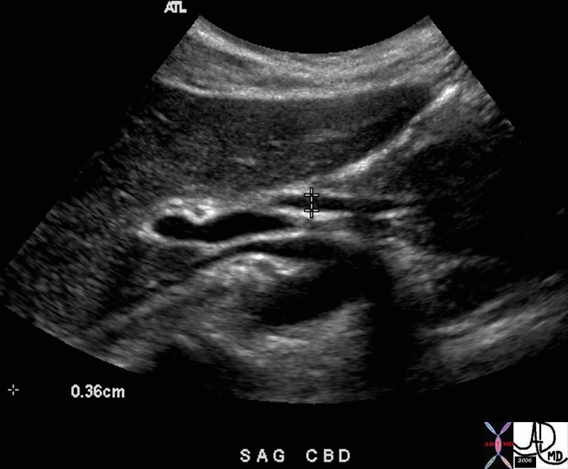
The Porta Hepatis –  Ultrasound


bile duct CBD portal vein right hepatic artery common bile duct common hepatic duct extrahepatic pancreas normal size position anatomy USscan
Copyright 2012 Courtesy Ashley Davidoff MD 47020


    [nodeType] => 1
    [parentNode] => (object value omitted)
    [childNodes] => (object value omitted)
    [firstChild] => (object value omitted)
    [lastChild] => (object value omitted)
    [previousSibling] => (object value omitted)
    [nextSibling] => (object value omitted)
    [attributes] => (object value omitted)
    [ownerDocument] => (object value omitted)
    [namespaceURI] =>
    [prefix] =>
    [localName] => table
    [baseURI] =>
    [textContent] =>


The Porta Hepatis –  Ultrasound


bile duct CBD portal vein right hepatic artery common bile duct common hepatic duct extrahepatic pancreas normal size position anatomy USscan
Copyright 2012 Courtesy Ashley Davidoff MD 47020


)
DOMElement Object
(
    [schemaTypeInfo] =>
    [tagName] => td
    [firstElementChild] => (object value omitted)
    [lastElementChild] => (object value omitted)
    [childElementCount] => 2
    [previousElementSibling] =>
    [nextElementSibling] =>
    [nodeName] => td
    [nodeValue] =>
bile duct CBD portal vein right hepatic artery common bile duct common hepatic duct extrahepatic pancreas normal size position anatomy USscan
Copyright 2012 Courtesy Ashley Davidoff MD 47020

    [nodeType] => 1
    [parentNode] => (object value omitted)
    [childNodes] => (object value omitted)
    [firstChild] => (object value omitted)
    [lastChild] => (object value omitted)
    [previousSibling] => (object value omitted)
    [nextSibling] => (object value omitted)
    [attributes] => (object value omitted)
    [ownerDocument] => (object value omitted)
    [namespaceURI] =>
    [prefix] =>
    [localName] => td
    [baseURI] =>
    [textContent] =>
bile duct CBD portal vein right hepatic artery common bile duct common hepatic duct extrahepatic pancreas normal size position anatomy USscan
Copyright 2012 Courtesy Ashley Davidoff MD 47020

)
DOMElement Object
(
    [schemaTypeInfo] =>
    [tagName] => td
    [firstElementChild] => (object value omitted)
    [lastElementChild] => (object value omitted)
    [childElementCount] => 2
    [previousElementSibling] =>
    [nextElementSibling] =>
    [nodeName] => td
    [nodeValue] =>
The Porta Hepatis –  Ultrasound

    [nodeType] => 1
    [parentNode] => (object value omitted)
    [childNodes] => (object value omitted)
    [firstChild] => (object value omitted)
    [lastChild] => (object value omitted)
    [previousSibling] => (object value omitted)
    [nextSibling] => (object value omitted)
    [attributes] => (object value omitted)
    [ownerDocument] => (object value omitted)
    [namespaceURI] =>
    [prefix] =>
    [localName] => td
    [baseURI] =>
    [textContent] =>
The Porta Hepatis –  Ultrasound

)
DOMElement Object
(
    [schemaTypeInfo] =>
    [tagName] => table
    [firstElementChild] => (object value omitted)
    [lastElementChild] => (object value omitted)
    [childElementCount] => 1
    [previousElementSibling] => (object value omitted)
    [nextElementSibling] => (object value omitted)
    [nodeName] => table
    [nodeValue] =>


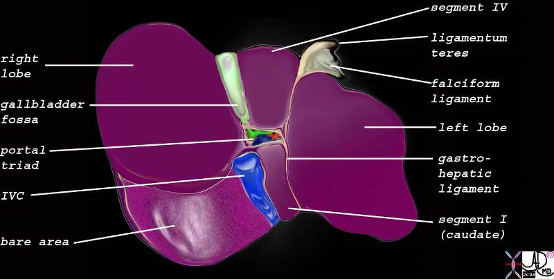
The Porta Hepatis


liver gallbladder porta hepatis hepatic artery portal vein IVC inferior vena cava falciform ligament ligamentum teres bare area of the liver left lobe segment IV segment I caudate lobe quadrate lobe gastrohepatic ligament gallbladder right lobe hepatic IVC Davidoff art
Copyright 2012 Courtesy Ashley Davidoff MD 82220b06.8s


    [nodeType] => 1
    [parentNode] => (object value omitted)
    [childNodes] => (object value omitted)
    [firstChild] => (object value omitted)
    [lastChild] => (object value omitted)
    [previousSibling] => (object value omitted)
    [nextSibling] => (object value omitted)
    [attributes] => (object value omitted)
    [ownerDocument] => (object value omitted)
    [namespaceURI] =>
    [prefix] =>
    [localName] => table
    [baseURI] =>
    [textContent] =>


The Porta Hepatis


liver gallbladder porta hepatis hepatic artery portal vein IVC inferior vena cava falciform ligament ligamentum teres bare area of the liver left lobe segment IV segment I caudate lobe quadrate lobe gastrohepatic ligament gallbladder right lobe hepatic IVC Davidoff art
Copyright 2012 Courtesy Ashley Davidoff MD 82220b06.8s


)
DOMElement Object
(
    [schemaTypeInfo] =>
    [tagName] => td
    [firstElementChild] => (object value omitted)
    [lastElementChild] => (object value omitted)
    [childElementCount] => 2
    [previousElementSibling] =>
    [nextElementSibling] =>
    [nodeName] => td
    [nodeValue] =>
liver gallbladder porta hepatis hepatic artery portal vein IVC inferior vena cava falciform ligament ligamentum teres bare area of the liver left lobe segment IV segment I caudate lobe quadrate lobe gastrohepatic ligament gallbladder right lobe hepatic IVC Davidoff art
Copyright 2012 Courtesy Ashley Davidoff MD 82220b06.8s
    [nodeType] => 1
    [parentNode] => (object value omitted)
    [childNodes] => (object value omitted)
    [firstChild] => (object value omitted)
    [lastChild] => (object value omitted)
    [previousSibling] => (object value omitted)
    [nextSibling] => (object value omitted)
    [attributes] => (object value omitted)
    [ownerDocument] => (object value omitted)
    [namespaceURI] =>
    [prefix] =>
    [localName] => td
    [baseURI] =>
    [textContent] =>
liver gallbladder porta hepatis hepatic artery portal vein IVC inferior vena cava falciform ligament ligamentum teres bare area of the liver left lobe segment IV segment I caudate lobe quadrate lobe gastrohepatic ligament gallbladder right lobe hepatic IVC Davidoff art
Copyright 2012 Courtesy Ashley Davidoff MD 82220b06.8s
)
DOMElement Object
(
    [schemaTypeInfo] =>
    [tagName] => td
    [firstElementChild] => (object value omitted)
    [lastElementChild] => (object value omitted)
    [childElementCount] => 2
    [previousElementSibling] =>
    [nextElementSibling] =>
    [nodeName] => td
    [nodeValue] =>
The Porta Hepatis
    [nodeType] => 1
    [parentNode] => (object value omitted)
    [childNodes] => (object value omitted)
    [firstChild] => (object value omitted)
    [lastChild] => (object value omitted)
    [previousSibling] => (object value omitted)
    [nextSibling] => (object value omitted)
    [attributes] => (object value omitted)
    [ownerDocument] => (object value omitted)
    [namespaceURI] =>
    [prefix] =>
    [localName] => td
    [baseURI] =>
    [textContent] =>
The Porta Hepatis
)
DOMElement Object
(
    [schemaTypeInfo] =>
    [tagName] => table
    [firstElementChild] => (object value omitted)
    [lastElementChild] => (object value omitted)
    [childElementCount] => 1
    [previousElementSibling] => (object value omitted)
    [nextElementSibling] => (object value omitted)
    [nodeName] => table
    [nodeValue] =>


The Portal vein of Vesalius


liver small bowel ? portal vein liver anatomy
Vesalius historical drawing 13190


    [nodeType] => 1
    [parentNode] => (object value omitted)
    [childNodes] => (object value omitted)
    [firstChild] => (object value omitted)
    [lastChild] => (object value omitted)
    [previousSibling] => (object value omitted)
    [nextSibling] => (object value omitted)
    [attributes] => (object value omitted)
    [ownerDocument] => (object value omitted)
    [namespaceURI] =>
    [prefix] =>
    [localName] => table
    [baseURI] =>
    [textContent] =>


The Portal vein of Vesalius


liver small bowel ? portal vein liver anatomy
Vesalius historical drawing 13190


)
DOMElement Object
(
    [schemaTypeInfo] =>
    [tagName] => td
    [firstElementChild] => (object value omitted)
    [lastElementChild] => (object value omitted)
    [childElementCount] => 2
    [previousElementSibling] =>
    [nextElementSibling] =>
    [nodeName] => td
    [nodeValue] =>
liver small bowel ? portal vein liver anatomy
Vesalius historical drawing 13190

    [nodeType] => 1
    [parentNode] => (object value omitted)
    [childNodes] => (object value omitted)
    [firstChild] => (object value omitted)
    [lastChild] => (object value omitted)
    [previousSibling] => (object value omitted)
    [nextSibling] => (object value omitted)
    [attributes] => (object value omitted)
    [ownerDocument] => (object value omitted)
    [namespaceURI] =>
    [prefix] =>
    [localName] => td
    [baseURI] =>
    [textContent] =>
liver small bowel ? portal vein liver anatomy
Vesalius historical drawing 13190

)
DOMElement Object
(
    [schemaTypeInfo] =>
    [tagName] => td
    [firstElementChild] => (object value omitted)
    [lastElementChild] => (object value omitted)
    [childElementCount] => 2
    [previousElementSibling] =>
    [nextElementSibling] =>
    [nodeName] => td
    [nodeValue] =>
The Portal vein of Vesalius

    [nodeType] => 1
    [parentNode] => (object value omitted)
    [childNodes] => (object value omitted)
    [firstChild] => (object value omitted)
    [lastChild] => (object value omitted)
    [previousSibling] => (object value omitted)
    [nextSibling] => (object value omitted)
    [attributes] => (object value omitted)
    [ownerDocument] => (object value omitted)
    [namespaceURI] =>
    [prefix] =>
    [localName] => td
    [baseURI] =>
    [textContent] =>
The Portal vein of Vesalius

)
DOMElement Object
(
    [schemaTypeInfo] =>
    [tagName] => table
    [firstElementChild] => (object value omitted)
    [lastElementChild] => (object value omitted)
    [childElementCount] => 1
    [previousElementSibling] => (object value omitted)
    [nextElementSibling] => (object value omitted)
    [nodeName] => table
    [nodeValue] =>


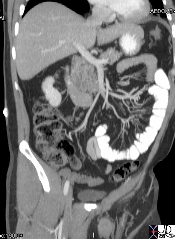
Superior mesenteric Vein to the Portal Vein


colon large bowel middle coloc vein mesenteric vein jejunun jeunal veins portal vein SMV superior mesenteric vein small bowel mesentery ascending colon venoud drainage normal anatomy
Copyright 2012 Courtesy Ashley Davidoff MD 45444


    [nodeType] => 1
    [parentNode] => (object value omitted)
    [childNodes] => (object value omitted)
    [firstChild] => (object value omitted)
    [lastChild] => (object value omitted)
    [previousSibling] => (object value omitted)
    [nextSibling] => (object value omitted)
    [attributes] => (object value omitted)
    [ownerDocument] => (object value omitted)
    [namespaceURI] =>
    [prefix] =>
    [localName] => table
    [baseURI] =>
    [textContent] =>


Superior mesenteric Vein to the Portal Vein


colon large bowel middle coloc vein mesenteric vein jejunun jeunal veins portal vein SMV superior mesenteric vein small bowel mesentery ascending colon venoud drainage normal anatomy
Copyright 2012 Courtesy Ashley Davidoff MD 45444


)
DOMElement Object
(
    [schemaTypeInfo] =>
    [tagName] => td
    [firstElementChild] => (object value omitted)
    [lastElementChild] => (object value omitted)
    [childElementCount] => 2
    [previousElementSibling] =>
    [nextElementSibling] =>
    [nodeName] => td
    [nodeValue] =>
colon large bowel middle coloc vein mesenteric vein jejunun jeunal veins portal vein SMV superior mesenteric vein small bowel mesentery ascending colon venoud drainage normal anatomy
Copyright 2012 Courtesy Ashley Davidoff MD 45444

    [nodeType] => 1
    [parentNode] => (object value omitted)
    [childNodes] => (object value omitted)
    [firstChild] => (object value omitted)
    [lastChild] => (object value omitted)
    [previousSibling] => (object value omitted)
    [nextSibling] => (object value omitted)
    [attributes] => (object value omitted)
    [ownerDocument] => (object value omitted)
    [namespaceURI] =>
    [prefix] =>
    [localName] => td
    [baseURI] =>
    [textContent] =>
colon large bowel middle coloc vein mesenteric vein jejunun jeunal veins portal vein SMV superior mesenteric vein small bowel mesentery ascending colon venoud drainage normal anatomy
Copyright 2012 Courtesy Ashley Davidoff MD 45444

)
DOMElement Object
(
    [schemaTypeInfo] =>
    [tagName] => td
    [firstElementChild] => (object value omitted)
    [lastElementChild] => (object value omitted)
    [childElementCount] => 2
    [previousElementSibling] =>
    [nextElementSibling] =>
    [nodeName] => td
    [nodeValue] =>
Superior mesenteric Vein to the Portal Vein

    [nodeType] => 1
    [parentNode] => (object value omitted)
    [childNodes] => (object value omitted)
    [firstChild] => (object value omitted)
    [lastChild] => (object value omitted)
    [previousSibling] => (object value omitted)
    [nextSibling] => (object value omitted)
    [attributes] => (object value omitted)
    [ownerDocument] => (object value omitted)
    [namespaceURI] =>
    [prefix] =>
    [localName] => td
    [baseURI] =>
    [textContent] =>
Superior mesenteric Vein to the Portal Vein

)
DOMElement Object
(
    [schemaTypeInfo] =>
    [tagName] => table
    [firstElementChild] => (object value omitted)
    [lastElementChild] => (object value omitted)
    [childElementCount] => 1
    [previousElementSibling] => (object value omitted)
    [nextElementSibling] => (object value omitted)
    [nodeName] => table
    [nodeValue] =>


SMV and Portal Vein


small bowel liver colon vein venous drainage SMV portal vein fx normal angiography anatomy
Copyright 2012 Courtesy Ashley Davidoff MD 26567


    [nodeType] => 1
    [parentNode] => (object value omitted)
    [childNodes] => (object value omitted)
    [firstChild] => (object value omitted)
    [lastChild] => (object value omitted)
    [previousSibling] => (object value omitted)
    [nextSibling] => (object value omitted)
    [attributes] => (object value omitted)
    [ownerDocument] => (object value omitted)
    [namespaceURI] =>
    [prefix] =>
    [localName] => table
    [baseURI] =>
    [textContent] =>


SMV and Portal Vein


small bowel liver colon vein venous drainage SMV portal vein fx normal angiography anatomy
Copyright 2012 Courtesy Ashley Davidoff MD 26567


)
DOMElement Object
(
    [schemaTypeInfo] =>
    [tagName] => td
    [firstElementChild] => (object value omitted)
    [lastElementChild] => (object value omitted)
    [childElementCount] => 2
    [previousElementSibling] =>
    [nextElementSibling] =>
    [nodeName] => td
    [nodeValue] =>
small bowel liver colon vein venous drainage SMV portal vein fx normal angiography anatomy
Copyright 2012 Courtesy Ashley Davidoff MD 26567

    [nodeType] => 1
    [parentNode] => (object value omitted)
    [childNodes] => (object value omitted)
    [firstChild] => (object value omitted)
    [lastChild] => (object value omitted)
    [previousSibling] => (object value omitted)
    [nextSibling] => (object value omitted)
    [attributes] => (object value omitted)
    [ownerDocument] => (object value omitted)
    [namespaceURI] =>
    [prefix] =>
    [localName] => td
    [baseURI] =>
    [textContent] =>
small bowel liver colon vein venous drainage SMV portal vein fx normal angiography anatomy
Copyright 2012 Courtesy Ashley Davidoff MD 26567

)
DOMElement Object
(
    [schemaTypeInfo] =>
    [tagName] => td
    [firstElementChild] => (object value omitted)
    [lastElementChild] => (object value omitted)
    [childElementCount] => 2
    [previousElementSibling] =>
    [nextElementSibling] =>
    [nodeName] => td
    [nodeValue] =>
SMV and Portal Vein

    [nodeType] => 1
    [parentNode] => (object value omitted)
    [childNodes] => (object value omitted)
    [firstChild] => (object value omitted)
    [lastChild] => (object value omitted)
    [previousSibling] => (object value omitted)
    [nextSibling] => (object value omitted)
    [attributes] => (object value omitted)
    [ownerDocument] => (object value omitted)
    [namespaceURI] =>
    [prefix] =>
    [localName] => td
    [baseURI] =>
    [textContent] =>
SMV and Portal Vein

)
DOMElement Object
(
    [schemaTypeInfo] =>
    [tagName] => table
    [firstElementChild] => (object value omitted)
    [lastElementChild] => (object value omitted)
    [childElementCount] => 1
    [previousElementSibling] => (object value omitted)
    [nextElementSibling] => (object value omitted)
    [nodeName] => table
    [nodeValue] =>


Splenic Vein and Portal Vein


26625 liver + vein + portal + fx normal + anatomy angiography
Copyright 2012 Courtesy Ashley Davidoff MD 26625


    [nodeType] => 1
    [parentNode] => (object value omitted)
    [childNodes] => (object value omitted)
    [firstChild] => (object value omitted)
    [lastChild] => (object value omitted)
    [previousSibling] => (object value omitted)
    [nextSibling] => (object value omitted)
    [attributes] => (object value omitted)
    [ownerDocument] => (object value omitted)
    [namespaceURI] =>
    [prefix] =>
    [localName] => table
    [baseURI] =>
    [textContent] =>


Splenic Vein and Portal Vein


26625 liver + vein + portal + fx normal + anatomy angiography
Copyright 2012 Courtesy Ashley Davidoff MD 26625


)
DOMElement Object
(
    [schemaTypeInfo] =>
    [tagName] => td
    [firstElementChild] => (object value omitted)
    [lastElementChild] => (object value omitted)
    [childElementCount] => 2
    [previousElementSibling] =>
    [nextElementSibling] =>
    [nodeName] => td
    [nodeValue] =>
26625 liver + vein + portal + fx normal + anatomy angiography
Copyright 2012 Courtesy Ashley Davidoff MD 26625

    [nodeType] => 1
    [parentNode] => (object value omitted)
    [childNodes] => (object value omitted)
    [firstChild] => (object value omitted)
    [lastChild] => (object value omitted)
    [previousSibling] => (object value omitted)
    [nextSibling] => (object value omitted)
    [attributes] => (object value omitted)
    [ownerDocument] => (object value omitted)
    [namespaceURI] =>
    [prefix] =>
    [localName] => td
    [baseURI] =>
    [textContent] =>
26625 liver + vein + portal + fx normal + anatomy angiography
Copyright 2012 Courtesy Ashley Davidoff MD 26625

)
DOMElement Object
(
    [schemaTypeInfo] =>
    [tagName] => td
    [firstElementChild] => (object value omitted)
    [lastElementChild] => (object value omitted)
    [childElementCount] => 2
    [previousElementSibling] =>
    [nextElementSibling] =>
    [nodeName] => td
    [nodeValue] =>
Splenic Vein and Portal Vein

    [nodeType] => 1
    [parentNode] => (object value omitted)
    [childNodes] => (object value omitted)
    [firstChild] => (object value omitted)
    [lastChild] => (object value omitted)
    [previousSibling] => (object value omitted)
    [nextSibling] => (object value omitted)
    [attributes] => (object value omitted)
    [ownerDocument] => (object value omitted)
    [namespaceURI] =>
    [prefix] =>
    [localName] => td
    [baseURI] =>
    [textContent] =>
Splenic Vein and Portal Vein

)
DOMElement Object
(
    [schemaTypeInfo] =>
    [tagName] => table
    [firstElementChild] => (object value omitted)
    [lastElementChild] => (object value omitted)
    [childElementCount] => 1
    [previousElementSibling] => (object value omitted)
    [nextElementSibling] => (object value omitted)
    [nodeName] => table
    [nodeValue] =>


The Porta Hepatis – Portl Vein – A Major Component


liver bile duct gallbladder pancreas spleen splenic vein splenic artery portal vein hepatic artery fx normal anatomy drawing art
Copyright 2012 Courtesy Ashley Davidoff MD 41774


    [nodeType] => 1
    [parentNode] => (object value omitted)
    [childNodes] => (object value omitted)
    [firstChild] => (object value omitted)
    [lastChild] => (object value omitted)
    [previousSibling] => (object value omitted)
    [nextSibling] => (object value omitted)
    [attributes] => (object value omitted)
    [ownerDocument] => (object value omitted)
    [namespaceURI] =>
    [prefix] =>
    [localName] => table
    [baseURI] =>
    [textContent] =>


The Porta Hepatis – Portl Vein – A Major Component


liver bile duct gallbladder pancreas spleen splenic vein splenic artery portal vein hepatic artery fx normal anatomy drawing art
Copyright 2012 Courtesy Ashley Davidoff MD 41774


)
DOMElement Object
(
    [schemaTypeInfo] =>
    [tagName] => td
    [firstElementChild] => (object value omitted)
    [lastElementChild] => (object value omitted)
    [childElementCount] => 2
    [previousElementSibling] =>
    [nextElementSibling] =>
    [nodeName] => td
    [nodeValue] =>
liver bile duct gallbladder pancreas spleen splenic vein splenic artery portal vein hepatic artery fx normal anatomy drawing art
Copyright 2012 Courtesy Ashley Davidoff MD 41774

    [nodeType] => 1
    [parentNode] => (object value omitted)
    [childNodes] => (object value omitted)
    [firstChild] => (object value omitted)
    [lastChild] => (object value omitted)
    [previousSibling] => (object value omitted)
    [nextSibling] => (object value omitted)
    [attributes] => (object value omitted)
    [ownerDocument] => (object value omitted)
    [namespaceURI] =>
    [prefix] =>
    [localName] => td
    [baseURI] =>
    [textContent] =>
liver bile duct gallbladder pancreas spleen splenic vein splenic artery portal vein hepatic artery fx normal anatomy drawing art
Copyright 2012 Courtesy Ashley Davidoff MD 41774

)
DOMElement Object
(
    [schemaTypeInfo] =>
    [tagName] => td
    [firstElementChild] => (object value omitted)
    [lastElementChild] => (object value omitted)
    [childElementCount] => 2
    [previousElementSibling] =>
    [nextElementSibling] =>
    [nodeName] => td
    [nodeValue] =>
The Porta Hepatis – Portl Vein – A Major Component

    [nodeType] => 1
    [parentNode] => (object value omitted)
    [childNodes] => (object value omitted)
    [firstChild] => (object value omitted)
    [lastChild] => (object value omitted)
    [previousSibling] => (object value omitted)
    [nextSibling] => (object value omitted)
    [attributes] => (object value omitted)
    [ownerDocument] => (object value omitted)
    [namespaceURI] =>
    [prefix] =>
    [localName] => td
    [baseURI] =>
    [textContent] =>
The Porta Hepatis – Portl Vein – A Major Component

)

