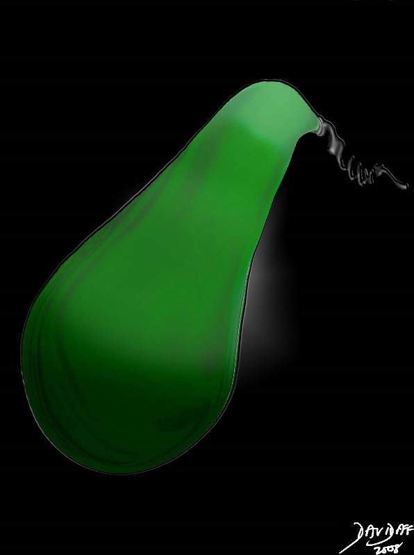
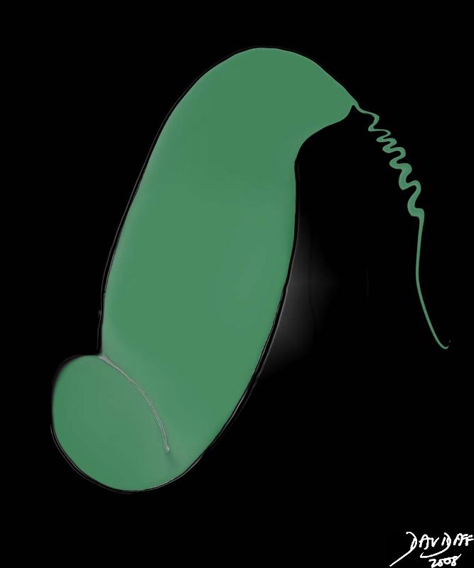
The gallbladder is characteristically pear-shaped (pyriform), oval, or cone shaped structure, but may be rounded, elongated, or angulated. The fundus has the largest diameter and is in general spherical, while the shape of the body of the gallbladder varies between tubular and ovoid , and the neck tends to be triangular as it becomes narrow and tapered with an overall sigmoid curve with the terminating with a downward turn to meet the cystic duct. There is a progressive diminution of diameter from the fundus to the neck.
When viewed macroscopically the internal surface has fine mucosal folds, but when viewed in the distended state by imaging techniques it is smooth. The many normal variations of the shape of the gallbladder will be discussed as we delve its depths in health and disease.
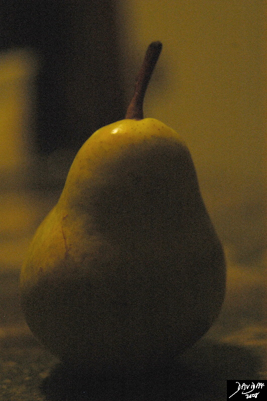 |
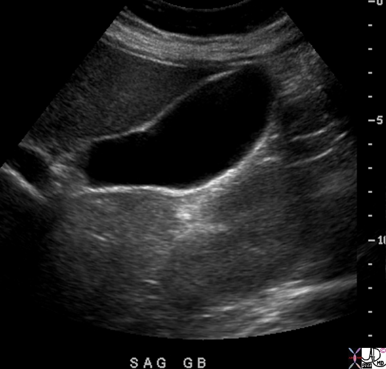
The classical pear shaped form is not the classical shape seen in imaging. The normal gallbladder by ultrasound is usually more elongated and its shape is coser to a zuchini than a pear.
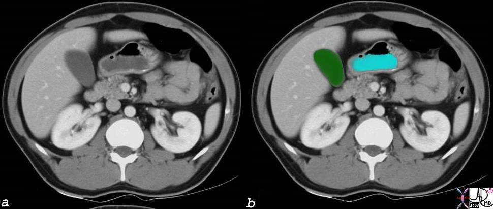
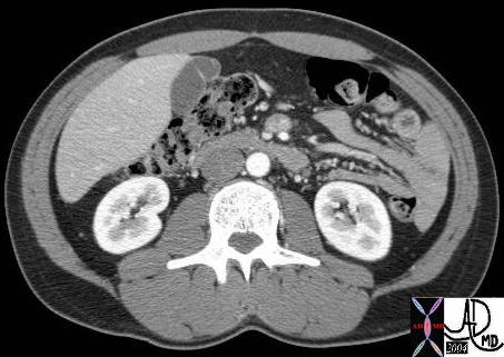
CT scanning is not performed along the long and true axis of the galbladder and sometimes therefore the pear shape is apparent as an artifact of the inaccurate plane acquired.
In the transverse view of the gallbladder as depicted by ultrasound below, it is seen as ovoid in shape. The intraluminal pressure of the gallbladder is normally relatively low and therefore the surface of the galbladder is flattened by normal structures that exert a higher pressure. In the example below the gallbladder is flattened by the normal liver resulting in an oval shape, inferring a low pressure state. We therefore use the shape of the gallbladder to infer intraluminal pressure.
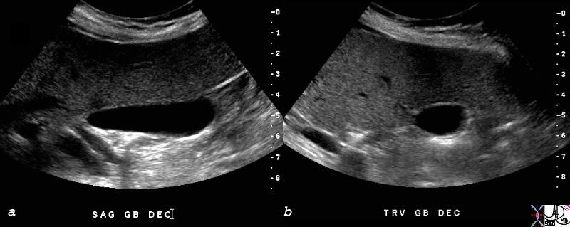
The Shape of the Gallbladder in Transverse Section |
| 82428c01.8s gallbladder small normal transverse oval shape normal anatomy USscan ultrasound copyright 2008 Courtesy Ashley DAvidoff MD |
Variations
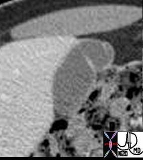
Phrygian cap
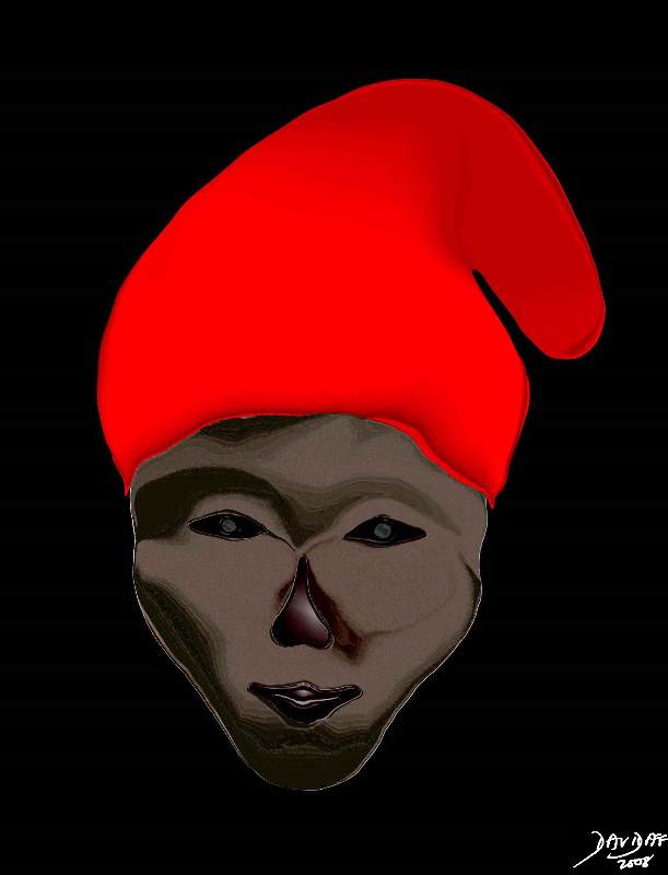
The phrygian cap is the most interesting shape of the galbladder with a fascinating cultural history. (Wiki) It is a normal variant of gallbladder shape occurring in 4% of the population and is characterized by a fundus that is folded forward. It was first described by Bartel in 1916. The Phrygians were a primitive race that migrated into northern and central Asia Minor in the second millenium BC. The Greeks knew of the cap as an Oriental headgear that was tight fitting and conical in shape and was depicted in their art. (Ober)
The cap was a symbol of freedom and liberty. It was worn by King Midas who was cursed with donkey ears by Apollo. He used the Phrygian cap to hide his ears. It was subsequently worn by the Trojan hero Paris, was a symbol of freedom in the French and American revolutions, was depicted on coinage in the US, and also depicted on variety of seals and flags throughout the world.
|
Phrygian cap |
|
The CT scan shows a fundal fold creating a phrygian cap 39696 39696b Courtesy Ashley Davidoff MD gallbladder normal anatomy shape phrygian cap |
Other Folds
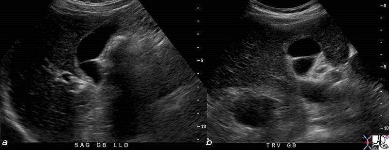
Folds between the body and neck are also relatively common.
Unusual but Normal
There are a variety of shapes that are normal variants. The sigmoid shaped gallbladder , and the Zuchini gallbladder shown below are two of examples.
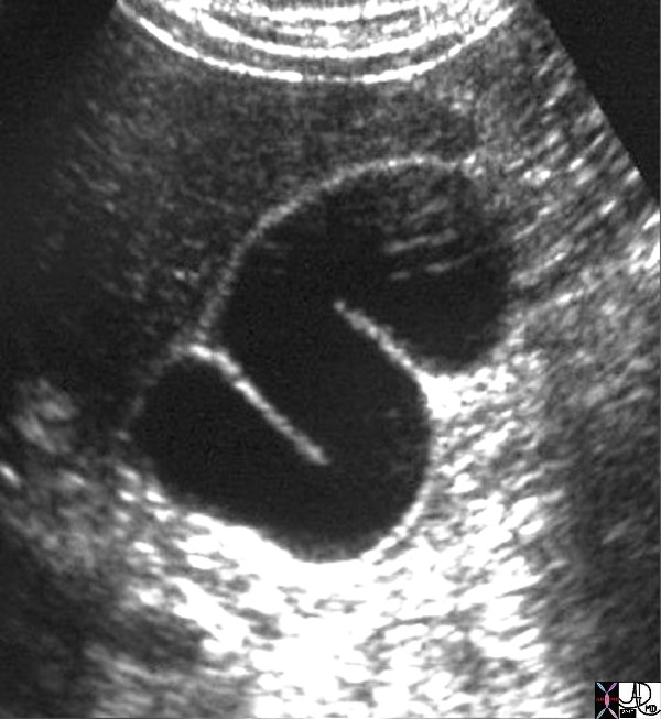
?S? shaped Gallbladder |
| 28470 gallbladder normal shape S-shape anatomy USscan Davidoff MD |
bnormal Shapes
Rotund Gallbladder (Chunky)
When the gallbladder truly starts to look like a pear, with a rotund fundus it is usually a sign of enlargement. The diameter of the fundus is the largest diameter and therefore by Lapalace?s law will have the highest tension on its walls and will therefore dilate first.
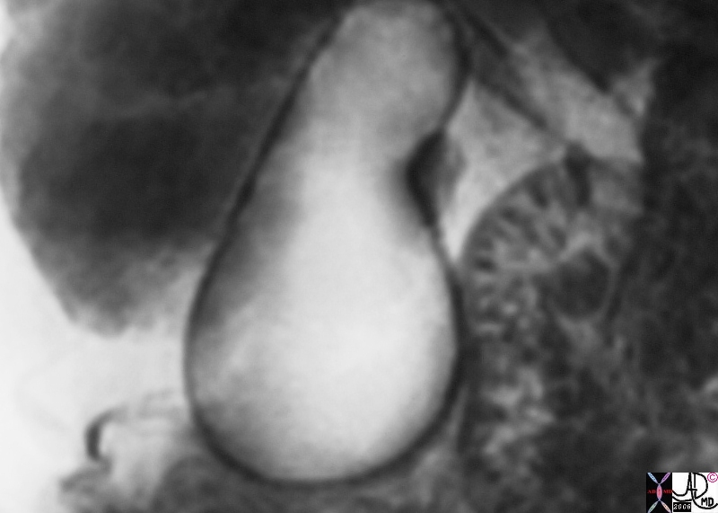 Pear Shaped Gallbladder Pear Shaped Gallbladder |
| 17082b01.8s gallbladder + fx distended + wall edema dx distal common bile duct stone imaging radiology MRI |
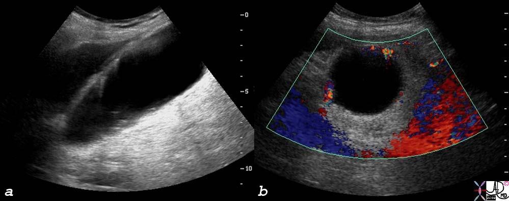
The rounded shape of enlargement reflecting increased pressure is seen also in the body of the gallbladder in transverse projection in the case of acute cholecystitis below.
Figure of 8
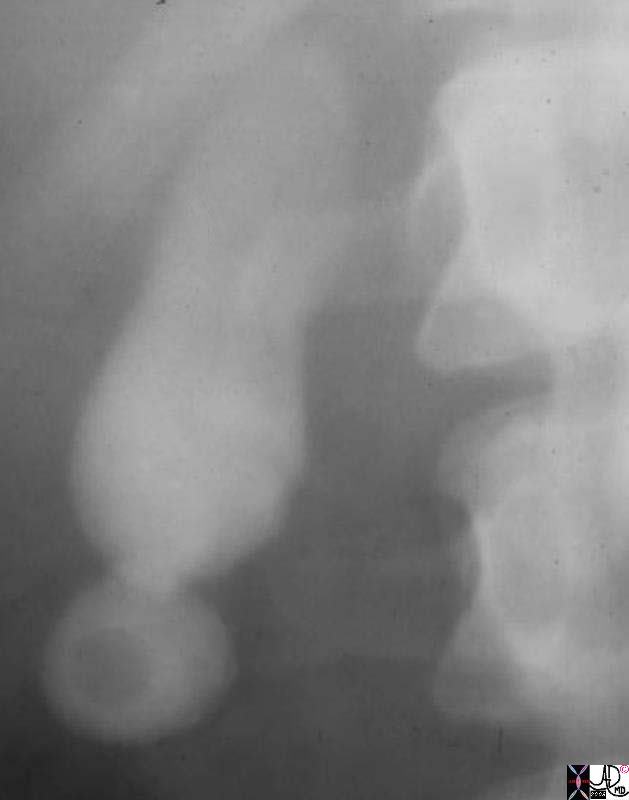
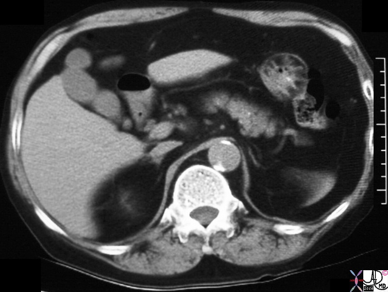
A gallstone sometimes lodges in the fundus, indices inflammation and fibrosis and gets trapped in a funfal pocket resulting in a ?figure of 8? shape to the gallbladder.
References
Block Berthold The Practice Of Ultrasound Thieme 2004
Ober WB, Wharton RN On the ?phrygian cap.? 255:571-572 New England Journal of Medicine 1956
DOMElement Object
(
    [schemaTypeInfo] =>
    [tagName] => table
    [firstElementChild] => (object value omitted)
    [lastElementChild] => (object value omitted)
    [childElementCount] => 1
    [previousElementSibling] => (object value omitted)
    [nextElementSibling] => (object value omitted)
    [nodeName] => table
    [nodeValue] =>


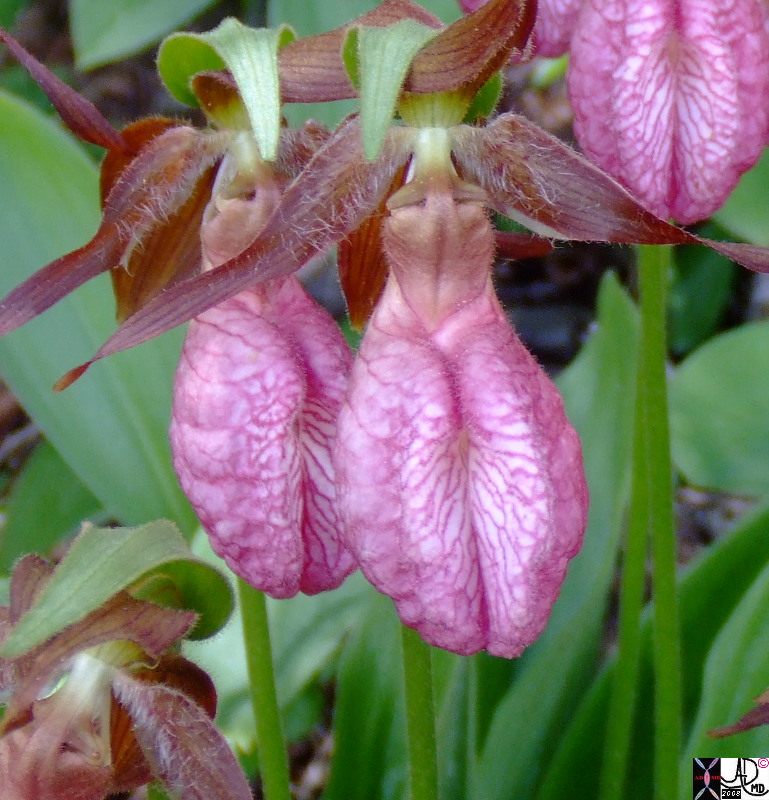
Pyriform Shape of the Lady?s Slipper


The Lady?s slipper (aka moccasin flower) is characterized by its slipper-shaped pouches, which are adapted to  trap insects so that the insects transport the  pollen of the flower.  It has a striking resemblance to the shape of the gallbladder as well ? admittedly the name lady?s slipper is far more elegant than gallbladder flower!89698p.8s flower lady?s slipper pear shape pyriform shape Courtesy Jamie Armstrong PA Copyright 2008


    [nodeType] => 1
    [parentNode] => (object value omitted)
    [childNodes] => (object value omitted)
    [firstChild] => (object value omitted)
    [lastChild] => (object value omitted)
    [previousSibling] => (object value omitted)
    [nextSibling] => (object value omitted)
    [attributes] => (object value omitted)
    [ownerDocument] => (object value omitted)
    [namespaceURI] =>
    [prefix] =>
    [localName] => table
    [baseURI] =>
    [textContent] =>


Pyriform Shape of the Lady?s Slipper


The Lady?s slipper (aka moccasin flower) is characterized by its slipper-shaped pouches, which are adapted to  trap insects so that the insects transport the  pollen of the flower.  It has a striking resemblance to the shape of the gallbladder as well ? admittedly the name lady?s slipper is far more elegant than gallbladder flower!89698p.8s flower lady?s slipper pear shape pyriform shape Courtesy Jamie Armstrong PA Copyright 2008


)
DOMElement Object
(
    [schemaTypeInfo] =>
    [tagName] => td
    [firstElementChild] =>
    [lastElementChild] =>
    [childElementCount] => 0
    [previousElementSibling] =>
    [nextElementSibling] =>
    [nodeName] => td
    [nodeValue] => The Lady?s slipper (aka moccasin flower) is characterized by its slipper-shaped pouches, which are adapted to  trap insects so that the insects transport the  pollen of the flower.  It has a striking resemblance to the shape of the gallbladder as well ? admittedly the name lady?s slipper is far more elegant than gallbladder flower!89698p.8s flower lady?s slipper pear shape pyriform shape Courtesy Jamie Armstrong PA Copyright 2008
    [nodeType] => 1
    [parentNode] => (object value omitted)
    [childNodes] => (object value omitted)
    [firstChild] => (object value omitted)
    [lastChild] => (object value omitted)
    [previousSibling] => (object value omitted)
    [nextSibling] => (object value omitted)
    [attributes] => (object value omitted)
    [ownerDocument] => (object value omitted)
    [namespaceURI] =>
    [prefix] =>
    [localName] => td
    [baseURI] =>
    [textContent] => The Lady?s slipper (aka moccasin flower) is characterized by its slipper-shaped pouches, which are adapted to  trap insects so that the insects transport the  pollen of the flower.  It has a striking resemblance to the shape of the gallbladder as well ? admittedly the name lady?s slipper is far more elegant than gallbladder flower!89698p.8s flower lady?s slipper pear shape pyriform shape Courtesy Jamie Armstrong PA Copyright 2008
)
DOMElement Object
(
    [schemaTypeInfo] =>
    [tagName] => td
    [firstElementChild] => (object value omitted)
    [lastElementChild] => (object value omitted)
    [childElementCount] => 2
    [previousElementSibling] =>
    [nextElementSibling] =>
    [nodeName] => td
    [nodeValue] => Pyriform Shape of the Lady?s Slipper
    [nodeType] => 1
    [parentNode] => (object value omitted)
    [childNodes] => (object value omitted)
    [firstChild] => (object value omitted)
    [lastChild] => (object value omitted)
    [previousSibling] => (object value omitted)
    [nextSibling] => (object value omitted)
    [attributes] => (object value omitted)
    [ownerDocument] => (object value omitted)
    [namespaceURI] =>
    [prefix] =>
    [localName] => td
    [baseURI] =>
    [textContent] => Pyriform Shape of the Lady?s Slipper
)
DOMElement Object
(
    [schemaTypeInfo] =>
    [tagName] => table
    [firstElementChild] => (object value omitted)
    [lastElementChild] => (object value omitted)
    [childElementCount] => 1
    [previousElementSibling] => (object value omitted)
    [nextElementSibling] => (object value omitted)
    [nodeName] => table
    [nodeValue] =>


Chronic Cholecystitis


The CTscan shows a similar figure of 8 appearance of thebgallbladder, but no radioopaque stone is visible
31285.8s 64 male gallbladder fibrosis across the fundus filling defect chronic cholecystitis cholelithiasis stone in fundus Ctscan figure of eight stricture Courtesy Ashley Davidoff MD copyright 2008


    [nodeType] => 1
    [parentNode] => (object value omitted)
    [childNodes] => (object value omitted)
    [firstChild] => (object value omitted)
    [lastChild] => (object value omitted)
    [previousSibling] => (object value omitted)
    [nextSibling] => (object value omitted)
    [attributes] => (object value omitted)
    [ownerDocument] => (object value omitted)
    [namespaceURI] =>
    [prefix] =>
    [localName] => table
    [baseURI] =>
    [textContent] =>


Chronic Cholecystitis


The CTscan shows a similar figure of 8 appearance of thebgallbladder, but no radioopaque stone is visible
31285.8s 64 male gallbladder fibrosis across the fundus filling defect chronic cholecystitis cholelithiasis stone in fundus Ctscan figure of eight stricture Courtesy Ashley Davidoff MD copyright 2008


)
DOMElement Object
(
    [schemaTypeInfo] =>
    [tagName] => td
    [firstElementChild] => (object value omitted)
    [lastElementChild] => (object value omitted)
    [childElementCount] => 2
    [previousElementSibling] =>
    [nextElementSibling] =>
    [nodeName] => td
    [nodeValue] =>
The CTscan shows a similar figure of 8 appearance of thebgallbladder, but no radioopaque stone is visible
31285.8s 64 male gallbladder fibrosis across the fundus filling defect chronic cholecystitis cholelithiasis stone in fundus Ctscan figure of eight stricture Courtesy Ashley Davidoff MD copyright 2008
    [nodeType] => 1
    [parentNode] => (object value omitted)
    [childNodes] => (object value omitted)
    [firstChild] => (object value omitted)
    [lastChild] => (object value omitted)
    [previousSibling] => (object value omitted)
    [nextSibling] => (object value omitted)
    [attributes] => (object value omitted)
    [ownerDocument] => (object value omitted)
    [namespaceURI] =>
    [prefix] =>
    [localName] => td
    [baseURI] =>
    [textContent] =>
The CTscan shows a similar figure of 8 appearance of thebgallbladder, but no radioopaque stone is visible
31285.8s 64 male gallbladder fibrosis across the fundus filling defect chronic cholecystitis cholelithiasis stone in fundus Ctscan figure of eight stricture Courtesy Ashley Davidoff MD copyright 2008
)
DOMElement Object
(
    [schemaTypeInfo] =>
    [tagName] => td
    [firstElementChild] => (object value omitted)
    [lastElementChild] => (object value omitted)
    [childElementCount] => 2
    [previousElementSibling] =>
    [nextElementSibling] =>
    [nodeName] => td
    [nodeValue] =>
Chronic Cholecystitis

    [nodeType] => 1
    [parentNode] => (object value omitted)
    [childNodes] => (object value omitted)
    [firstChild] => (object value omitted)
    [lastChild] => (object value omitted)
    [previousSibling] => (object value omitted)
    [nextSibling] => (object value omitted)
    [attributes] => (object value omitted)
    [ownerDocument] => (object value omitted)
    [namespaceURI] =>
    [prefix] =>
    [localName] => td
    [baseURI] =>
    [textContent] =>
Chronic Cholecystitis

)
DOMElement Object
(
    [schemaTypeInfo] =>
    [tagName] => table
    [firstElementChild] => (object value omitted)
    [lastElementChild] => (object value omitted)
    [childElementCount] => 1
    [previousElementSibling] => (object value omitted)
    [nextElementSibling] => (object value omitted)
    [nodeName] => table
    [nodeValue] =>


Figure of 8
Chronic Cholecystitis


Sometimes a stone is trapped in the fundus of the gallbladder and it induces a chronic inflammatory process resulting in inflammation and subsequent fibrosis.  A separate fundal chamber evolves, giving the gallbladder a figure of 8 appearance.  In this oral cholecystogram a filling defect in the fundus of the gallbladder represents a stone and it is trapped by a ring of fibrosis it has induced.
11530.81s gallbladder fibrosis across the fundus filling defect chronic cholecystitis cholelithiasis stone in fundus OCG historical stricture Courtesy Ashley Davidoff MD copyright 2008


    [nodeType] => 1
    [parentNode] => (object value omitted)
    [childNodes] => (object value omitted)
    [firstChild] => (object value omitted)
    [lastChild] => (object value omitted)
    [previousSibling] => (object value omitted)
    [nextSibling] => (object value omitted)
    [attributes] => (object value omitted)
    [ownerDocument] => (object value omitted)
    [namespaceURI] =>
    [prefix] =>
    [localName] => table
    [baseURI] =>
    [textContent] =>


Figure of 8
Chronic Cholecystitis


Sometimes a stone is trapped in the fundus of the gallbladder and it induces a chronic inflammatory process resulting in inflammation and subsequent fibrosis.  A separate fundal chamber evolves, giving the gallbladder a figure of 8 appearance.  In this oral cholecystogram a filling defect in the fundus of the gallbladder represents a stone and it is trapped by a ring of fibrosis it has induced.
11530.81s gallbladder fibrosis across the fundus filling defect chronic cholecystitis cholelithiasis stone in fundus OCG historical stricture Courtesy Ashley Davidoff MD copyright 2008


)
DOMElement Object
(
    [schemaTypeInfo] =>
    [tagName] => td
    [firstElementChild] => (object value omitted)
    [lastElementChild] => (object value omitted)
    [childElementCount] => 2
    [previousElementSibling] =>
    [nextElementSibling] =>
    [nodeName] => td
    [nodeValue] =>
Sometimes a stone is trapped in the fundus of the gallbladder and it induces a chronic inflammatory process resulting in inflammation and subsequent fibrosis.  A separate fundal chamber evolves, giving the gallbladder a figure of 8 appearance.  In this oral cholecystogram a filling defect in the fundus of the gallbladder represents a stone and it is trapped by a ring of fibrosis it has induced.
11530.81s gallbladder fibrosis across the fundus filling defect chronic cholecystitis cholelithiasis stone in fundus OCG historical stricture Courtesy Ashley Davidoff MD copyright 2008

    [nodeType] => 1
    [parentNode] => (object value omitted)
    [childNodes] => (object value omitted)
    [firstChild] => (object value omitted)
    [lastChild] => (object value omitted)
    [previousSibling] => (object value omitted)
    [nextSibling] => (object value omitted)
    [attributes] => (object value omitted)
    [ownerDocument] => (object value omitted)
    [namespaceURI] =>
    [prefix] =>
    [localName] => td
    [baseURI] =>
    [textContent] =>
Sometimes a stone is trapped in the fundus of the gallbladder and it induces a chronic inflammatory process resulting in inflammation and subsequent fibrosis.  A separate fundal chamber evolves, giving the gallbladder a figure of 8 appearance.  In this oral cholecystogram a filling defect in the fundus of the gallbladder represents a stone and it is trapped by a ring of fibrosis it has induced.
11530.81s gallbladder fibrosis across the fundus filling defect chronic cholecystitis cholelithiasis stone in fundus OCG historical stricture Courtesy Ashley Davidoff MD copyright 2008

)
DOMElement Object
(
    [schemaTypeInfo] =>
    [tagName] => td
    [firstElementChild] => (object value omitted)
    [lastElementChild] => (object value omitted)
    [childElementCount] => 3
    [previousElementSibling] =>
    [nextElementSibling] =>
    [nodeName] => td
    [nodeValue] =>
Figure of 8
Chronic Cholecystitis

    [nodeType] => 1
    [parentNode] => (object value omitted)
    [childNodes] => (object value omitted)
    [firstChild] => (object value omitted)
    [lastChild] => (object value omitted)
    [previousSibling] => (object value omitted)
    [nextSibling] => (object value omitted)
    [attributes] => (object value omitted)
    [ownerDocument] => (object value omitted)
    [namespaceURI] =>
    [prefix] =>
    [localName] => td
    [baseURI] =>
    [textContent] =>
Figure of 8
Chronic Cholecystitis

)
DOMElement Object
(
    [schemaTypeInfo] =>
    [tagName] => table
    [firstElementChild] => (object value omitted)
    [lastElementChild] => (object value omitted)
    [childElementCount] => 1
    [previousElementSibling] => (object value omitted)
    [nextElementSibling] => (object value omitted)
    [nodeName] => table
    [nodeValue] =>


Hyperplastic Cholecystosis with Acute Cholecystitis filled with stones in the Fundus


In this ultrasound of the gallbladder is from a patient with right upper quadrant pain the gallbladder in the transverse plane measures 5cms and is round in shape rather than oval.  The diagnosis was acute cholecystitis.
78434c01s right upper quadrant pain positive Murphy?s sign gallbladder outpouchings diverticula prominent Aschoff Rokitansky stones ring down artifact thickened wall hyperemic wall by doppler hyperplastic cholecystosis hyperplastic cholecystoses adenomyomatosis acute cholecystitis USscan ultrasound courtesy Ashley Davidoff MD copyright 2008


    [nodeType] => 1
    [parentNode] => (object value omitted)
    [childNodes] => (object value omitted)
    [firstChild] => (object value omitted)
    [lastChild] => (object value omitted)
    [previousSibling] => (object value omitted)
    [nextSibling] => (object value omitted)
    [attributes] => (object value omitted)
    [ownerDocument] => (object value omitted)
    [namespaceURI] =>
    [prefix] =>
    [localName] => table
    [baseURI] =>
    [textContent] =>


Hyperplastic Cholecystosis with Acute Cholecystitis filled with stones in the Fundus


In this ultrasound of the gallbladder is from a patient with right upper quadrant pain the gallbladder in the transverse plane measures 5cms and is round in shape rather than oval.  The diagnosis was acute cholecystitis.
78434c01s right upper quadrant pain positive Murphy?s sign gallbladder outpouchings diverticula prominent Aschoff Rokitansky stones ring down artifact thickened wall hyperemic wall by doppler hyperplastic cholecystosis hyperplastic cholecystoses adenomyomatosis acute cholecystitis USscan ultrasound courtesy Ashley Davidoff MD copyright 2008


)
DOMElement Object
(
    [schemaTypeInfo] =>
    [tagName] => td
    [firstElementChild] => (object value omitted)
    [lastElementChild] => (object value omitted)
    [childElementCount] => 2
    [previousElementSibling] =>
    [nextElementSibling] =>
    [nodeName] => td
    [nodeValue] =>
In this ultrasound of the gallbladder is from a patient with right upper quadrant pain the gallbladder in the transverse plane measures 5cms and is round in shape rather than oval.  The diagnosis was acute cholecystitis.
78434c01s right upper quadrant pain positive Murphy?s sign gallbladder outpouchings diverticula prominent Aschoff Rokitansky stones ring down artifact thickened wall hyperemic wall by doppler hyperplastic cholecystosis hyperplastic cholecystoses adenomyomatosis acute cholecystitis USscan ultrasound courtesy Ashley Davidoff MD copyright 2008

    [nodeType] => 1
    [parentNode] => (object value omitted)
    [childNodes] => (object value omitted)
    [firstChild] => (object value omitted)
    [lastChild] => (object value omitted)
    [previousSibling] => (object value omitted)
    [nextSibling] => (object value omitted)
    [attributes] => (object value omitted)
    [ownerDocument] => (object value omitted)
    [namespaceURI] =>
    [prefix] =>
    [localName] => td
    [baseURI] =>
    [textContent] =>
In this ultrasound of the gallbladder is from a patient with right upper quadrant pain the gallbladder in the transverse plane measures 5cms and is round in shape rather than oval.  The diagnosis was acute cholecystitis.
78434c01s right upper quadrant pain positive Murphy?s sign gallbladder outpouchings diverticula prominent Aschoff Rokitansky stones ring down artifact thickened wall hyperemic wall by doppler hyperplastic cholecystosis hyperplastic cholecystoses adenomyomatosis acute cholecystitis USscan ultrasound courtesy Ashley Davidoff MD copyright 2008

)
DOMElement Object
(
    [schemaTypeInfo] =>
    [tagName] => td
    [firstElementChild] => (object value omitted)
    [lastElementChild] => (object value omitted)
    [childElementCount] => 2
    [previousElementSibling] =>
    [nextElementSibling] =>
    [nodeName] => td
    [nodeValue] =>
Hyperplastic Cholecystosis with Acute Cholecystitis filled with stones in the Fundus

    [nodeType] => 1
    [parentNode] => (object value omitted)
    [childNodes] => (object value omitted)
    [firstChild] => (object value omitted)
    [lastChild] => (object value omitted)
    [previousSibling] => (object value omitted)
    [nextSibling] => (object value omitted)
    [attributes] => (object value omitted)
    [ownerDocument] => (object value omitted)
    [namespaceURI] =>
    [prefix] =>
    [localName] => td
    [baseURI] =>
    [textContent] =>
Hyperplastic Cholecystosis with Acute Cholecystitis filled with stones in the Fundus

)
DOMElement Object
(
    [schemaTypeInfo] =>
    [tagName] => table
    [firstElementChild] => (object value omitted)
    [lastElementChild] => (object value omitted)
    [childElementCount] => 1
    [previousElementSibling] => (object value omitted)
    [nextElementSibling] => (object value omitted)
    [nodeName] => table
    [nodeValue] =>


Pear Shaped Gallbladder


17082b01.8s gallbladder + fx distended + wall edema dx distal common bile duct stone imaging radiology MRI


    [nodeType] => 1
    [parentNode] => (object value omitted)
    [childNodes] => (object value omitted)
    [firstChild] => (object value omitted)
    [lastChild] => (object value omitted)
    [previousSibling] => (object value omitted)
    [nextSibling] => (object value omitted)
    [attributes] => (object value omitted)
    [ownerDocument] => (object value omitted)
    [namespaceURI] =>
    [prefix] =>
    [localName] => table
    [baseURI] =>
    [textContent] =>


Pear Shaped Gallbladder


17082b01.8s gallbladder + fx distended + wall edema dx distal common bile duct stone imaging radiology MRI


)
DOMElement Object
(
    [schemaTypeInfo] =>
    [tagName] => td
    [firstElementChild] =>
    [lastElementChild] =>
    [childElementCount] => 0
    [previousElementSibling] =>
    [nextElementSibling] =>
    [nodeName] => td
    [nodeValue] => 17082b01.8s gallbladder + fx distended + wall edema dx distal common bile duct stone imaging radiology MRI
    [nodeType] => 1
    [parentNode] => (object value omitted)
    [childNodes] => (object value omitted)
    [firstChild] => (object value omitted)
    [lastChild] => (object value omitted)
    [previousSibling] => (object value omitted)
    [nextSibling] => (object value omitted)
    [attributes] => (object value omitted)
    [ownerDocument] => (object value omitted)
    [namespaceURI] =>
    [prefix] =>
    [localName] => td
    [baseURI] =>
    [textContent] => 17082b01.8s gallbladder + fx distended + wall edema dx distal common bile duct stone imaging radiology MRI
)
DOMElement Object
(
    [schemaTypeInfo] =>
    [tagName] => td
    [firstElementChild] => (object value omitted)
    [lastElementChild] => (object value omitted)
    [childElementCount] => 2
    [previousElementSibling] =>
    [nextElementSibling] =>
    [nodeName] => td
    [nodeValue] => Pear Shaped Gallbladder
    [nodeType] => 1
    [parentNode] => (object value omitted)
    [childNodes] => (object value omitted)
    [firstChild] => (object value omitted)
    [lastChild] => (object value omitted)
    [previousSibling] => (object value omitted)
    [nextSibling] => (object value omitted)
    [attributes] => (object value omitted)
    [ownerDocument] => (object value omitted)
    [namespaceURI] =>
    [prefix] =>
    [localName] => td
    [baseURI] =>
    [textContent] => Pear Shaped Gallbladder
)
DOMElement Object
(
    [schemaTypeInfo] =>
    [tagName] => table
    [firstElementChild] => (object value omitted)
    [lastElementChild] => (object value omitted)
    [childElementCount] => 1
    [previousElementSibling] => (object value omitted)
    [nextElementSibling] => (object value omitted)
    [nodeName] => table
    [nodeValue] =>


Elongated but not Dilated ? The Aging Gallbladder
Zuchini Gallbladder


With the aging process elasticity and compliance is diminished and the gallbladder can become voluminous, usually in the long axis.  In the normally atonic bladderseen in the elderly and diabetics,  the transsverse dimension is not usually larger than 4 cms.
76764c.81s elderly female gallbladder elongated sausage shape zuchini shape transverse dimension 4.5 cms longitudinal dimension 12.5cms aging gallbladder vs enlarged? food in the body CTscan Courtesy Ashley DAvidoff MD Davidoff art Davidoff photography copyright 2008


    [nodeType] => 1
    [parentNode] => (object value omitted)
    [childNodes] => (object value omitted)
    [firstChild] => (object value omitted)
    [lastChild] => (object value omitted)
    [previousSibling] => (object value omitted)
    [nextSibling] => (object value omitted)
    [attributes] => (object value omitted)
    [ownerDocument] => (object value omitted)
    [namespaceURI] =>
    [prefix] =>
    [localName] => table
    [baseURI] =>
    [textContent] =>


Elongated but not Dilated ? The Aging Gallbladder
Zuchini Gallbladder


With the aging process elasticity and compliance is diminished and the gallbladder can become voluminous, usually in the long axis.  In the normally atonic bladderseen in the elderly and diabetics,  the transsverse dimension is not usually larger than 4 cms.
76764c.81s elderly female gallbladder elongated sausage shape zuchini shape transverse dimension 4.5 cms longitudinal dimension 12.5cms aging gallbladder vs enlarged? food in the body CTscan Courtesy Ashley DAvidoff MD Davidoff art Davidoff photography copyright 2008


)
DOMElement Object
(
    [schemaTypeInfo] =>
    [tagName] => td
    [firstElementChild] => (object value omitted)
    [lastElementChild] => (object value omitted)
    [childElementCount] => 2
    [previousElementSibling] =>
    [nextElementSibling] =>
    [nodeName] => td
    [nodeValue] =>
With the aging process elasticity and compliance is diminished and the gallbladder can become voluminous, usually in the long axis.  In the normally atonic bladderseen in the elderly and diabetics,  the transsverse dimension is not usually larger than 4 cms.
76764c.81s elderly female gallbladder elongated sausage shape zuchini shape transverse dimension 4.5 cms longitudinal dimension 12.5cms aging gallbladder vs enlarged? food in the body CTscan Courtesy Ashley DAvidoff MD Davidoff art Davidoff photography copyright 2008
    [nodeType] => 1
    [parentNode] => (object value omitted)
    [childNodes] => (object value omitted)
    [firstChild] => (object value omitted)
    [lastChild] => (object value omitted)
    [previousSibling] => (object value omitted)
    [nextSibling] => (object value omitted)
    [attributes] => (object value omitted)
    [ownerDocument] => (object value omitted)
    [namespaceURI] =>
    [prefix] =>
    [localName] => td
    [baseURI] =>
    [textContent] =>
With the aging process elasticity and compliance is diminished and the gallbladder can become voluminous, usually in the long axis.  In the normally atonic bladderseen in the elderly and diabetics,  the transsverse dimension is not usually larger than 4 cms.
76764c.81s elderly female gallbladder elongated sausage shape zuchini shape transverse dimension 4.5 cms longitudinal dimension 12.5cms aging gallbladder vs enlarged? food in the body CTscan Courtesy Ashley DAvidoff MD Davidoff art Davidoff photography copyright 2008
)
DOMElement Object
(
    [schemaTypeInfo] =>
    [tagName] => td
    [firstElementChild] => (object value omitted)
    [lastElementChild] => (object value omitted)
    [childElementCount] => 3
    [previousElementSibling] =>
    [nextElementSibling] =>
    [nodeName] => td
    [nodeValue] =>
Elongated but not Dilated ? The Aging Gallbladder
Zuchini Gallbladder

    [nodeType] => 1
    [parentNode] => (object value omitted)
    [childNodes] => (object value omitted)
    [firstChild] => (object value omitted)
    [lastChild] => (object value omitted)
    [previousSibling] => (object value omitted)
    [nextSibling] => (object value omitted)
    [attributes] => (object value omitted)
    [ownerDocument] => (object value omitted)
    [namespaceURI] =>
    [prefix] =>
    [localName] => td
    [baseURI] =>
    [textContent] =>
Elongated but not Dilated ? The Aging Gallbladder
Zuchini Gallbladder

)
DOMElement Object
(
    [schemaTypeInfo] =>
    [tagName] => table
    [firstElementChild] => (object value omitted)
    [lastElementChild] => (object value omitted)
    [childElementCount] => 1
    [previousElementSibling] => (object value omitted)
    [nextElementSibling] => (object value omitted)
    [nodeName] => table
    [nodeValue] =>


?S? shaped Gallbladder


28470 gallbladder normal shape S-shape anatomy USscan Davidoff MD


    [nodeType] => 1
    [parentNode] => (object value omitted)
    [childNodes] => (object value omitted)
    [firstChild] => (object value omitted)
    [lastChild] => (object value omitted)
    [previousSibling] => (object value omitted)
    [nextSibling] => (object value omitted)
    [attributes] => (object value omitted)
    [ownerDocument] => (object value omitted)
    [namespaceURI] =>
    [prefix] =>
    [localName] => table
    [baseURI] =>
    [textContent] =>


?S? shaped Gallbladder


28470 gallbladder normal shape S-shape anatomy USscan Davidoff MD


)
DOMElement Object
(
    [schemaTypeInfo] =>
    [tagName] => td
    [firstElementChild] =>
    [lastElementChild] =>
    [childElementCount] => 0
    [previousElementSibling] =>
    [nextElementSibling] =>
    [nodeName] => td
    [nodeValue] => 28470 gallbladder normal shape S-shape anatomy USscan Davidoff MD
    [nodeType] => 1
    [parentNode] => (object value omitted)
    [childNodes] => (object value omitted)
    [firstChild] => (object value omitted)
    [lastChild] => (object value omitted)
    [previousSibling] => (object value omitted)
    [nextSibling] => (object value omitted)
    [attributes] => (object value omitted)
    [ownerDocument] => (object value omitted)
    [namespaceURI] =>
    [prefix] =>
    [localName] => td
    [baseURI] =>
    [textContent] => 28470 gallbladder normal shape S-shape anatomy USscan Davidoff MD
)
DOMElement Object
(
    [schemaTypeInfo] =>
    [tagName] => td
    [firstElementChild] => (object value omitted)
    [lastElementChild] => (object value omitted)
    [childElementCount] => 2
    [previousElementSibling] =>
    [nextElementSibling] =>
    [nodeName] => td
    [nodeValue] =>
?S? shaped Gallbladder

    [nodeType] => 1
    [parentNode] => (object value omitted)
    [childNodes] => (object value omitted)
    [firstChild] => (object value omitted)
    [lastChild] => (object value omitted)
    [previousSibling] => (object value omitted)
    [nextSibling] => (object value omitted)
    [attributes] => (object value omitted)
    [ownerDocument] => (object value omitted)
    [namespaceURI] =>
    [prefix] =>
    [localName] => td
    [baseURI] =>
    [textContent] =>
?S? shaped Gallbladder

)
DOMElement Object
(
    [schemaTypeInfo] =>
    [tagName] => table
    [firstElementChild] => (object value omitted)
    [lastElementChild] => (object value omitted)
    [childElementCount] => 1
    [previousElementSibling] => (object value omitted)
    [nextElementSibling] => (object value omitted)
    [nodeName] => table
    [nodeValue] =>


Folds in the Body Neck Area


The ultrasound examination in both the sagittal and transverse plane shows a fold  between the body and the neck of the gallbladder.
82163c02.8s 70 m gallbladder fold between body and neck normal anatomy USscan ultrasound Courtesy Ashley DAvidoff MD copyright 2008


    [nodeType] => 1
    [parentNode] => (object value omitted)
    [childNodes] => (object value omitted)
    [firstChild] => (object value omitted)
    [lastChild] => (object value omitted)
    [previousSibling] => (object value omitted)
    [nextSibling] => (object value omitted)
    [attributes] => (object value omitted)
    [ownerDocument] => (object value omitted)
    [namespaceURI] =>
    [prefix] =>
    [localName] => table
    [baseURI] =>
    [textContent] =>


Folds in the Body Neck Area


The ultrasound examination in both the sagittal and transverse plane shows a fold  between the body and the neck of the gallbladder.
82163c02.8s 70 m gallbladder fold between body and neck normal anatomy USscan ultrasound Courtesy Ashley DAvidoff MD copyright 2008


)
DOMElement Object
(
    [schemaTypeInfo] =>
    [tagName] => td
    [firstElementChild] => (object value omitted)
    [lastElementChild] => (object value omitted)
    [childElementCount] => 2
    [previousElementSibling] =>
    [nextElementSibling] =>
    [nodeName] => td
    [nodeValue] =>
The ultrasound examination in both the sagittal and transverse plane shows a fold  between the body and the neck of the gallbladder.
82163c02.8s 70 m gallbladder fold between body and neck normal anatomy USscan ultrasound Courtesy Ashley DAvidoff MD copyright 2008
    [nodeType] => 1
    [parentNode] => (object value omitted)
    [childNodes] => (object value omitted)
    [firstChild] => (object value omitted)
    [lastChild] => (object value omitted)
    [previousSibling] => (object value omitted)
    [nextSibling] => (object value omitted)
    [attributes] => (object value omitted)
    [ownerDocument] => (object value omitted)
    [namespaceURI] =>
    [prefix] =>
    [localName] => td
    [baseURI] =>
    [textContent] =>
The ultrasound examination in both the sagittal and transverse plane shows a fold  between the body and the neck of the gallbladder.
82163c02.8s 70 m gallbladder fold between body and neck normal anatomy USscan ultrasound Courtesy Ashley DAvidoff MD copyright 2008
)
DOMElement Object
(
    [schemaTypeInfo] =>
    [tagName] => td
    [firstElementChild] => (object value omitted)
    [lastElementChild] => (object value omitted)
    [childElementCount] => 2
    [previousElementSibling] =>
    [nextElementSibling] =>
    [nodeName] => td
    [nodeValue] =>
Folds in the Body Neck Area

    [nodeType] => 1
    [parentNode] => (object value omitted)
    [childNodes] => (object value omitted)
    [firstChild] => (object value omitted)
    [lastChild] => (object value omitted)
    [previousSibling] => (object value omitted)
    [nextSibling] => (object value omitted)
    [attributes] => (object value omitted)
    [ownerDocument] => (object value omitted)
    [namespaceURI] =>
    [prefix] =>
    [localName] => td
    [baseURI] =>
    [textContent] =>
Folds in the Body Neck Area

)
DOMElement Object
(
    [schemaTypeInfo] =>
    [tagName] => table
    [firstElementChild] => (object value omitted)
    [lastElementChild] => (object value omitted)
    [childElementCount] => 1
    [previousElementSibling] => (object value omitted)
    [nextElementSibling] => (object value omitted)
    [nodeName] => table
    [nodeValue] =>


Phrygian cap


The CT scan shows a fundal fold creating a phrygian cap
39696 39696b Courtesy Ashley Davidoff MD gallbladder normal anatomy shape phrygian cap


    [nodeType] => 1
    [parentNode] => (object value omitted)
    [childNodes] => (object value omitted)
    [firstChild] => (object value omitted)
    [lastChild] => (object value omitted)
    [previousSibling] => (object value omitted)
    [nextSibling] => (object value omitted)
    [attributes] => (object value omitted)
    [ownerDocument] => (object value omitted)
    [namespaceURI] =>
    [prefix] =>
    [localName] => table
    [baseURI] =>
    [textContent] =>


Phrygian cap


The CT scan shows a fundal fold creating a phrygian cap
39696 39696b Courtesy Ashley Davidoff MD gallbladder normal anatomy shape phrygian cap


)
DOMElement Object
(
    [schemaTypeInfo] =>
    [tagName] => td
    [firstElementChild] => (object value omitted)
    [lastElementChild] => (object value omitted)
    [childElementCount] => 2
    [previousElementSibling] =>
    [nextElementSibling] =>
    [nodeName] => td
    [nodeValue] =>
The CT scan shows a fundal fold creating a phrygian cap
39696 39696b Courtesy Ashley Davidoff MD gallbladder normal anatomy shape phrygian cap

    [nodeType] => 1
    [parentNode] => (object value omitted)
    [childNodes] => (object value omitted)
    [firstChild] => (object value omitted)
    [lastChild] => (object value omitted)
    [previousSibling] => (object value omitted)
    [nextSibling] => (object value omitted)
    [attributes] => (object value omitted)
    [ownerDocument] => (object value omitted)
    [namespaceURI] =>
    [prefix] =>
    [localName] => td
    [baseURI] =>
    [textContent] =>
The CT scan shows a fundal fold creating a phrygian cap
39696 39696b Courtesy Ashley Davidoff MD gallbladder normal anatomy shape phrygian cap

)
DOMElement Object
(
    [schemaTypeInfo] =>
    [tagName] => td
    [firstElementChild] => (object value omitted)
    [lastElementChild] => (object value omitted)
    [childElementCount] => 2
    [previousElementSibling] =>
    [nextElementSibling] =>
    [nodeName] => td
    [nodeValue] =>

Phrygian cap

    [nodeType] => 1
    [parentNode] => (object value omitted)
    [childNodes] => (object value omitted)
    [firstChild] => (object value omitted)
    [lastChild] => (object value omitted)
    [previousSibling] => (object value omitted)
    [nextSibling] => (object value omitted)
    [attributes] => (object value omitted)
    [ownerDocument] => (object value omitted)
    [namespaceURI] =>
    [prefix] =>
    [localName] => td
    [baseURI] =>
    [textContent] =>

Phrygian cap

)
DOMElement Object
(
    [schemaTypeInfo] =>
    [tagName] => table
    [firstElementChild] => (object value omitted)
    [lastElementChild] => (object value omitted)
    [childElementCount] => 1
    [previousElementSibling] => (object value omitted)
    [nextElementSibling] => (object value omitted)
    [nodeName] => table
    [nodeValue] =>


Phrygians of Northern and Central Asia Minor 1000-2000BC


The Phrygians were a primitive people that migrated to northern and central Asia Minor between 1000 and 2000BC.  They stood for liberty and freedom and wore a tight fitting red cap that was folded at its apex.  The artistic rendition above reflects the cap.  4% of the population have a gallbladder fundus that is folded on the body of the gallbladder and was named a phrygian cap by Bartel in 1916.   The size of the fold and the cap is quite variable.
82407b14.8s gallbladder Phrygian cap freedom liberty shape symbol Davidoff art Copyright 2008
04487.84s.8s gallbladder fundus fundal fold fold normal variant anatomy shape cystic duct phrygian cap Davidoff art copyright 2008


    [nodeType] => 1
    [parentNode] => (object value omitted)
    [childNodes] => (object value omitted)
    [firstChild] => (object value omitted)
    [lastChild] => (object value omitted)
    [previousSibling] => (object value omitted)
    [nextSibling] => (object value omitted)
    [attributes] => (object value omitted)
    [ownerDocument] => (object value omitted)
    [namespaceURI] =>
    [prefix] =>
    [localName] => table
    [baseURI] =>
    [textContent] =>


Phrygians of Northern and Central Asia Minor 1000-2000BC


The Phrygians were a primitive people that migrated to northern and central Asia Minor between 1000 and 2000BC.  They stood for liberty and freedom and wore a tight fitting red cap that was folded at its apex.  The artistic rendition above reflects the cap.  4% of the population have a gallbladder fundus that is folded on the body of the gallbladder and was named a phrygian cap by Bartel in 1916.   The size of the fold and the cap is quite variable.
82407b14.8s gallbladder Phrygian cap freedom liberty shape symbol Davidoff art Copyright 2008
04487.84s.8s gallbladder fundus fundal fold fold normal variant anatomy shape cystic duct phrygian cap Davidoff art copyright 2008


)
DOMElement Object
(
    [schemaTypeInfo] =>
    [tagName] => td
    [firstElementChild] => (object value omitted)
    [lastElementChild] => (object value omitted)
    [childElementCount] => 3
    [previousElementSibling] =>
    [nextElementSibling] =>
    [nodeName] => td
    [nodeValue] =>
The Phrygians were a primitive people that migrated to northern and central Asia Minor between 1000 and 2000BC.  They stood for liberty and freedom and wore a tight fitting red cap that was folded at its apex.  The artistic rendition above reflects the cap.  4% of the population have a gallbladder fundus that is folded on the body of the gallbladder and was named a phrygian cap by Bartel in 1916.   The size of the fold and the cap is quite variable.
82407b14.8s gallbladder Phrygian cap freedom liberty shape symbol Davidoff art Copyright 2008
04487.84s.8s gallbladder fundus fundal fold fold normal variant anatomy shape cystic duct phrygian cap Davidoff art copyright 2008

    [nodeType] => 1
    [parentNode] => (object value omitted)
    [childNodes] => (object value omitted)
    [firstChild] => (object value omitted)
    [lastChild] => (object value omitted)
    [previousSibling] => (object value omitted)
    [nextSibling] => (object value omitted)
    [attributes] => (object value omitted)
    [ownerDocument] => (object value omitted)
    [namespaceURI] =>
    [prefix] =>
    [localName] => td
    [baseURI] =>
    [textContent] =>
The Phrygians were a primitive people that migrated to northern and central Asia Minor between 1000 and 2000BC.  They stood for liberty and freedom and wore a tight fitting red cap that was folded at its apex.  The artistic rendition above reflects the cap.  4% of the population have a gallbladder fundus that is folded on the body of the gallbladder and was named a phrygian cap by Bartel in 1916.   The size of the fold and the cap is quite variable.
82407b14.8s gallbladder Phrygian cap freedom liberty shape symbol Davidoff art Copyright 2008
04487.84s.8s gallbladder fundus fundal fold fold normal variant anatomy shape cystic duct phrygian cap Davidoff art copyright 2008

)
DOMElement Object
(
    [schemaTypeInfo] =>
    [tagName] => td
    [firstElementChild] => (object value omitted)
    [lastElementChild] => (object value omitted)
    [childElementCount] => 3
    [previousElementSibling] =>
    [nextElementSibling] =>
    [nodeName] => td
    [nodeValue] =>
Phrygians of Northern and Central Asia Minor 1000-2000BC

    [nodeType] => 1
    [parentNode] => (object value omitted)
    [childNodes] => (object value omitted)
    [firstChild] => (object value omitted)
    [lastChild] => (object value omitted)
    [previousSibling] => (object value omitted)
    [nextSibling] => (object value omitted)
    [attributes] => (object value omitted)
    [ownerDocument] => (object value omitted)
    [namespaceURI] =>
    [prefix] =>
    [localName] => td
    [baseURI] =>
    [textContent] =>
Phrygians of Northern and Central Asia Minor 1000-2000BC

)
DOMElement Object
(
    [schemaTypeInfo] =>
    [tagName] => table
    [firstElementChild] => (object value omitted)
    [lastElementChild] => (object value omitted)
    [childElementCount] => 1
    [previousElementSibling] => (object value omitted)
    [nextElementSibling] => (object value omitted)
    [nodeName] => table
    [nodeValue] =>


The Shape of the Gallbladder in Transverse Section


82428c01.8s gallbladder small normal transverse oval shape normal anatomy USscan ultrasound copyright 2008 Courtesy Ashley DAvidoff MD


    [nodeType] => 1
    [parentNode] => (object value omitted)
    [childNodes] => (object value omitted)
    [firstChild] => (object value omitted)
    [lastChild] => (object value omitted)
    [previousSibling] => (object value omitted)
    [nextSibling] => (object value omitted)
    [attributes] => (object value omitted)
    [ownerDocument] => (object value omitted)
    [namespaceURI] =>
    [prefix] =>
    [localName] => table
    [baseURI] =>
    [textContent] =>


The Shape of the Gallbladder in Transverse Section


82428c01.8s gallbladder small normal transverse oval shape normal anatomy USscan ultrasound copyright 2008 Courtesy Ashley DAvidoff MD


)
DOMElement Object
(
    [schemaTypeInfo] =>
    [tagName] => td
    [firstElementChild] =>
    [lastElementChild] =>
    [childElementCount] => 0
    [previousElementSibling] =>
    [nextElementSibling] =>
    [nodeName] => td
    [nodeValue] => 82428c01.8s gallbladder small normal transverse oval shape normal anatomy USscan ultrasound copyright 2008 Courtesy Ashley DAvidoff MD
    [nodeType] => 1
    [parentNode] => (object value omitted)
    [childNodes] => (object value omitted)
    [firstChild] => (object value omitted)
    [lastChild] => (object value omitted)
    [previousSibling] => (object value omitted)
    [nextSibling] => (object value omitted)
    [attributes] => (object value omitted)
    [ownerDocument] => (object value omitted)
    [namespaceURI] =>
    [prefix] =>
    [localName] => td
    [baseURI] =>
    [textContent] => 82428c01.8s gallbladder small normal transverse oval shape normal anatomy USscan ultrasound copyright 2008 Courtesy Ashley DAvidoff MD
)
DOMElement Object
(
    [schemaTypeInfo] =>
    [tagName] => td
    [firstElementChild] => (object value omitted)
    [lastElementChild] => (object value omitted)
    [childElementCount] => 2
    [previousElementSibling] =>
    [nextElementSibling] =>
    [nodeName] => td
    [nodeValue] =>
The Shape of the Gallbladder in Transverse Section

    [nodeType] => 1
    [parentNode] => (object value omitted)
    [childNodes] => (object value omitted)
    [firstChild] => (object value omitted)
    [lastChild] => (object value omitted)
    [previousSibling] => (object value omitted)
    [nextSibling] => (object value omitted)
    [attributes] => (object value omitted)
    [ownerDocument] => (object value omitted)
    [namespaceURI] =>
    [prefix] =>
    [localName] => td
    [baseURI] =>
    [textContent] =>
The Shape of the Gallbladder in Transverse Section

)
DOMElement Object
(
    [schemaTypeInfo] =>
    [tagName] => table
    [firstElementChild] => (object value omitted)
    [lastElementChild] => (object value omitted)
    [childElementCount] => 1
    [previousElementSibling] => (object value omitted)
    [nextElementSibling] => (object value omitted)
    [nodeName] => table
    [nodeValue] =>


Normal Fasting Gallbladder by CTscan


The CT scan reflects an orthogonal view of the gallbladder which is not necessarily along its true axis.  In this CT image the characteristic pear shape is noted.
18138c.81s gallbladder fasting normal size about 100ccs fluid secretions in the stomach anatomy size physiology function shape position character water density CTscan Courtesy Ashley Davidoff MD copyright 2008


    [nodeType] => 1
    [parentNode] => (object value omitted)
    [childNodes] => (object value omitted)
    [firstChild] => (object value omitted)
    [lastChild] => (object value omitted)
    [previousSibling] => (object value omitted)
    [nextSibling] => (object value omitted)
    [attributes] => (object value omitted)
    [ownerDocument] => (object value omitted)
    [namespaceURI] =>
    [prefix] =>
    [localName] => table
    [baseURI] =>
    [textContent] =>


Normal Fasting Gallbladder by CTscan


The CT scan reflects an orthogonal view of the gallbladder which is not necessarily along its true axis.  In this CT image the characteristic pear shape is noted.
18138c.81s gallbladder fasting normal size about 100ccs fluid secretions in the stomach anatomy size physiology function shape position character water density CTscan Courtesy Ashley Davidoff MD copyright 2008


)
DOMElement Object
(
    [schemaTypeInfo] =>
    [tagName] => td
    [firstElementChild] => (object value omitted)
    [lastElementChild] => (object value omitted)
    [childElementCount] => 2
    [previousElementSibling] =>
    [nextElementSibling] =>
    [nodeName] => td
    [nodeValue] =>
The CT scan reflects an orthogonal view of the gallbladder which is not necessarily along its true axis.  In this CT image the characteristic pear shape is noted.
18138c.81s gallbladder fasting normal size about 100ccs fluid secretions in the stomach anatomy size physiology function shape position character water density CTscan Courtesy Ashley Davidoff MD copyright 2008

    [nodeType] => 1
    [parentNode] => (object value omitted)
    [childNodes] => (object value omitted)
    [firstChild] => (object value omitted)
    [lastChild] => (object value omitted)
    [previousSibling] => (object value omitted)
    [nextSibling] => (object value omitted)
    [attributes] => (object value omitted)
    [ownerDocument] => (object value omitted)
    [namespaceURI] =>
    [prefix] =>
    [localName] => td
    [baseURI] =>
    [textContent] =>
The CT scan reflects an orthogonal view of the gallbladder which is not necessarily along its true axis.  In this CT image the characteristic pear shape is noted.
18138c.81s gallbladder fasting normal size about 100ccs fluid secretions in the stomach anatomy size physiology function shape position character water density CTscan Courtesy Ashley Davidoff MD copyright 2008

)
DOMElement Object
(
    [schemaTypeInfo] =>
    [tagName] => td
    [firstElementChild] => (object value omitted)
    [lastElementChild] => (object value omitted)
    [childElementCount] => 2
    [previousElementSibling] =>
    [nextElementSibling] =>
    [nodeName] => td
    [nodeValue] =>
Normal Fasting Gallbladder by CTscan

    [nodeType] => 1
    [parentNode] => (object value omitted)
    [childNodes] => (object value omitted)
    [firstChild] => (object value omitted)
    [lastChild] => (object value omitted)
    [previousSibling] => (object value omitted)
    [nextSibling] => (object value omitted)
    [attributes] => (object value omitted)
    [ownerDocument] => (object value omitted)
    [namespaceURI] =>
    [prefix] =>
    [localName] => td
    [baseURI] =>
    [textContent] =>
Normal Fasting Gallbladder by CTscan

)
DOMElement Object
(
    [schemaTypeInfo] =>
    [tagName] => table
    [firstElementChild] => (object value omitted)
    [lastElementChild] => (object value omitted)
    [childElementCount] => 1
    [previousElementSibling] => (object value omitted)
    [nextElementSibling] => (object value omitted)
    [nodeName] => table
    [nodeValue] =>


Normal Shape  of the Fasting Gallbladder on Ultrasound


 This ultrasound reveals a longitudinal view of the gallbladder in the fasting state.  The classical pear (pyriform) shape of the gallbladder is not readily apparent and it looks more like a zuchini than a pear.  However when the gallbladder enlarges, the fundus rounds out and the the shape then becomes more reminiscent of a pear.
82021.8s gallbladder normal anatomy size shape position character width 3.5cms length = 10cms pear shape pyriform shape fundus body neck fundus anterior neck posterior USscan ultrasound Courtesy Ashley Davidoff MD copyright 2008 pear shape


    [nodeType] => 1
    [parentNode] => (object value omitted)
    [childNodes] => (object value omitted)
    [firstChild] => (object value omitted)
    [lastChild] => (object value omitted)
    [previousSibling] => (object value omitted)
    [nextSibling] => (object value omitted)
    [attributes] => (object value omitted)
    [ownerDocument] => (object value omitted)
    [namespaceURI] =>
    [prefix] =>
    [localName] => table
    [baseURI] =>
    [textContent] =>


Normal Shape  of the Fasting Gallbladder on Ultrasound


 This ultrasound reveals a longitudinal view of the gallbladder in the fasting state.  The classical pear (pyriform) shape of the gallbladder is not readily apparent and it looks more like a zuchini than a pear.  However when the gallbladder enlarges, the fundus rounds out and the the shape then becomes more reminiscent of a pear.
82021.8s gallbladder normal anatomy size shape position character width 3.5cms length = 10cms pear shape pyriform shape fundus body neck fundus anterior neck posterior USscan ultrasound Courtesy Ashley Davidoff MD copyright 2008 pear shape


)
DOMElement Object
(
    [schemaTypeInfo] =>
    [tagName] => td
    [firstElementChild] => (object value omitted)
    [lastElementChild] => (object value omitted)
    [childElementCount] => 2
    [previousElementSibling] =>
    [nextElementSibling] =>
    [nodeName] => td
    [nodeValue] =>
 This ultrasound reveals a longitudinal view of the gallbladder in the fasting state.  The classical pear (pyriform) shape of the gallbladder is not readily apparent and it looks more like a zuchini than a pear.  However when the gallbladder enlarges, the fundus rounds out and the the shape then becomes more reminiscent of a pear.
82021.8s gallbladder normal anatomy size shape position character width 3.5cms length = 10cms pear shape pyriform shape fundus body neck fundus anterior neck posterior USscan ultrasound Courtesy Ashley Davidoff MD copyright 2008 pear shape
    [nodeType] => 1
    [parentNode] => (object value omitted)
    [childNodes] => (object value omitted)
    [firstChild] => (object value omitted)
    [lastChild] => (object value omitted)
    [previousSibling] => (object value omitted)
    [nextSibling] => (object value omitted)
    [attributes] => (object value omitted)
    [ownerDocument] => (object value omitted)
    [namespaceURI] =>
    [prefix] =>
    [localName] => td
    [baseURI] =>
    [textContent] =>
 This ultrasound reveals a longitudinal view of the gallbladder in the fasting state.  The classical pear (pyriform) shape of the gallbladder is not readily apparent and it looks more like a zuchini than a pear.  However when the gallbladder enlarges, the fundus rounds out and the the shape then becomes more reminiscent of a pear.
82021.8s gallbladder normal anatomy size shape position character width 3.5cms length = 10cms pear shape pyriform shape fundus body neck fundus anterior neck posterior USscan ultrasound Courtesy Ashley Davidoff MD copyright 2008 pear shape
)
DOMElement Object
(
    [schemaTypeInfo] =>
    [tagName] => td
    [firstElementChild] => (object value omitted)
    [lastElementChild] => (object value omitted)
    [childElementCount] => 2
    [previousElementSibling] =>
    [nextElementSibling] =>
    [nodeName] => td
    [nodeValue] => Normal Shape  of the Fasting Gallbladder on Ultrasound
    [nodeType] => 1
    [parentNode] => (object value omitted)
    [childNodes] => (object value omitted)
    [firstChild] => (object value omitted)
    [lastChild] => (object value omitted)
    [previousSibling] => (object value omitted)
    [nextSibling] => (object value omitted)
    [attributes] => (object value omitted)
    [ownerDocument] => (object value omitted)
    [namespaceURI] =>
    [prefix] =>
    [localName] => td
    [baseURI] =>
    [textContent] => Normal Shape  of the Fasting Gallbladder on Ultrasound
)
DOMElement Object
(
    [schemaTypeInfo] =>
    [tagName] => table
    [firstElementChild] => (object value omitted)
    [lastElementChild] => (object value omitted)
    [childElementCount] => 1
    [previousElementSibling] => (object value omitted)
    [nextElementSibling] => (object value omitted)
    [nodeName] => table
    [nodeValue] =>


Pear


Pear or pyriform shape is the most common adjective used to describe the gallbladder.
85370pb01.8s pear fruit food gallbladder shape Davidoff photography Davidoff art Copyright 2008
04766b05b10.2k.8s gallbladder parts fundus = dark green body = green neck = light green normal anatomy Davidoff Art Copyright 2008


    [nodeType] => 1
    [parentNode] => (object value omitted)
    [childNodes] => (object value omitted)
    [firstChild] => (object value omitted)
    [lastChild] => (object value omitted)
    [previousSibling] => (object value omitted)
    [nextSibling] => (object value omitted)
    [attributes] => (object value omitted)
    [ownerDocument] => (object value omitted)
    [namespaceURI] =>
    [prefix] =>
    [localName] => table
    [baseURI] =>
    [textContent] =>


Pear


Pear or pyriform shape is the most common adjective used to describe the gallbladder.
85370pb01.8s pear fruit food gallbladder shape Davidoff photography Davidoff art Copyright 2008
04766b05b10.2k.8s gallbladder parts fundus = dark green body = green neck = light green normal anatomy Davidoff Art Copyright 2008


)
DOMElement Object
(
    [schemaTypeInfo] =>
    [tagName] => td
    [firstElementChild] => (object value omitted)
    [lastElementChild] => (object value omitted)
    [childElementCount] => 3
    [previousElementSibling] =>
    [nextElementSibling] =>
    [nodeName] => td
    [nodeValue] =>
Pear or pyriform shape is the most common adjective used to describe the gallbladder.
85370pb01.8s pear fruit food gallbladder shape Davidoff photography Davidoff art Copyright 2008
04766b05b10.2k.8s gallbladder parts fundus = dark green body = green neck = light green normal anatomy Davidoff Art Copyright 2008
    [nodeType] => 1
    [parentNode] => (object value omitted)
    [childNodes] => (object value omitted)
    [firstChild] => (object value omitted)
    [lastChild] => (object value omitted)
    [previousSibling] => (object value omitted)
    [nextSibling] => (object value omitted)
    [attributes] => (object value omitted)
    [ownerDocument] => (object value omitted)
    [namespaceURI] =>
    [prefix] =>
    [localName] => td
    [baseURI] =>
    [textContent] =>
Pear or pyriform shape is the most common adjective used to describe the gallbladder.
85370pb01.8s pear fruit food gallbladder shape Davidoff photography Davidoff art Copyright 2008
04766b05b10.2k.8s gallbladder parts fundus = dark green body = green neck = light green normal anatomy Davidoff Art Copyright 2008
)
DOMElement Object
(
    [schemaTypeInfo] =>
    [tagName] => td
    [firstElementChild] => (object value omitted)
    [lastElementChild] => (object value omitted)
    [childElementCount] => 1
    [previousElementSibling] =>
    [nextElementSibling] =>
    [nodeName] => td
    [nodeValue] =>
Pear

    [nodeType] => 1
    [parentNode] => (object value omitted)
    [childNodes] => (object value omitted)
    [firstChild] => (object value omitted)
    [lastChild] => (object value omitted)
    [previousSibling] => (object value omitted)
    [nextSibling] => (object value omitted)
    [attributes] => (object value omitted)
    [ownerDocument] => (object value omitted)
    [namespaceURI] =>
    [prefix] =>
    [localName] => td
    [baseURI] =>
    [textContent] =>
Pear

)
DOMElement Object
(
    [schemaTypeInfo] =>
    [tagName] => table
    [firstElementChild] => (object value omitted)
    [lastElementChild] => (object value omitted)
    [childElementCount] => 1
    [previousElementSibling] => (object value omitted)
    [nextElementSibling] => (object value omitted)
    [nodeName] => table
    [nodeValue] =>


    [nodeType] => 1
    [parentNode] => (object value omitted)
    [childNodes] => (object value omitted)
    [firstChild] => (object value omitted)
    [lastChild] => (object value omitted)
    [previousSibling] => (object value omitted)
    [nextSibling] => (object value omitted)
    [attributes] => (object value omitted)
    [ownerDocument] => (object value omitted)
    [namespaceURI] =>
    [prefix] =>
    [localName] => table
    [baseURI] =>
    [textContent] =>


)
DOMElement Object
(
    [schemaTypeInfo] =>
    [tagName] => td
    [firstElementChild] => (object value omitted)
    [lastElementChild] => (object value omitted)
    [childElementCount] => 1
    [previousElementSibling] =>
    [nextElementSibling] =>
    [nodeName] => td
    [nodeValue] =>
    [nodeType] => 1
    [parentNode] => (object value omitted)
    [childNodes] => (object value omitted)
    [firstChild] => (object value omitted)
    [lastChild] => (object value omitted)
    [previousSibling] => (object value omitted)
    [nextSibling] => (object value omitted)
    [attributes] => (object value omitted)
    [ownerDocument] => (object value omitted)
    [namespaceURI] =>
    [prefix] =>
    [localName] => td
    [baseURI] =>
    [textContent] =>
)


