The Common Vein Copyright 2008
The gallbladder arises from the primitive endoderm at the junction of foregut with the midgut from tissue called the hepatic diverticulum. As noted in the embryology section, the gallbladder starts as a solid cord, canalizes, becomes solid again, only to recanalize once again.
Our earliest clinical glimpses of the gallbladder are through fetal ultrasound at 14-16weeks gestation and occasionally via MRI.

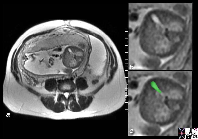
Gallbladder – 24 week Pregnancy
|
| The T2 weighted MRI shows a 24 week pregnancy with a normal gallbladder. The gallbladder at this time is about 2cms long and 8mms wide.
egallbladder 47754c01.8s 24 week pregnancy OB fetus amniotic sac placenta gallbladder pregnancy character water T2 bright MRI scan Davidoff MD concepts copyright 2008
|
Size Changes
At birth the gall bladder is approximately 2.5cms long and 1cms wide. By 12-16 yeras the length is about 6cms long and 2cms wide and by adulthood it is about 8-10cms long and 3-5cms wide. In the elderly, elasticity and muscular tone may be lost, causing elongation of the gallbladder. The transverse dimension of the body of the gallbladder is however maintained remaining at 4-5cms in diameter. Wall thickness is constant throughout life ranging between 1-3mm thick. The common bile duct in neonates measures about 1.6mms increasing to 2.5-3mm in childhood, and to 5mm in adulthood. The bile duct may also lose elasticity in the elderly and for each decade beyond 60 years the duct enlarges by 1mms. Thus a 60 year old is allowed a CBD of 6mms, a 70 year old a CBD of 7mm, and an 80 year old a CBD of 8mms.
|
 
Neonatal Gallbladder
|
|
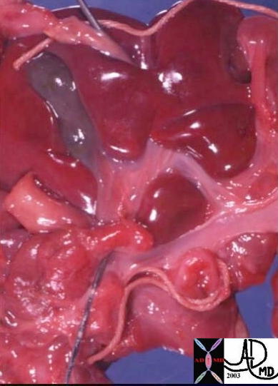
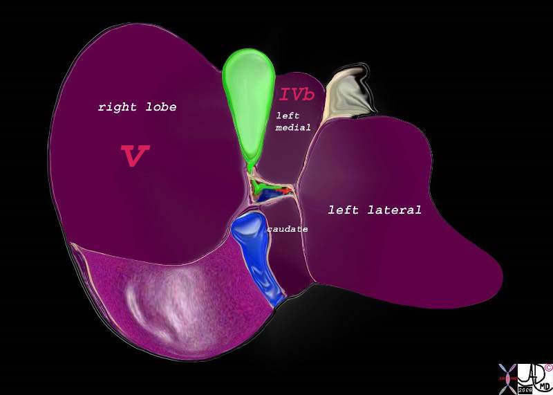
The anatomic specimen of a neonate shows the undersurface of the liver with the gallbladder lying between the right and left lobes in the interlobar fissure. The moist peritoneal surface creates the glistening texture. Glisson’s capsule (with unseen portal triad lies posterior to the gallbladder and the caudate lobe lies posterior to the porta. A string lies around umbilicus. The probe lies in the portal vein. A diagram of the undersurface of the liver serves as orientation.
02101 liver gallbladder newborn ligamentum venosum probe in IVC falciform ligament ligamentum teres unmbilicus
82220b01.2k05.82s liver gallbladder porta hepatis hepatic artery portal vein IVC inferior vena cava falciform ligament ligamentum teres bare area of the liver left lobe segment IV segment I caudate lobe quadrate lobe medial segment left ;obe lateral segment left lobe gastrohepatic ligament gallbladder right lobe hepatic Davidoff art copyright 2008
|
The aging process varies among individuals with maintenance of full functionality and structural integrity, but usually with some slowing of activity. There is a tendency among some to lose contractile strength and elasticity and this results in bile stasis.

Elongated but not Dilated – The Aging Gallbladder
|
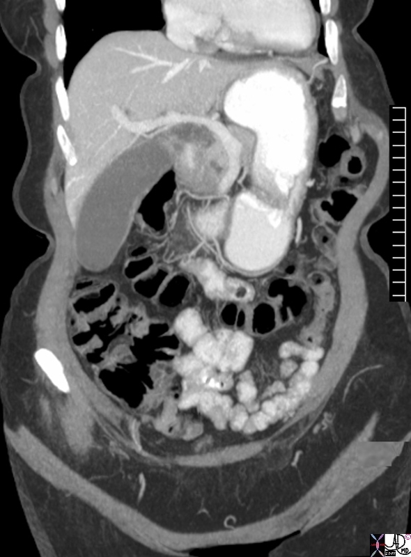
| The gallbladder in this asymptomatic octagenerian showed elongation but without associated increase in the transverse dimension. The gallbladder measured 12.5cms long and 4.5cms wide. We have called this the Zuchini gallbladder.
individual 76764.8s elderly femal gallbladder elongated sausage shape zuchini shape transverse dimension 4.5 cms longitudinal dimension 12.5cms aging gallbladder vs enlarged? CTscan Courtesy Ashley DAvidoff MD copyright 2008 |
Applied Biology
As a result of advancing cholestasis with the aging process, predisposition to gallstone formation results.
References
Haller Textbook of Neonatal Ultrasound By Jack O. Haller Published by Informa Health Care, 1998
DOMElement Object
(
    [schemaTypeInfo] =>
    [tagName] => table
    [firstElementChild] => (object value omitted)
    [lastElementChild] => (object value omitted)
    [childElementCount] => 1
    [previousElementSibling] => (object value omitted)
    [nextElementSibling] => (object value omitted)
    [nodeName] => table
    [nodeValue] =>


Elongated but not Dilated – The Aging Gallbladder


The gallbladder in this asymptomatic octagenerian showed elongation but without associated increase in the transverse dimension.  The gallbladder measured 12.5cms long and 4.5cms wide.  We have called this the Zuchini gallbladder.
individual 76764.8s elderly femal gallbladder elongated sausage shape zuchini shape transverse dimension 4.5 cms longitudinal dimension 12.5cms aging gallbladder vs enlarged? CTscan Courtesy Ashley DAvidoff MD copyright 2008


    [nodeType] => 1
    [parentNode] => (object value omitted)
    [childNodes] => (object value omitted)
    [firstChild] => (object value omitted)
    [lastChild] => (object value omitted)
    [previousSibling] => (object value omitted)
    [nextSibling] => (object value omitted)
    [attributes] => (object value omitted)
    [ownerDocument] => (object value omitted)
    [namespaceURI] =>
    [prefix] =>
    [localName] => table
    [baseURI] =>
    [textContent] =>


Elongated but not Dilated – The Aging Gallbladder


The gallbladder in this asymptomatic octagenerian showed elongation but without associated increase in the transverse dimension.  The gallbladder measured 12.5cms long and 4.5cms wide.  We have called this the Zuchini gallbladder.
individual 76764.8s elderly femal gallbladder elongated sausage shape zuchini shape transverse dimension 4.5 cms longitudinal dimension 12.5cms aging gallbladder vs enlarged? CTscan Courtesy Ashley DAvidoff MD copyright 2008


)
DOMElement Object
(
    [schemaTypeInfo] =>
    [tagName] => td
    [firstElementChild] => (object value omitted)
    [lastElementChild] => (object value omitted)
    [childElementCount] => 2
    [previousElementSibling] =>
    [nextElementSibling] =>
    [nodeName] => td
    [nodeValue] => The gallbladder in this asymptomatic octagenerian showed elongation but without associated increase in the transverse dimension.  The gallbladder measured 12.5cms long and 4.5cms wide.  We have called this the Zuchini gallbladder.
individual 76764.8s elderly femal gallbladder elongated sausage shape zuchini shape transverse dimension 4.5 cms longitudinal dimension 12.5cms aging gallbladder vs enlarged? CTscan Courtesy Ashley DAvidoff MD copyright 2008
    [nodeType] => 1
    [parentNode] => (object value omitted)
    [childNodes] => (object value omitted)
    [firstChild] => (object value omitted)
    [lastChild] => (object value omitted)
    [previousSibling] => (object value omitted)
    [nextSibling] => (object value omitted)
    [attributes] => (object value omitted)
    [ownerDocument] => (object value omitted)
    [namespaceURI] =>
    [prefix] =>
    [localName] => td
    [baseURI] =>
    [textContent] => The gallbladder in this asymptomatic octagenerian showed elongation but without associated increase in the transverse dimension.  The gallbladder measured 12.5cms long and 4.5cms wide.  We have called this the Zuchini gallbladder.
individual 76764.8s elderly femal gallbladder elongated sausage shape zuchini shape transverse dimension 4.5 cms longitudinal dimension 12.5cms aging gallbladder vs enlarged? CTscan Courtesy Ashley DAvidoff MD copyright 2008
)
DOMElement Object
(
    [schemaTypeInfo] =>
    [tagName] => td
    [firstElementChild] => (object value omitted)
    [lastElementChild] => (object value omitted)
    [childElementCount] => 2
    [previousElementSibling] =>
    [nextElementSibling] =>
    [nodeName] => td
    [nodeValue] =>
Elongated but not Dilated – The Aging Gallbladder

    [nodeType] => 1
    [parentNode] => (object value omitted)
    [childNodes] => (object value omitted)
    [firstChild] => (object value omitted)
    [lastChild] => (object value omitted)
    [previousSibling] => (object value omitted)
    [nextSibling] => (object value omitted)
    [attributes] => (object value omitted)
    [ownerDocument] => (object value omitted)
    [namespaceURI] =>
    [prefix] =>
    [localName] => td
    [baseURI] =>
    [textContent] =>
Elongated but not Dilated – The Aging Gallbladder

)
DOMElement Object
(
    [schemaTypeInfo] =>
    [tagName] => table
    [firstElementChild] => (object value omitted)
    [lastElementChild] => (object value omitted)
    [childElementCount] => 1
    [previousElementSibling] => (object value omitted)
    [nextElementSibling] => (object value omitted)
    [nodeName] => table
    [nodeValue] =>


Neonatal Gallbladder


The anatomic specimen of a neonate shows the undersurface of the liver with the gallbladder lying between the right and left lobes in the interlobar fissure.  The moist peritoneal surface creates the glistening texture.  Glisson’s capsule (with unseen portal triad lies posterior to the gallbladder and the caudate lobe lies posterior to the porta.  A string lies around umbilicus.  The probe lies in the portal vein.   A diagram of the undersurface of the liver serves as orientation.
02101 liver gallbladder newborn ligamentum venosum probe in IVC falciform ligament ligamentum teres unmbilicus
82220b01.2k05.82s liver gallbladder porta hepatis hepatic artery portal vein IVC inferior vena cava falciform ligament ligamentum teres bare area of the liver left lobe segment IV segment I caudate lobe quadrate lobe medial segment left ;obe lateral segment left lobe gastrohepatic ligament gallbladder right lobe hepatic Davidoff art copyright 2008


    [nodeType] => 1
    [parentNode] => (object value omitted)
    [childNodes] => (object value omitted)
    [firstChild] => (object value omitted)
    [lastChild] => (object value omitted)
    [previousSibling] => (object value omitted)
    [nextSibling] => (object value omitted)
    [attributes] => (object value omitted)
    [ownerDocument] => (object value omitted)
    [namespaceURI] =>
    [prefix] =>
    [localName] => table
    [baseURI] =>
    [textContent] =>


Neonatal Gallbladder


The anatomic specimen of a neonate shows the undersurface of the liver with the gallbladder lying between the right and left lobes in the interlobar fissure.  The moist peritoneal surface creates the glistening texture.  Glisson’s capsule (with unseen portal triad lies posterior to the gallbladder and the caudate lobe lies posterior to the porta.  A string lies around umbilicus.  The probe lies in the portal vein.   A diagram of the undersurface of the liver serves as orientation.
02101 liver gallbladder newborn ligamentum venosum probe in IVC falciform ligament ligamentum teres unmbilicus
82220b01.2k05.82s liver gallbladder porta hepatis hepatic artery portal vein IVC inferior vena cava falciform ligament ligamentum teres bare area of the liver left lobe segment IV segment I caudate lobe quadrate lobe medial segment left ;obe lateral segment left lobe gastrohepatic ligament gallbladder right lobe hepatic Davidoff art copyright 2008


)
DOMElement Object
(
    [schemaTypeInfo] =>
    [tagName] => td
    [firstElementChild] => (object value omitted)
    [lastElementChild] => (object value omitted)
    [childElementCount] => 3
    [previousElementSibling] =>
    [nextElementSibling] =>
    [nodeName] => td
    [nodeValue] =>
The anatomic specimen of a neonate shows the undersurface of the liver with the gallbladder lying between the right and left lobes in the interlobar fissure.  The moist peritoneal surface creates the glistening texture.  Glisson’s capsule (with unseen portal triad lies posterior to the gallbladder and the caudate lobe lies posterior to the porta.  A string lies around umbilicus.  The probe lies in the portal vein.   A diagram of the undersurface of the liver serves as orientation.
02101 liver gallbladder newborn ligamentum venosum probe in IVC falciform ligament ligamentum teres unmbilicus
82220b01.2k05.82s liver gallbladder porta hepatis hepatic artery portal vein IVC inferior vena cava falciform ligament ligamentum teres bare area of the liver left lobe segment IV segment I caudate lobe quadrate lobe medial segment left ;obe lateral segment left lobe gastrohepatic ligament gallbladder right lobe hepatic Davidoff art copyright 2008

    [nodeType] => 1
    [parentNode] => (object value omitted)
    [childNodes] => (object value omitted)
    [firstChild] => (object value omitted)
    [lastChild] => (object value omitted)
    [previousSibling] => (object value omitted)
    [nextSibling] => (object value omitted)
    [attributes] => (object value omitted)
    [ownerDocument] => (object value omitted)
    [namespaceURI] =>
    [prefix] =>
    [localName] => td
    [baseURI] =>
    [textContent] =>
The anatomic specimen of a neonate shows the undersurface of the liver with the gallbladder lying between the right and left lobes in the interlobar fissure.  The moist peritoneal surface creates the glistening texture.  Glisson’s capsule (with unseen portal triad lies posterior to the gallbladder and the caudate lobe lies posterior to the porta.  A string lies around umbilicus.  The probe lies in the portal vein.   A diagram of the undersurface of the liver serves as orientation.
02101 liver gallbladder newborn ligamentum venosum probe in IVC falciform ligament ligamentum teres unmbilicus
82220b01.2k05.82s liver gallbladder porta hepatis hepatic artery portal vein IVC inferior vena cava falciform ligament ligamentum teres bare area of the liver left lobe segment IV segment I caudate lobe quadrate lobe medial segment left ;obe lateral segment left lobe gastrohepatic ligament gallbladder right lobe hepatic Davidoff art copyright 2008

)
DOMElement Object
(
    [schemaTypeInfo] =>
    [tagName] => td
    [firstElementChild] => (object value omitted)
    [lastElementChild] => (object value omitted)
    [childElementCount] => 2
    [previousElementSibling] =>
    [nextElementSibling] =>
    [nodeName] => td
    [nodeValue] =>

Neonatal Gallbladder

    [nodeType] => 1
    [parentNode] => (object value omitted)
    [childNodes] => (object value omitted)
    [firstChild] => (object value omitted)
    [lastChild] => (object value omitted)
    [previousSibling] => (object value omitted)
    [nextSibling] => (object value omitted)
    [attributes] => (object value omitted)
    [ownerDocument] => (object value omitted)
    [namespaceURI] =>
    [prefix] =>
    [localName] => td
    [baseURI] =>
    [textContent] =>

Neonatal Gallbladder

)
DOMElement Object
(
    [schemaTypeInfo] =>
    [tagName] => table
    [firstElementChild] => (object value omitted)
    [lastElementChild] => (object value omitted)
    [childElementCount] => 1
    [previousElementSibling] => (object value omitted)
    [nextElementSibling] => (object value omitted)
    [nodeName] => table
    [nodeValue] =>


Gallbladder – 24 week Pregnancy


The T2 weighted MRI  shows a 24 week pregnancy with a normal gallbladder. The gallbladder at this time is about 2cms long and 8mms wide.
egallbladder 47754c01.8s 24 week pregnancy OB fetus amniotic sac placenta gallbladder pregnancy character water T2 bright MRI scan Davidoff MD concepts copyright 2008


    [nodeType] => 1
    [parentNode] => (object value omitted)
    [childNodes] => (object value omitted)
    [firstChild] => (object value omitted)
    [lastChild] => (object value omitted)
    [previousSibling] => (object value omitted)
    [nextSibling] => (object value omitted)
    [attributes] => (object value omitted)
    [ownerDocument] => (object value omitted)
    [namespaceURI] =>
    [prefix] =>
    [localName] => table
    [baseURI] =>
    [textContent] =>


Gallbladder – 24 week Pregnancy


The T2 weighted MRI  shows a 24 week pregnancy with a normal gallbladder. The gallbladder at this time is about 2cms long and 8mms wide.
egallbladder 47754c01.8s 24 week pregnancy OB fetus amniotic sac placenta gallbladder pregnancy character water T2 bright MRI scan Davidoff MD concepts copyright 2008


)
DOMElement Object
(
    [schemaTypeInfo] =>
    [tagName] => td
    [firstElementChild] => (object value omitted)
    [lastElementChild] => (object value omitted)
    [childElementCount] => 3
    [previousElementSibling] =>
    [nextElementSibling] =>
    [nodeName] => td
    [nodeValue] => The T2 weighted MRI  shows a 24 week pregnancy with a normal gallbladder. The gallbladder at this time is about 2cms long and 8mms wide.
egallbladder 47754c01.8s 24 week pregnancy OB fetus amniotic sac placenta gallbladder pregnancy character water T2 bright MRI scan Davidoff MD concepts copyright 2008

    [nodeType] => 1
    [parentNode] => (object value omitted)
    [childNodes] => (object value omitted)
    [firstChild] => (object value omitted)
    [lastChild] => (object value omitted)
    [previousSibling] => (object value omitted)
    [nextSibling] => (object value omitted)
    [attributes] => (object value omitted)
    [ownerDocument] => (object value omitted)
    [namespaceURI] =>
    [prefix] =>
    [localName] => td
    [baseURI] =>
    [textContent] => The T2 weighted MRI  shows a 24 week pregnancy with a normal gallbladder. The gallbladder at this time is about 2cms long and 8mms wide.
egallbladder 47754c01.8s 24 week pregnancy OB fetus amniotic sac placenta gallbladder pregnancy character water T2 bright MRI scan Davidoff MD concepts copyright 2008

)
DOMElement Object
(
    [schemaTypeInfo] =>
    [tagName] => td
    [firstElementChild] => (object value omitted)
    [lastElementChild] => (object value omitted)
    [childElementCount] => 2
    [previousElementSibling] =>
    [nextElementSibling] =>
    [nodeName] => td
    [nodeValue] =>
Gallbladder – 24 week Pregnancy

    [nodeType] => 1
    [parentNode] => (object value omitted)
    [childNodes] => (object value omitted)
    [firstChild] => (object value omitted)
    [lastChild] => (object value omitted)
    [previousSibling] => (object value omitted)
    [nextSibling] => (object value omitted)
    [attributes] => (object value omitted)
    [ownerDocument] => (object value omitted)
    [namespaceURI] =>
    [prefix] =>
    [localName] => td
    [baseURI] =>
    [textContent] =>
Gallbladder – 24 week Pregnancy

)


