Physiology
Basic Principles: Receive, Process, and Export
The function of every biological unit in the body is based on its ability to receive a product, process it, and deliver the new product efficiently and effectively to where it is used and needed.
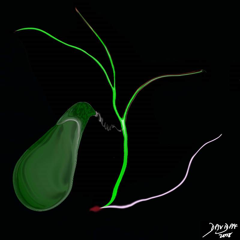
The gallbladder receives bile produced by the liver, processes it by concentration and storage, and delivers it on demand to the duodenum via the bile duct, so that the digestion of fat can be optimized.
The process of bile production occurs in two stages;
primary production
concentration
The gallbladder has no part in the primary production but is essential for the concentration. Bile is produced 24 hours a day, but seemingly is only needed for 3 short periods during mealtimes. On the other hand it is also provides a route for the excretion of end products of hemoglobin breakdown and to some degree excretion of cholesterol.
With its simple design of a musculomembranous bag, and single duct it is able to receive and concentrate 24 hours a day and deliver the bile via seemingly complex contraction patterns. Although it is not an essential organ, it does help optimize the function of the digestive process. People who have had a cholecystectomy do and can live an otherwise normal life, and seemingly have no untoward effects without it.
Bile
Bile is a yellow or green bitter fluid that is smooth, sterile, relatively thick, and sticky, with “spinnbarkeit” character. Spinnbarkeit is a German word that means ability to be spun and it infers that when bile is pulled apart it forms a continuous thin thread much like thin nasal mucus. The specific gravity ranges between 1.010 to 1.040 but can be as high as 1.059. (Yeh)
Structurally it consists mainly of water, cholesterol, bile pigments, and bile salts. It is continually produced by the hepatocytes. It contains no enzymes and it is only the bile salts that are important in digestion. The end products of cholesterol are the bile acids (aka bile salts) which are synthesized by oxidation of cholesterol in the liver, and then conjugated with taurine or glycine. The major primary bile acids are chenodeoxycholic acid and cholic acid. When the bile acids mix with the alkaline milieu of the duodenum they become bile salts. About 20-30gms of bile acids are produced per day, but 90% are reabsorbed via the enterohepatic circulation where they are reused and recycled 10-12 times per day.
Bile has two main functions: It aids the in the solubilization and digestion of fats including cholesterol and fat soluble vitamins, and it eliminates certain waste products – mainly hemoglobin and excess cholesterol from the body. Within the bile itself, the bile acids and phospholipids solubilize the cholesterol, thus preventing its precipitation.
While bile acids are important in fat digestion, they do not actually digest it. Rather, they help emulsify fat particles into smaller particles to facilitate the digestive process. The net effect is to expose active receptors that enable the pancreatic lipases (enzymes) to act. They also continue to facilitate polar-non-polar molecular interactions by aiding in the absorption of these digested fat products through the intestinal mucosa and into the into the lymphatics of the small bowel.
Bile salts are also an important vehicle for excretion. Two important excreted solutes are bilirubin (which once conjugated is not water-soluble and thus can not be excreted through the kidneys) in appreciable amounts, as well as cholesterol.
Bile is a noxious substance when not confined to the tubes of the digestive tract. Bile in the peritoneal cavity causes bile peritonitis which is a potentially fatal disease. Jaundice is a clinical sign indicating excessive bilirubin in the serum. This has many causes but in general is obstructive, hepatocellular or hemolytic in origin.
From a diagnostic point of view, excessive bilirubin in the body is diagnosed clinically by looking at the sclera and skin color, and by evaluation of serum bilirubin. Ultrasound is helpful to exclude obstructive causes.
The treatment of jaundice depends on the cause, with surgical treatments usually indicated for obstructive causes, and medical treatments for hepatocellular or hemolytic causes.
The gallbladder is central to bile concentration and delivery. As we have learned in the module on structure, its design provides certain features that optimize its function. It consists of an incomplete muscular wall, which at a histological level contains mucosal fronds that absorb water and concentrate the bile. It has an unusual spiral cystic duct that optimises the transport of bile to and from the gallbladder. The size shape and position of its component parts are optimized to enable the receipt, concentration storage and transportation of bile.
Receiving
As the bile secretion flows from the hepatocytes to the bile ducts via the bile canaliculi, a secondary secretion of water containing sodium and bicarbonate ions from the epithelial cells lining ductules occurs resulting a total volume for an average human adult of between 600-1000mL daily. The bile ducts are gracile and of low volume with a maximum normal pressure of about 20cms of water when the sphincter is closed, and which falls to 10cms of water or less as the sphincter opens. With the sphincter of Oddi closed and the cystic duct open overflow of bile into the cystic duct occurs as a result of the small rise in pressure.
The cystic duct has an upward course to reach the neck of the gallbladder since in the upright position, the neck of the gallbladder is more cranial than the cystic duct/bile duct junction. During the day we are upright, whether we are walking, standing, or sitting, it is an uphill climb for bile to reach the cystic duct.
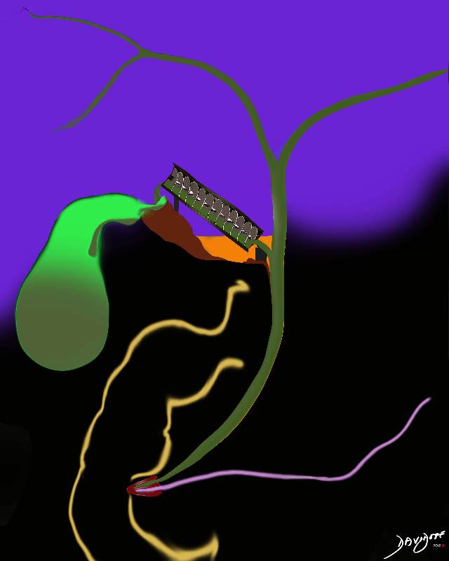
The spiral valves of Heister have been a puzzle for many years. We theorise that they have been ingeniously designed to hold and pass on the bile during this ascent against the forces of gravity, in much the same way that Archimedes design of the waterscrew helped the delivery of water from a low source to a high receptacle, also going against the force of gravity, and with the least energy expenditure.
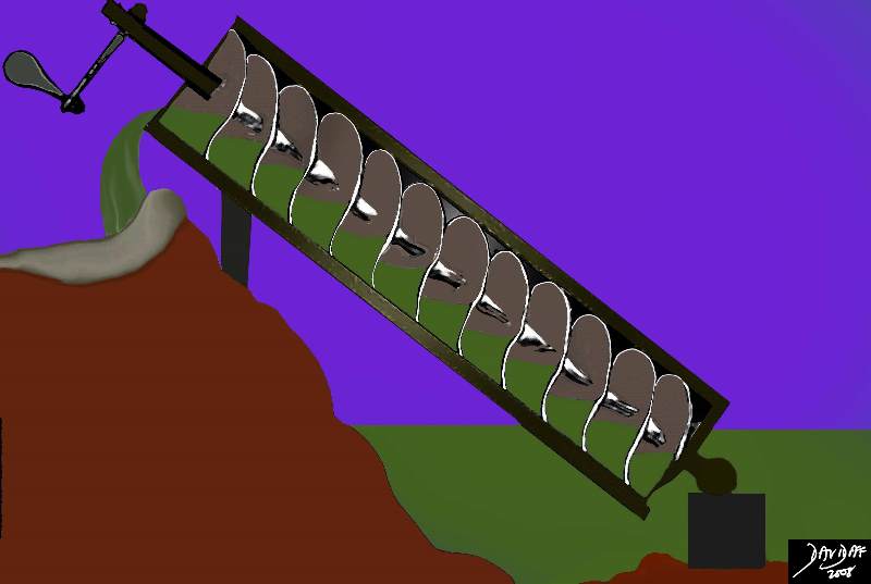
Archimedes Water Screw |
| The design of the water screw has been attributed to Archimedes from 3rd century BC though some designate the originator of the screw to be Nebuchadnezzar II of 7th century BC who purportedly used the screw to deliver water to the Hanging Gardens of Babylon. Water at a low level is scooped up into the spiral mechanism. As the screw rotates, the water advances upward and gets delivered to a higher level until it finally reaches its destination and is delivered to a repository.
has been 82656b15.8s Archimedes water screw elevation gravity screw delivery cystic duct spiral valves of Heister anatomy physiology function uploading Davidoff art copyright 2008 The image was modified from Wikipedia; detail of image: Archimedes’ screw. Public domain, from Chambers’s Encyclopedia (Philadelphia: J. B. Lippincott Company, 1875). Added to illustrate article :Archimedes. |
In the body there is no handle to turn the valves (screws ) of Heister. There are hydrostatic forces from below caused by the continued 24hour production of bile by the liver at about the rate of 1/2ccs per minute. (approximately 800ccs in 24 hours, = approximately 1/2ccs per minute)
The maximum secreting pressures of bile from the hepatocystes is approximately 30cms of water. The resting pressure of the sphincter of Oddi is 12-15cms of water, the opening pressure of the cystic duct is 8cms of water and the opening pressure of the gallbladder is 10cms of water. Thus when the sphincter is closed the lowest pressure is in the cystic duct and hence filling of the cystic duct occurs. (Clavien)
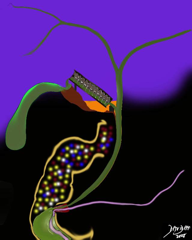
As the new drop of bile spills into the bile ducts it will cause a minute rise in pressure against the closed sphincter of Oddi. Flow in the system is directed toward the cystic duct offers the least resistance as described above. Bile will therefore fill the first valve which will push the bile upward to the second valve and so on. With this repeated many times, each valve is filled until the bile reaches the top of the ladder after which it will spill over like a waterfall into the neck, and then body, and finally into the fundus.
The sphincter of Oddi is composed of a ring of circular and longitudinal muscle at the distal end of the common bile duct which has a phasic resting pressure of about 13mmHg. It has a cyclical pattern during fasting that works in concordance with the intermittant myoelectric migratory complex of the intestinal tract (IMMC), as well as a pattern specific to the presence of CCK and the parasympathetic nerve supply.
The common bile duct unites with with the pancreatic duct to form a common channel called the ampulla of Vater which empties into the duodenum at the papilla which is seen as a small nipple in the middle of the descending duodenum.
Mechanisms of Control
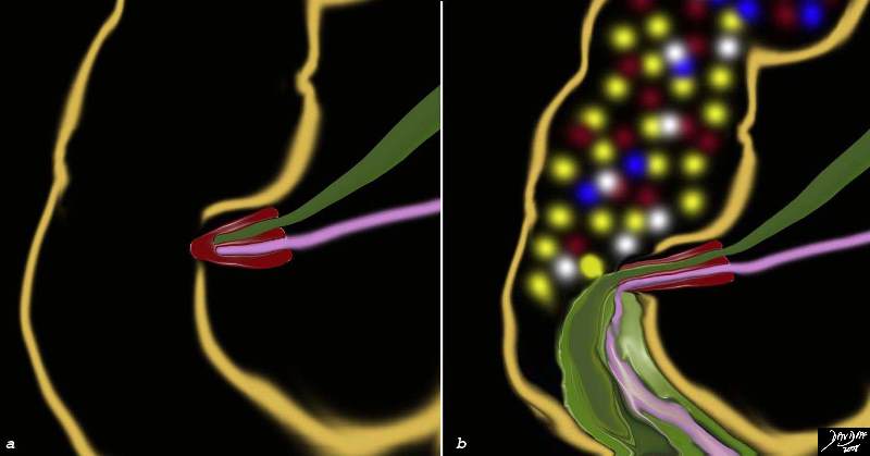
In between meals the sympathetic nervous system governing the gallbladder is relatively active and the parasympathetic system and CCK hormone are inactive. Hence the sphincter of Oddi under the sympathetic control of the greater splanchnic nerves is contracted, and the gallbladder smooth muscle is relaxed. With a fatty meal release of CCK is instituted and it acts as the primary mechanism of smooth muscle and sphincter control. The parasympathetic system provides accessory control with the right (posterior) vagus causing the sphincter to relax and the left vagus causing the gallbladder wall to relax.
It is becoming more apparent that the the neural and hormonal control of bile release into the duodenum is not as is not as tightly regulated by meals and that the sphincter of Oddi has periods between meals when it is relaxed and the gallbladder has phases when it contracts and partly empties between meals as well. (Kusano, Rodes, Toouli ) It is also well known that only half the bile produced by the liver enters the gallbladder and so it infers that the ampulla opens intermittantly between meals to allow unconcentrated bile into the duodenum. We alluded to the intermittant myoelectric migratory complex of the intestinal tract (IMMC) which is a cyclical contraction pattern that coordinates muscular activity of the bowel and has emptying effects on the biliary system.
Processing
Concentration of bile
The shape and positon of the gallbladder are well designed to enable bile concentration. The bile in the fundus and body is mostly “old bile” and represents about 90% of the gallbladder volume since they are the most capacious parts of the gallbladder, most dependant in position, and contain the heavier, already concentrated bile.
The new bile which is delivered to the gallbladder is small in volume, relatively dilute, has a relatively low specific gravity, and consists of water (97% ), bile acids (1-2%), and other metabolites and electrolytes that make up the remaining 1-2%. The “other” components include cholesterol, phospholipids, bile pigments, sodium and chloride. The major cation is sodium and the dominant anion is chloride. Small amounts of potassium, calcium, and approximately 20?50 mM bicarbonate are present. Bile acid concentration is approximately 30-50mM. Hepatic bile is isotonic with plasma.
Once the bile comes into contact with the mucosal surface, about 90% of the water is reabsorbed, allowing the large volume of biliary secretions to be stored and concentrated in a space that can accomodate only 60-80ccs. The concentration is achieved by the active absorbtion of electrolytes and passive absorbtion of water. Typically the gallbladder can concentrate the bile from 5-6 fold, but this can incrase to 10-12 fold. Thus for every 100ccs of dilute bile received, between 10 and 20ccs of concentrated bile is produced. It is now becoming apparent that only half of the bile production goes through the gallbladder so the gallbladder may only “see” and concentrate between 250-500ccs per 24 hour period. The concentration of bile acids increases by 10 fold, and the sodium and and chloride concentrations are reduced significantly. The concentration of divalent calcium although low, is relatively high when compared to monovalent anions because phenomenon called the Gibbs-Donnan effect which favors the relative accumulation of divalent anions in the proteinaceous mileu that exists.
Applied Biology
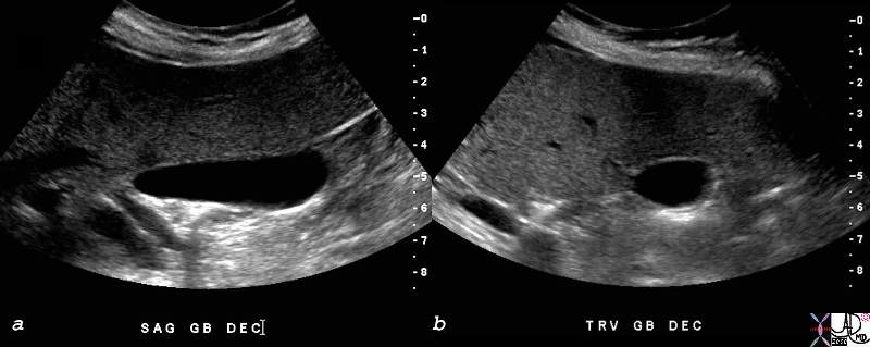
From a practical perspective, it is routine to have a patient fast (usually overnight) for 8-12 hours before an ultrasound exam when the gallbladder is optimally distended without being dilated . The wall is better evaluated in full distension, and stones that may hide in the neck or infundibulum are seen to better advantage as they are surrounded by the anechoic bile.
Storage
The storage of bile is not as static as one would imagine. During fasting, there is continued cyclical myoelectric activity in synchrony with duodenal activity called intermittant myoelectric migratory complex of the intestinal tract (IMMC) which we have discussed above. This results in gallbladder contraction between meals, with an an open sphincter, and with 15-20% emptying. Motilin is a polypeptide secreted by the small intestine probably as a result of an alkaline PH in the duodenum and is associated with the IMMC. Despite the cyclical contractions, there are still prolonged periods of “down time” of muscular activity where storage of the bile occurs enabling the process of bile concentration to occur.
Applied Biology
Static bile accumulates in the fundus and spends the majority of time in the fundus since we are in the upright position most the time. Sediments of bile salts, stones and inflammation are therefore dominant in the fundus and it is not surprising therefore to find that 60% of gallbladder cancers occur in the fundus, 30% in the body, and the least (10%) originating in the neck.
Export Function
While we have stressed the apparent simplicity of gallbladder function the muscular function of the gallbladder brings this notion to a screeching halt. Even during the fasting state bile is delivered through the ampulla at about the rate of about .5-1ml/minute. At peak flow post prandially, this may increase to 2-3mls per minute.
The engines of delivery start to rev up even as we think of the meal we are about to partake in. This is called the cephalic phase of digestion and it affects the gallbladder as well. The mere thought, sight, or smell of food stimulates the parasympathetic neural axis to the gallbladder resulting in stimulation of the vagus nerve and its effects on the smooth muscle.
When the fat-rich or protein rich foods reach the duodenum (typically 30 minutes post-prandially), CCK, the primary regulator of gallbladder contraction and Oddi function is released by the duodenum into the blood stream resulting in a serum CCK that rises over 15-30 minutes and then slowly plateaus. The response is not all contraction by gallbladder muscle nor all relaxation by the sphincter. Instead there is a contraction and relaxation pattern designed to provide a sustained, slow and steady stream of bile. (Howard) (Rodes), with emptying occuring over two hours.
The contraction pattern is even more complex it seems. During the post prandial phase a tripahsic pattern; early emptying, early refilling, and late emptying seems to be an overall pattern. duration of eachof the triphasic pghases includes emptying (40 minutes) refilling (10minutes) and emptying again (100minutes) and it tales over two hours to occur during following a meal.
In addition to the triphasic pattern there also appear to be contraction and relaxation phases, that occur minute to minute and are of a rhythmic nature.
The spiral valves of Heister prevent the rapid overdistension of the bile duct during the phases of contraction and also play a part of providing the staed slow and steady stream of bile into the bile duct and therefpre into the duodenum.
CCK also affects the acinar cells of the pancreas and stimulates the productoion and transport of pancreatic juices to the duodenum at the same time. these juices among many enzymes also contain lipases.
Applied Biology
The gallbladder in the fasting state is distended amd its ability to deliver bile and contract can be evaluated by ultrasound or by nuclear medicine techniques. Pre (fasting) and post prandial volumes can be assessed.
Using ultrasound the volume diferences between the fasting and post prandial volume can be calculated, but this study is rarely performed since the nuclear study usinng CCK is more accurate.
|
The Size of the Gallbladder in the Fasting State |
| The ultrasound of the gallbladder in the fasting state is taken in the longitudinal plane in a, and the transverse plane in b, showing a gallbladder that is of a relatively small size but still distende showing a single wall, characteristic of the distended gallbladder.
82428c01.8s gallbladder small normal transverse oval shape normal anatomy USscan ultrasound copyright 2008 Courtesy Ashley DAvidoff MD |
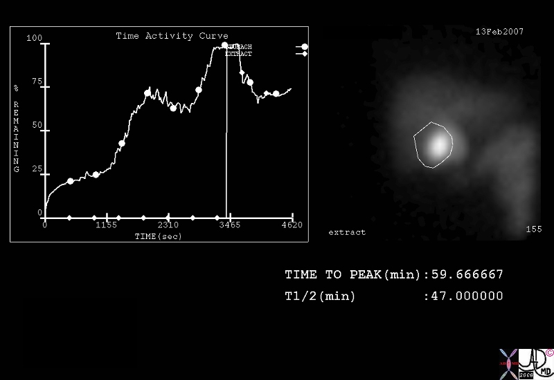
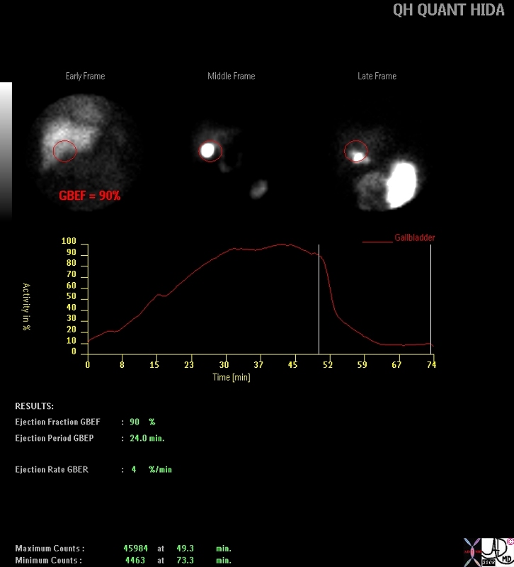
Nuclear medicine offers the most accurate assessment of gallbladder contractile function since it is based on isotope counts which can be accurately measured in the fasting state and following the intravenous injection of CCK. The normal ejection fraction ranges between 35-75 percent.
Nuclear medicine offers the most accurate assessment of gallbladder contractile function since it is based on isotope counts which can be accurately measured in the fasting state and following the intravenous injection of CCK. The normal ejection fraction ranges between 35-75 percent.
Conclusion and Summary
In summary therefore the receiving, processing and delivery of bile by the gallbladder is not a simple black and white process. It requires complex interactions of neural, hormonal and muscular actions exerted on a structure that is more than a musculomebranous sac with a duct.
References
Afdhal Nezam Gallbladder and Biliary Diseases Published by Informa Health Care, 2000
Clavien Diseases of the Gallbladder and Bile Ducts: Diagnosis and Treatment By Pierre-Alain Clavien, John Baillie Published by Blackwell Publishing, 2006
du Fresne Marlène, Seva Catherine and Fourmy, Daniel Cholecystokinin and Gastrin Receptors Physiol. Rev. 86: 805-847, 2006
Kusano M, Sekiguchi T, Nishioka T, Kawamura O, et al The relationship between interdigestive gallbladder and gastroduodenal motility in man.Gastroenterol Jpn. 1990 Oct;25(5):568-74
Li et al One-Dimensional Models of the Human Biliary System J. Biomech. Eng. Volume 129 : pp. 164
Marzio L, Neri M, Capone F, Di Felice F, et al Gallbladder contraction and its relationship to interdigestive duodenal motor activity in normal human subjects. Dig Dis Sci. 1988 May;33(5):540-4.
Norton Jeffrey A. Norton, R. Randall Bollinger, Alfred E. Chang, M. K. Shirazi Essential Practice of Surgery: Basic Science and Clinical Evidence Published by Springer, 2003
Rodes The Textbook of Hepatology: From Basic Science to Clinical Practice By Juan Rodes, Jean-Pierre Benhamou, Mario Rizzetto Published by Blackwell Publishing, 2007
Toouli J, Bushell M, Stevenson G, Dent J, Wycherley A, Iannos J. Gallbladder emptying in man related to fasting duodenal migrating motor contractions. Aust N Z J Surg. 1986 Feb;56(2):147-51.
Yeh Hsu-Chong Yeh, Joan Goodman, Jack G. Rabinowitz Floating Gallstones in Bile without Added Contrast Material AJR 146:49-50, January 1986
Ziessman , Harvey A.,. Fahey, Frederic H , and Hixson Donald J. Calculation of a Gallbladder Ejection Fraction: dvantage of Continuous Sincalide Infusion over the Three-Minute Infusion Method J NucIMed1992;33:537- 541
DOMElement Object
(
    [schemaTypeInfo] =>
    [tagName] => table
    [firstElementChild] => (object value omitted)
    [lastElementChild] => (object value omitted)
    [childElementCount] => 1
    [previousElementSibling] => (object value omitted)
    [nextElementSibling] => (object value omitted)
    [nodeName] => table
    [nodeValue] =>


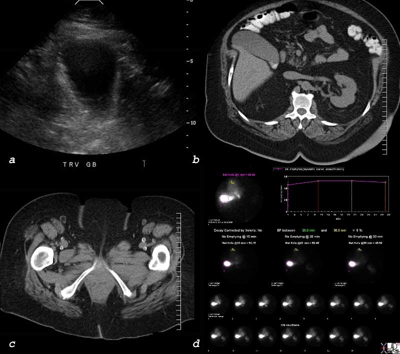
Diabetic Arteriopathy – Gallbladder Atony


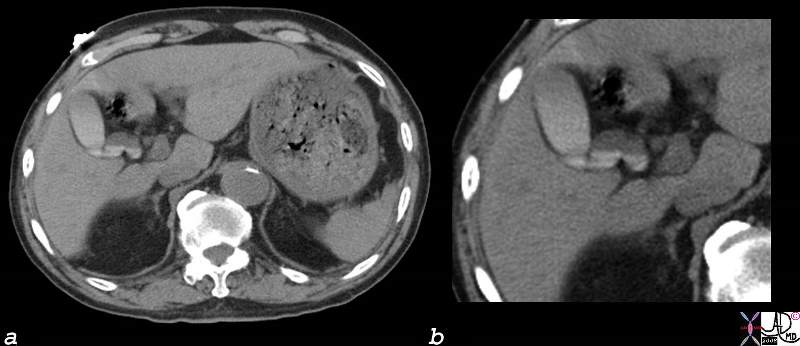
In this case of a 68 year old malewith diabetes, the CTscan (a) shows an enlarged gallbladder with   The image of the seminal vesicles (b) shows calcified vasa deferentia characteristic of diabetes.  Image c and d show calcified medium and  small arteries (anterior and posterior tibials and peroneals) bilaterally which is characteristic of diabetes. Atonic gallbladder characteristic of diabetes is present.
82719c.8s 68M diabetes peripheral vascular disease diabetic arteriopathy artery small vessel disease gallbladder atonic enlarged vas deferens calcification calcified Courtesy Ashley Davidoff MD copyright 2008


    [nodeType] => 1
    [parentNode] => (object value omitted)
    [childNodes] => (object value omitted)
    [firstChild] => (object value omitted)
    [lastChild] => (object value omitted)
    [previousSibling] => (object value omitted)
    [nextSibling] => (object value omitted)
    [attributes] => (object value omitted)
    [ownerDocument] => (object value omitted)
    [namespaceURI] =>
    [prefix] =>
    [localName] => table
    [baseURI] =>
    [textContent] =>


Diabetic Arteriopathy – Gallbladder Atony


In this case of a 68 year old malewith diabetes, the CTscan (a) shows an enlarged gallbladder with   The image of the seminal vesicles (b) shows calcified vasa deferentia characteristic of diabetes.  Image c and d show calcified medium and  small arteries (anterior and posterior tibials and peroneals) bilaterally which is characteristic of diabetes. Atonic gallbladder characteristic of diabetes is present.
82719c.8s 68M diabetes peripheral vascular disease diabetic arteriopathy artery small vessel disease gallbladder atonic enlarged vas deferens calcification calcified Courtesy Ashley Davidoff MD copyright 2008


)
DOMElement Object
(
    [schemaTypeInfo] =>
    [tagName] => td
    [firstElementChild] => (object value omitted)
    [lastElementChild] => (object value omitted)
    [childElementCount] => 2
    [previousElementSibling] =>
    [nextElementSibling] =>
    [nodeName] => td
    [nodeValue] =>
In this case of a 68 year old malewith diabetes, the CTscan (a) shows an enlarged gallbladder with   The image of the seminal vesicles (b) shows calcified vasa deferentia characteristic of diabetes.  Image c and d show calcified medium and  small arteries (anterior and posterior tibials and peroneals) bilaterally which is characteristic of diabetes. Atonic gallbladder characteristic of diabetes is present.
82719c.8s 68M diabetes peripheral vascular disease diabetic arteriopathy artery small vessel disease gallbladder atonic enlarged vas deferens calcification calcified Courtesy Ashley Davidoff MD copyright 2008
    [nodeType] => 1
    [parentNode] => (object value omitted)
    [childNodes] => (object value omitted)
    [firstChild] => (object value omitted)
    [lastChild] => (object value omitted)
    [previousSibling] => (object value omitted)
    [nextSibling] => (object value omitted)
    [attributes] => (object value omitted)
    [ownerDocument] => (object value omitted)
    [namespaceURI] =>
    [prefix] =>
    [localName] => td
    [baseURI] =>
    [textContent] =>
In this case of a 68 year old malewith diabetes, the CTscan (a) shows an enlarged gallbladder with   The image of the seminal vesicles (b) shows calcified vasa deferentia characteristic of diabetes.  Image c and d show calcified medium and  small arteries (anterior and posterior tibials and peroneals) bilaterally which is characteristic of diabetes. Atonic gallbladder characteristic of diabetes is present.
82719c.8s 68M diabetes peripheral vascular disease diabetic arteriopathy artery small vessel disease gallbladder atonic enlarged vas deferens calcification calcified Courtesy Ashley Davidoff MD copyright 2008
)
DOMElement Object
(
    [schemaTypeInfo] =>
    [tagName] => td
    [firstElementChild] => (object value omitted)
    [lastElementChild] => (object value omitted)
    [childElementCount] => 2
    [previousElementSibling] =>
    [nextElementSibling] =>
    [nodeName] => td
    [nodeValue] =>
Diabetic Arteriopathy – Gallbladder Atony

    [nodeType] => 1
    [parentNode] => (object value omitted)
    [childNodes] => (object value omitted)
    [firstChild] => (object value omitted)
    [lastChild] => (object value omitted)
    [previousSibling] => (object value omitted)
    [nextSibling] => (object value omitted)
    [attributes] => (object value omitted)
    [ownerDocument] => (object value omitted)
    [namespaceURI] =>
    [prefix] =>
    [localName] => td
    [baseURI] =>
    [textContent] =>
Diabetic Arteriopathy – Gallbladder Atony

)
DOMElement Object
(
    [schemaTypeInfo] =>
    [tagName] => td
    [firstElementChild] => (object value omitted)
    [lastElementChild] => (object value omitted)
    [childElementCount] => 2
    [previousElementSibling] =>
    [nextElementSibling] =>
    [nodeName] => td
    [nodeValue] =>
Diabetic Atony

    [nodeType] => 1
    [parentNode] => (object value omitted)
    [childNodes] => (object value omitted)
    [firstChild] => (object value omitted)
    [lastChild] => (object value omitted)
    [previousSibling] => (object value omitted)
    [nextSibling] => (object value omitted)
    [attributes] => (object value omitted)
    [ownerDocument] => (object value omitted)
    [namespaceURI] =>
    [prefix] =>
    [localName] => td
    [baseURI] =>
    [textContent] =>
Diabetic Atony

)
DOMElement Object
(
    [schemaTypeInfo] =>
    [tagName] => table
    [firstElementChild] => (object value omitted)
    [lastElementChild] => (object value omitted)
    [childElementCount] => 1
    [previousElementSibling] => (object value omitted)
    [nextElementSibling] => (object value omitted)
    [nodeName] => table
    [nodeValue] =>


Hypercontractile Gallbladder


These images are from the case described above.  Note that on image 10 (X5minutes = 50minutes) the gallbladder is maximally distended and after CCK the gallbladder is barely visible by image 15.  Note also the maximal filling of the small bowel after CCK injection.
82760.8s 44F gallbladder hypercontractile EF 90percent NM US CTscan Courtesy Ashley Davidoff copyright 2008 5Mci Mci Tc99m Choletec gb normal viz by 10 min and 20 min in SB viz only after cck injection. 50min CCK over 3-10 min EF = 30%-90% suspicious for gallbladder hypercontractility


    [nodeType] => 1
    [parentNode] => (object value omitted)
    [childNodes] => (object value omitted)
    [firstChild] => (object value omitted)
    [lastChild] => (object value omitted)
    [previousSibling] => (object value omitted)
    [nextSibling] => (object value omitted)
    [attributes] => (object value omitted)
    [ownerDocument] => (object value omitted)
    [namespaceURI] =>
    [prefix] =>
    [localName] => table
    [baseURI] =>
    [textContent] =>


Hypercontractile Gallbladder


These images are from the case described above.  Note that on image 10 (X5minutes = 50minutes) the gallbladder is maximally distended and after CCK the gallbladder is barely visible by image 15.  Note also the maximal filling of the small bowel after CCK injection.
82760.8s 44F gallbladder hypercontractile EF 90percent NM US CTscan Courtesy Ashley Davidoff copyright 2008 5Mci Mci Tc99m Choletec gb normal viz by 10 min and 20 min in SB viz only after cck injection. 50min CCK over 3-10 min EF = 30%-90% suspicious for gallbladder hypercontractility


)
DOMElement Object
(
    [schemaTypeInfo] =>
    [tagName] => td
    [firstElementChild] => (object value omitted)
    [lastElementChild] => (object value omitted)
    [childElementCount] => 2
    [previousElementSibling] =>
    [nextElementSibling] =>
    [nodeName] => td
    [nodeValue] =>
These images are from the case described above.  Note that on image 10 (X5minutes = 50minutes) the gallbladder is maximally distended and after CCK the gallbladder is barely visible by image 15.  Note also the maximal filling of the small bowel after CCK injection.
82760.8s 44F gallbladder hypercontractile EF 90percent NM US CTscan Courtesy Ashley Davidoff copyright 2008 5Mci Mci Tc99m Choletec gb normal viz by 10 min and 20 min in SB viz only after cck injection. 50min CCK over 3-10 min EF = 30%-90% suspicious for gallbladder hypercontractility
    [nodeType] => 1
    [parentNode] => (object value omitted)
    [childNodes] => (object value omitted)
    [firstChild] => (object value omitted)
    [lastChild] => (object value omitted)
    [previousSibling] => (object value omitted)
    [nextSibling] => (object value omitted)
    [attributes] => (object value omitted)
    [ownerDocument] => (object value omitted)
    [namespaceURI] =>
    [prefix] =>
    [localName] => td
    [baseURI] =>
    [textContent] =>
These images are from the case described above.  Note that on image 10 (X5minutes = 50minutes) the gallbladder is maximally distended and after CCK the gallbladder is barely visible by image 15.  Note also the maximal filling of the small bowel after CCK injection.
82760.8s 44F gallbladder hypercontractile EF 90percent NM US CTscan Courtesy Ashley Davidoff copyright 2008 5Mci Mci Tc99m Choletec gb normal viz by 10 min and 20 min in SB viz only after cck injection. 50min CCK over 3-10 min EF = 30%-90% suspicious for gallbladder hypercontractility
)
DOMElement Object
(
    [schemaTypeInfo] =>
    [tagName] => td
    [firstElementChild] => (object value omitted)
    [lastElementChild] => (object value omitted)
    [childElementCount] => 2
    [previousElementSibling] =>
    [nextElementSibling] =>
    [nodeName] => td
    [nodeValue] =>
Hypercontractile Gallbladder

    [nodeType] => 1
    [parentNode] => (object value omitted)
    [childNodes] => (object value omitted)
    [firstChild] => (object value omitted)
    [lastChild] => (object value omitted)
    [previousSibling] => (object value omitted)
    [nextSibling] => (object value omitted)
    [attributes] => (object value omitted)
    [ownerDocument] => (object value omitted)
    [namespaceURI] =>
    [prefix] =>
    [localName] => td
    [baseURI] =>
    [textContent] =>
Hypercontractile Gallbladder

)
DOMElement Object
(
    [schemaTypeInfo] =>
    [tagName] => table
    [firstElementChild] => (object value omitted)
    [lastElementChild] => (object value omitted)
    [childElementCount] => 1
    [previousElementSibling] => (object value omitted)
    [nextElementSibling] => (object value omitted)
    [nodeName] => table
    [nodeValue] =>


Hypercontractile Gallbladder


The technetium study (Tc99m) showed normal visualization of the gallbladder at 10 minutes and visualization of the small bowel at 20 minutes. CCK was injected at 50 minutes over 3-10 minutes at the peak filling, and the gallbladder emptied 90% of its contents.  This represents a hypercontractile gallbladder.
82759.8s 44F gallbladder hypercontractile EF 90percent NM US CTscan Courtesy Ashley Davidoff copyright 2008 5Mci Mci Tc99m Choletec gb normal viz by 10 min and 20 min in SB viz only after cck injection. 50min CCK over 3-10 min EF = 30%-90% suspicious for gallbladder hypercontractility


    [nodeType] => 1
    [parentNode] => (object value omitted)
    [childNodes] => (object value omitted)
    [firstChild] => (object value omitted)
    [lastChild] => (object value omitted)
    [previousSibling] => (object value omitted)
    [nextSibling] => (object value omitted)
    [attributes] => (object value omitted)
    [ownerDocument] => (object value omitted)
    [namespaceURI] =>
    [prefix] =>
    [localName] => table
    [baseURI] =>
    [textContent] =>


Hypercontractile Gallbladder


The technetium study (Tc99m) showed normal visualization of the gallbladder at 10 minutes and visualization of the small bowel at 20 minutes. CCK was injected at 50 minutes over 3-10 minutes at the peak filling, and the gallbladder emptied 90% of its contents.  This represents a hypercontractile gallbladder.
82759.8s 44F gallbladder hypercontractile EF 90percent NM US CTscan Courtesy Ashley Davidoff copyright 2008 5Mci Mci Tc99m Choletec gb normal viz by 10 min and 20 min in SB viz only after cck injection. 50min CCK over 3-10 min EF = 30%-90% suspicious for gallbladder hypercontractility


)
DOMElement Object
(
    [schemaTypeInfo] =>
    [tagName] => td
    [firstElementChild] => (object value omitted)
    [lastElementChild] => (object value omitted)
    [childElementCount] => 2
    [previousElementSibling] =>
    [nextElementSibling] =>
    [nodeName] => td
    [nodeValue] =>
The technetium study (Tc99m) showed normal visualization of the gallbladder at 10 minutes and visualization of the small bowel at 20 minutes. CCK was injected at 50 minutes over 3-10 minutes at the peak filling, and the gallbladder emptied 90% of its contents.  This represents a hypercontractile gallbladder.
82759.8s 44F gallbladder hypercontractile EF 90percent NM US CTscan Courtesy Ashley Davidoff copyright 2008 5Mci Mci Tc99m Choletec gb normal viz by 10 min and 20 min in SB viz only after cck injection. 50min CCK over 3-10 min EF = 30%-90% suspicious for gallbladder hypercontractility
    [nodeType] => 1
    [parentNode] => (object value omitted)
    [childNodes] => (object value omitted)
    [firstChild] => (object value omitted)
    [lastChild] => (object value omitted)
    [previousSibling] => (object value omitted)
    [nextSibling] => (object value omitted)
    [attributes] => (object value omitted)
    [ownerDocument] => (object value omitted)
    [namespaceURI] =>
    [prefix] =>
    [localName] => td
    [baseURI] =>
    [textContent] =>
The technetium study (Tc99m) showed normal visualization of the gallbladder at 10 minutes and visualization of the small bowel at 20 minutes. CCK was injected at 50 minutes over 3-10 minutes at the peak filling, and the gallbladder emptied 90% of its contents.  This represents a hypercontractile gallbladder.
82759.8s 44F gallbladder hypercontractile EF 90percent NM US CTscan Courtesy Ashley Davidoff copyright 2008 5Mci Mci Tc99m Choletec gb normal viz by 10 min and 20 min in SB viz only after cck injection. 50min CCK over 3-10 min EF = 30%-90% suspicious for gallbladder hypercontractility
)
DOMElement Object
(
    [schemaTypeInfo] =>
    [tagName] => td
    [firstElementChild] => (object value omitted)
    [lastElementChild] => (object value omitted)
    [childElementCount] => 2
    [previousElementSibling] =>
    [nextElementSibling] =>
    [nodeName] => td
    [nodeValue] =>
Hypercontractile Gallbladder

    [nodeType] => 1
    [parentNode] => (object value omitted)
    [childNodes] => (object value omitted)
    [firstChild] => (object value omitted)
    [lastChild] => (object value omitted)
    [previousSibling] => (object value omitted)
    [nextSibling] => (object value omitted)
    [attributes] => (object value omitted)
    [ownerDocument] => (object value omitted)
    [namespaceURI] =>
    [prefix] =>
    [localName] => td
    [baseURI] =>
    [textContent] =>
Hypercontractile Gallbladder

)
DOMElement Object
(
    [schemaTypeInfo] =>
    [tagName] => table
    [firstElementChild] => (object value omitted)
    [lastElementChild] => (object value omitted)
    [childElementCount] => 1
    [previousElementSibling] => (object value omitted)
    [nextElementSibling] => (object value omitted)
    [nodeName] => table
    [nodeValue] =>


Hypokinetic Gallbladder Gallbladder


The technetium study (Tc99m) showed normal visualization of the gallbladder at 25 minutes and visualization of the small bowel at 40 minutes. CCK was injected at 60 minutes over 3-10 minutes at the peak filling, and the gallbladder was only able to empty 30% of its contents.  This represents an abnormally low ejection fraction, and represents a hypokinetic gallbladder.
82720.8s 43F gallbladder distended fundus ejection fraction 30% abnormal nuclear medicine Courtesy Ashley Davidoff MD copyright 2008 4.9Mci Tc99m Choletec gb normal viz by 25 min and 40 min in SB viz only after cck injection. 60min CCK 2.mmCi over 3-10 min EF depressed = 30% suspicious for gallbladder ato


    [nodeType] => 1
    [parentNode] => (object value omitted)
    [childNodes] => (object value omitted)
    [firstChild] => (object value omitted)
    [lastChild] => (object value omitted)
    [previousSibling] => (object value omitted)
    [nextSibling] => (object value omitted)
    [attributes] => (object value omitted)
    [ownerDocument] => (object value omitted)
    [namespaceURI] =>
    [prefix] =>
    [localName] => table
    [baseURI] =>
    [textContent] =>


Hypokinetic Gallbladder Gallbladder


The technetium study (Tc99m) showed normal visualization of the gallbladder at 25 minutes and visualization of the small bowel at 40 minutes. CCK was injected at 60 minutes over 3-10 minutes at the peak filling, and the gallbladder was only able to empty 30% of its contents.  This represents an abnormally low ejection fraction, and represents a hypokinetic gallbladder.
82720.8s 43F gallbladder distended fundus ejection fraction 30% abnormal nuclear medicine Courtesy Ashley Davidoff MD copyright 2008 4.9Mci Tc99m Choletec gb normal viz by 25 min and 40 min in SB viz only after cck injection. 60min CCK 2.mmCi over 3-10 min EF depressed = 30% suspicious for gallbladder ato


)
DOMElement Object
(
    [schemaTypeInfo] =>
    [tagName] => td
    [firstElementChild] => (object value omitted)
    [lastElementChild] => (object value omitted)
    [childElementCount] => 2
    [previousElementSibling] =>
    [nextElementSibling] =>
    [nodeName] => td
    [nodeValue] =>
The technetium study (Tc99m) showed normal visualization of the gallbladder at 25 minutes and visualization of the small bowel at 40 minutes. CCK was injected at 60 minutes over 3-10 minutes at the peak filling, and the gallbladder was only able to empty 30% of its contents.  This represents an abnormally low ejection fraction, and represents a hypokinetic gallbladder.
82720.8s 43F gallbladder distended fundus ejection fraction 30% abnormal nuclear medicine Courtesy Ashley Davidoff MD copyright 2008 4.9Mci Tc99m Choletec gb normal viz by 25 min and 40 min in SB viz only after cck injection. 60min CCK 2.mmCi over 3-10 min EF depressed = 30% suspicious for gallbladder ato
    [nodeType] => 1
    [parentNode] => (object value omitted)
    [childNodes] => (object value omitted)
    [firstChild] => (object value omitted)
    [lastChild] => (object value omitted)
    [previousSibling] => (object value omitted)
    [nextSibling] => (object value omitted)
    [attributes] => (object value omitted)
    [ownerDocument] => (object value omitted)
    [namespaceURI] =>
    [prefix] =>
    [localName] => td
    [baseURI] =>
    [textContent] =>
The technetium study (Tc99m) showed normal visualization of the gallbladder at 25 minutes and visualization of the small bowel at 40 minutes. CCK was injected at 60 minutes over 3-10 minutes at the peak filling, and the gallbladder was only able to empty 30% of its contents.  This represents an abnormally low ejection fraction, and represents a hypokinetic gallbladder.
82720.8s 43F gallbladder distended fundus ejection fraction 30% abnormal nuclear medicine Courtesy Ashley Davidoff MD copyright 2008 4.9Mci Tc99m Choletec gb normal viz by 25 min and 40 min in SB viz only after cck injection. 60min CCK 2.mmCi over 3-10 min EF depressed = 30% suspicious for gallbladder ato
)
DOMElement Object
(
    [schemaTypeInfo] =>
    [tagName] => td
    [firstElementChild] => (object value omitted)
    [lastElementChild] => (object value omitted)
    [childElementCount] => 2
    [previousElementSibling] =>
    [nextElementSibling] =>
    [nodeName] => td
    [nodeValue] =>
Hypokinetic Gallbladder Gallbladder

    [nodeType] => 1
    [parentNode] => (object value omitted)
    [childNodes] => (object value omitted)
    [firstChild] => (object value omitted)
    [lastChild] => (object value omitted)
    [previousSibling] => (object value omitted)
    [nextSibling] => (object value omitted)
    [attributes] => (object value omitted)
    [ownerDocument] => (object value omitted)
    [namespaceURI] =>
    [prefix] =>
    [localName] => td
    [baseURI] =>
    [textContent] =>
Hypokinetic Gallbladder Gallbladder

)
DOMElement Object
(
    [schemaTypeInfo] =>
    [tagName] => table
    [firstElementChild] => (object value omitted)
    [lastElementChild] => (object value omitted)
    [childElementCount] => 1
    [previousElementSibling] => (object value omitted)
    [nextElementSibling] => (object value omitted)
    [nodeName] => table
    [nodeValue] =>


Contracted Gallbladder Following a Fatty Meal


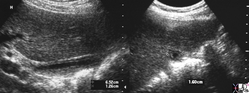
In this ultrasound a fatty meal has resulted in a contracted gallbladder and the the wall is now a 3 layered structure consisting of an inner echogenic mucosal layer, a middle hypoechoic muscular layer and an outer echogenic serosal/adventitial layer.  The volume of the gallbladder lumen is significantly reduced.
25902c.8s  gallbladder small normal post fatty meal normal physiology USscan ultrasound copyright 2008 Courtesy Ashley Davidoff MD


    [nodeType] => 1
    [parentNode] => (object value omitted)
    [childNodes] => (object value omitted)
    [firstChild] => (object value omitted)
    [lastChild] => (object value omitted)
    [previousSibling] => (object value omitted)
    [nextSibling] => (object value omitted)
    [attributes] => (object value omitted)
    [ownerDocument] => (object value omitted)
    [namespaceURI] =>
    [prefix] =>
    [localName] => table
    [baseURI] =>
    [textContent] =>


Contracted Gallbladder Following a Fatty Meal


In this ultrasound a fatty meal has resulted in a contracted gallbladder and the the wall is now a 3 layered structure consisting of an inner echogenic mucosal layer, a middle hypoechoic muscular layer and an outer echogenic serosal/adventitial layer.  The volume of the gallbladder lumen is significantly reduced.
25902c.8s  gallbladder small normal post fatty meal normal physiology USscan ultrasound copyright 2008 Courtesy Ashley Davidoff MD


)
DOMElement Object
(
    [schemaTypeInfo] =>
    [tagName] => td
    [firstElementChild] => (object value omitted)
    [lastElementChild] => (object value omitted)
    [childElementCount] => 2
    [previousElementSibling] =>
    [nextElementSibling] =>
    [nodeName] => td
    [nodeValue] => In this ultrasound a fatty meal has resulted in a contracted gallbladder and the the wall is now a 3 layered structure consisting of an inner echogenic mucosal layer, a middle hypoechoic muscular layer and an outer echogenic serosal/adventitial layer.  The volume of the gallbladder lumen is significantly reduced.
25902c.8s  gallbladder small normal post fatty meal normal physiology USscan ultrasound copyright 2008 Courtesy Ashley Davidoff MD
    [nodeType] => 1
    [parentNode] => (object value omitted)
    [childNodes] => (object value omitted)
    [firstChild] => (object value omitted)
    [lastChild] => (object value omitted)
    [previousSibling] => (object value omitted)
    [nextSibling] => (object value omitted)
    [attributes] => (object value omitted)
    [ownerDocument] => (object value omitted)
    [namespaceURI] =>
    [prefix] =>
    [localName] => td
    [baseURI] =>
    [textContent] => In this ultrasound a fatty meal has resulted in a contracted gallbladder and the the wall is now a 3 layered structure consisting of an inner echogenic mucosal layer, a middle hypoechoic muscular layer and an outer echogenic serosal/adventitial layer.  The volume of the gallbladder lumen is significantly reduced.
25902c.8s  gallbladder small normal post fatty meal normal physiology USscan ultrasound copyright 2008 Courtesy Ashley Davidoff MD
)
DOMElement Object
(
    [schemaTypeInfo] =>
    [tagName] => td
    [firstElementChild] => (object value omitted)
    [lastElementChild] => (object value omitted)
    [childElementCount] => 2
    [previousElementSibling] =>
    [nextElementSibling] =>
    [nodeName] => td
    [nodeValue] =>
Contracted Gallbladder Following a Fatty Meal

    [nodeType] => 1
    [parentNode] => (object value omitted)
    [childNodes] => (object value omitted)
    [firstChild] => (object value omitted)
    [lastChild] => (object value omitted)
    [previousSibling] => (object value omitted)
    [nextSibling] => (object value omitted)
    [attributes] => (object value omitted)
    [ownerDocument] => (object value omitted)
    [namespaceURI] =>
    [prefix] =>
    [localName] => td
    [baseURI] =>
    [textContent] =>
Contracted Gallbladder Following a Fatty Meal

)
DOMElement Object
(
    [schemaTypeInfo] =>
    [tagName] => table
    [firstElementChild] => (object value omitted)
    [lastElementChild] => (object value omitted)
    [childElementCount] => 1
    [previousElementSibling] => (object value omitted)
    [nextElementSibling] => (object value omitted)
    [nodeName] => table
    [nodeValue] =>


The Size of the Gallbladder in the Fasting State


The  ultrasound of the gallbladder in the fasting state is taken in the longitudinal plane in a, and the transverse plane in b, showing a gallbladder that is of a relatively small size but still distende showing a single wall, characteristic of the distended gallbladder.
82428c01.8s gallbladder small normal transverse oval shape normal anatomy USscan ultrasound copyright 2008 Courtesy Ashley DAvidoff MD


    [nodeType] => 1
    [parentNode] => (object value omitted)
    [childNodes] => (object value omitted)
    [firstChild] => (object value omitted)
    [lastChild] => (object value omitted)
    [previousSibling] => (object value omitted)
    [nextSibling] => (object value omitted)
    [attributes] => (object value omitted)
    [ownerDocument] => (object value omitted)
    [namespaceURI] =>
    [prefix] =>
    [localName] => table
    [baseURI] =>
    [textContent] =>


The Size of the Gallbladder in the Fasting State


The  ultrasound of the gallbladder in the fasting state is taken in the longitudinal plane in a, and the transverse plane in b, showing a gallbladder that is of a relatively small size but still distende showing a single wall, characteristic of the distended gallbladder.
82428c01.8s gallbladder small normal transverse oval shape normal anatomy USscan ultrasound copyright 2008 Courtesy Ashley DAvidoff MD


)
DOMElement Object
(
    [schemaTypeInfo] =>
    [tagName] => td
    [firstElementChild] => (object value omitted)
    [lastElementChild] => (object value omitted)
    [childElementCount] => 2
    [previousElementSibling] =>
    [nextElementSibling] =>
    [nodeName] => td
    [nodeValue] => The  ultrasound of the gallbladder in the fasting state is taken in the longitudinal plane in a, and the transverse plane in b, showing a gallbladder that is of a relatively small size but still distende showing a single wall, characteristic of the distended gallbladder.
82428c01.8s gallbladder small normal transverse oval shape normal anatomy USscan ultrasound copyright 2008 Courtesy Ashley DAvidoff MD
    [nodeType] => 1
    [parentNode] => (object value omitted)
    [childNodes] => (object value omitted)
    [firstChild] => (object value omitted)
    [lastChild] => (object value omitted)
    [previousSibling] => (object value omitted)
    [nextSibling] => (object value omitted)
    [attributes] => (object value omitted)
    [ownerDocument] => (object value omitted)
    [namespaceURI] =>
    [prefix] =>
    [localName] => td
    [baseURI] =>
    [textContent] => The  ultrasound of the gallbladder in the fasting state is taken in the longitudinal plane in a, and the transverse plane in b, showing a gallbladder that is of a relatively small size but still distende showing a single wall, characteristic of the distended gallbladder.
82428c01.8s gallbladder small normal transverse oval shape normal anatomy USscan ultrasound copyright 2008 Courtesy Ashley DAvidoff MD
)
DOMElement Object
(
    [schemaTypeInfo] =>
    [tagName] => td
    [firstElementChild] => (object value omitted)
    [lastElementChild] => (object value omitted)
    [childElementCount] => 2
    [previousElementSibling] =>
    [nextElementSibling] =>
    [nodeName] => td
    [nodeValue] =>

The Size of the Gallbladder in the Fasting State

    [nodeType] => 1
    [parentNode] => (object value omitted)
    [childNodes] => (object value omitted)
    [firstChild] => (object value omitted)
    [lastChild] => (object value omitted)
    [previousSibling] => (object value omitted)
    [nextSibling] => (object value omitted)
    [attributes] => (object value omitted)
    [ownerDocument] => (object value omitted)
    [namespaceURI] =>
    [prefix] =>
    [localName] => td
    [baseURI] =>
    [textContent] =>

The Size of the Gallbladder in the Fasting State

)
DOMElement Object
(
    [schemaTypeInfo] =>
    [tagName] => table
    [firstElementChild] => (object value omitted)
    [lastElementChild] => (object value omitted)
    [childElementCount] => 1
    [previousElementSibling] => (object value omitted)
    [nextElementSibling] => (object value omitted)
    [nodeName] => table
    [nodeValue] =>


Maximal Distension (fasting) and Maximal Emptying


82656b23c02.8s gallbladder sphincter of Oddi sympathetic system parasympathetic system juice gates fatty food closed open storage delivery contracted relaxed fat protein carbohydrates CCK cholecystokinin normal physiology function delivery davidoff art Copyright 2008


    [nodeType] => 1
    [parentNode] => (object value omitted)
    [childNodes] => (object value omitted)
    [firstChild] => (object value omitted)
    [lastChild] => (object value omitted)
    [previousSibling] => (object value omitted)
    [nextSibling] => (object value omitted)
    [attributes] => (object value omitted)
    [ownerDocument] => (object value omitted)
    [namespaceURI] =>
    [prefix] =>
    [localName] => table
    [baseURI] =>
    [textContent] =>


Maximal Distension (fasting) and Maximal Emptying


82656b23c02.8s gallbladder sphincter of Oddi sympathetic system parasympathetic system juice gates fatty food closed open storage delivery contracted relaxed fat protein carbohydrates CCK cholecystokinin normal physiology function delivery davidoff art Copyright 2008


)
DOMElement Object
(
    [schemaTypeInfo] =>
    [tagName] => td
    [firstElementChild] => (object value omitted)
    [lastElementChild] => (object value omitted)
    [childElementCount] => 1
    [previousElementSibling] =>
    [nextElementSibling] =>
    [nodeName] => td
    [nodeValue] => 82656b23c02.8s gallbladder sphincter of Oddi sympathetic system parasympathetic system juice gates fatty food closed open storage delivery contracted relaxed fat protein carbohydrates CCK cholecystokinin normal physiology function delivery davidoff art Copyright 2008
    [nodeType] => 1
    [parentNode] => (object value omitted)
    [childNodes] => (object value omitted)
    [firstChild] => (object value omitted)
    [lastChild] => (object value omitted)
    [previousSibling] => (object value omitted)
    [nextSibling] => (object value omitted)
    [attributes] => (object value omitted)
    [ownerDocument] => (object value omitted)
    [namespaceURI] =>
    [prefix] =>
    [localName] => td
    [baseURI] =>
    [textContent] => 82656b23c02.8s gallbladder sphincter of Oddi sympathetic system parasympathetic system juice gates fatty food closed open storage delivery contracted relaxed fat protein carbohydrates CCK cholecystokinin normal physiology function delivery davidoff art Copyright 2008
)
DOMElement Object
(
    [schemaTypeInfo] =>
    [tagName] => td
    [firstElementChild] => (object value omitted)
    [lastElementChild] => (object value omitted)
    [childElementCount] => 2
    [previousElementSibling] =>
    [nextElementSibling] =>
    [nodeName] => td
    [nodeValue] =>
Maximal Distension (fasting) and Maximal Emptying

    [nodeType] => 1
    [parentNode] => (object value omitted)
    [childNodes] => (object value omitted)
    [firstChild] => (object value omitted)
    [lastChild] => (object value omitted)
    [previousSibling] => (object value omitted)
    [nextSibling] => (object value omitted)
    [attributes] => (object value omitted)
    [ownerDocument] => (object value omitted)
    [namespaceURI] =>
    [prefix] =>
    [localName] => td
    [baseURI] =>
    [textContent] =>
Maximal Distension (fasting) and Maximal Emptying

)
DOMElement Object
(
    [schemaTypeInfo] =>
    [tagName] => table
    [firstElementChild] => (object value omitted)
    [lastElementChild] => (object value omitted)
    [childElementCount] => 1
    [previousElementSibling] => (object value omitted)
    [nextElementSibling] => (object value omitted)
    [nodeName] => table
    [nodeValue] =>


 Gallbladder Contraction


The delivery of bile and pancreatic secretions are slow and steady in response to fat (yellow) protein red) in the diet which causes the release of CCK into the blood which in turn acts on the smooth muscle of the gallbladder and sphincter resulting in open juice gates and steady flow of bile and pancreatic enzymes over two hours.  The spiral valves (downhill Archimedes waterscrew) prevent overdistension of the bile duct during the delivery phase.  Carbohydrates (white) have litttle effect on CCK
82656b23.8s gallbladder Archimedes water screw cystic duct gravity upright spiral valves of Heister filling ampulla of VAter closed neck body fundus distended parasympahtetic bile duct CCK cholecystokinin contraction relaxation of ampulla juice gates open delivery of bile and pancreatic juices to duodenum pancreatic duct Davidoff art Copyright 2008


    [nodeType] => 1
    [parentNode] => (object value omitted)
    [childNodes] => (object value omitted)
    [firstChild] => (object value omitted)
    [lastChild] => (object value omitted)
    [previousSibling] => (object value omitted)
    [nextSibling] => (object value omitted)
    [attributes] => (object value omitted)
    [ownerDocument] => (object value omitted)
    [namespaceURI] =>
    [prefix] =>
    [localName] => table
    [baseURI] =>
    [textContent] =>


 Gallbladder Contraction


The delivery of bile and pancreatic secretions are slow and steady in response to fat (yellow) protein red) in the diet which causes the release of CCK into the blood which in turn acts on the smooth muscle of the gallbladder and sphincter resulting in open juice gates and steady flow of bile and pancreatic enzymes over two hours.  The spiral valves (downhill Archimedes waterscrew) prevent overdistension of the bile duct during the delivery phase.  Carbohydrates (white) have litttle effect on CCK
82656b23.8s gallbladder Archimedes water screw cystic duct gravity upright spiral valves of Heister filling ampulla of VAter closed neck body fundus distended parasympahtetic bile duct CCK cholecystokinin contraction relaxation of ampulla juice gates open delivery of bile and pancreatic juices to duodenum pancreatic duct Davidoff art Copyright 2008


)
DOMElement Object
(
    [schemaTypeInfo] =>
    [tagName] => td
    [firstElementChild] => (object value omitted)
    [lastElementChild] => (object value omitted)
    [childElementCount] => 2
    [previousElementSibling] =>
    [nextElementSibling] =>
    [nodeName] => td
    [nodeValue] => The delivery of bile and pancreatic secretions are slow and steady in response to fat (yellow) protein red) in the diet which causes the release of CCK into the blood which in turn acts on the smooth muscle of the gallbladder and sphincter resulting in open juice gates and steady flow of bile and pancreatic enzymes over two hours.  The spiral valves (downhill Archimedes waterscrew) prevent overdistension of the bile duct during the delivery phase.  Carbohydrates (white) have litttle effect on CCK
82656b23.8s gallbladder Archimedes water screw cystic duct gravity upright spiral valves of Heister filling ampulla of VAter closed neck body fundus distended parasympahtetic bile duct CCK cholecystokinin contraction relaxation of ampulla juice gates open delivery of bile and pancreatic juices to duodenum pancreatic duct Davidoff art Copyright 2008
    [nodeType] => 1
    [parentNode] => (object value omitted)
    [childNodes] => (object value omitted)
    [firstChild] => (object value omitted)
    [lastChild] => (object value omitted)
    [previousSibling] => (object value omitted)
    [nextSibling] => (object value omitted)
    [attributes] => (object value omitted)
    [ownerDocument] => (object value omitted)
    [namespaceURI] =>
    [prefix] =>
    [localName] => td
    [baseURI] =>
    [textContent] => The delivery of bile and pancreatic secretions are slow and steady in response to fat (yellow) protein red) in the diet which causes the release of CCK into the blood which in turn acts on the smooth muscle of the gallbladder and sphincter resulting in open juice gates and steady flow of bile and pancreatic enzymes over two hours.  The spiral valves (downhill Archimedes waterscrew) prevent overdistension of the bile duct during the delivery phase.  Carbohydrates (white) have litttle effect on CCK
82656b23.8s gallbladder Archimedes water screw cystic duct gravity upright spiral valves of Heister filling ampulla of VAter closed neck body fundus distended parasympahtetic bile duct CCK cholecystokinin contraction relaxation of ampulla juice gates open delivery of bile and pancreatic juices to duodenum pancreatic duct Davidoff art Copyright 2008
)
DOMElement Object
(
    [schemaTypeInfo] =>
    [tagName] => td
    [firstElementChild] => (object value omitted)
    [lastElementChild] => (object value omitted)
    [childElementCount] => 2
    [previousElementSibling] =>
    [nextElementSibling] =>
    [nodeName] => td
    [nodeValue] =>
 Gallbladder Contraction

    [nodeType] => 1
    [parentNode] => (object value omitted)
    [childNodes] => (object value omitted)
    [firstChild] => (object value omitted)
    [lastChild] => (object value omitted)
    [previousSibling] => (object value omitted)
    [nextSibling] => (object value omitted)
    [attributes] => (object value omitted)
    [ownerDocument] => (object value omitted)
    [namespaceURI] =>
    [prefix] =>
    [localName] => td
    [baseURI] =>
    [textContent] =>
 Gallbladder Contraction

)
DOMElement Object
(
    [schemaTypeInfo] =>
    [tagName] => table
    [firstElementChild] => (object value omitted)
    [lastElementChild] => (object value omitted)
    [childElementCount] => 1
    [previousElementSibling] => (object value omitted)
    [nextElementSibling] => (object value omitted)
    [nodeName] => table
    [nodeValue] =>


Juice Gates Closed and Open


CCK acts on the sphincter of Oddi relaxing the muscle to enable stable steady flow.  Flow through the CBD during the fasting state is low being only about .5-1ml and following a meal it rises to 2-3mls per minute.
82656b23c01.8s gallbladder sphincter of Oddi sympathetic system parasympathetic system juice gates closed open spasm relaxed fat protein carbohydrates CCK cholecystokinin normal physiology function delivery davidoff art Copyright 2008


    [nodeType] => 1
    [parentNode] => (object value omitted)
    [childNodes] => (object value omitted)
    [firstChild] => (object value omitted)
    [lastChild] => (object value omitted)
    [previousSibling] => (object value omitted)
    [nextSibling] => (object value omitted)
    [attributes] => (object value omitted)
    [ownerDocument] => (object value omitted)
    [namespaceURI] =>
    [prefix] =>
    [localName] => table
    [baseURI] =>
    [textContent] =>


Juice Gates Closed and Open


CCK acts on the sphincter of Oddi relaxing the muscle to enable stable steady flow.  Flow through the CBD during the fasting state is low being only about .5-1ml and following a meal it rises to 2-3mls per minute.
82656b23c01.8s gallbladder sphincter of Oddi sympathetic system parasympathetic system juice gates closed open spasm relaxed fat protein carbohydrates CCK cholecystokinin normal physiology function delivery davidoff art Copyright 2008


)
DOMElement Object
(
    [schemaTypeInfo] =>
    [tagName] => td
    [firstElementChild] => (object value omitted)
    [lastElementChild] => (object value omitted)
    [childElementCount] => 2
    [previousElementSibling] =>
    [nextElementSibling] =>
    [nodeName] => td
    [nodeValue] => CCK acts on the sphincter of Oddi relaxing the muscle to enable stable steady flow.  Flow through the CBD during the fasting state is low being only about .5-1ml and following a meal it rises to 2-3mls per minute.
82656b23c01.8s gallbladder sphincter of Oddi sympathetic system parasympathetic system juice gates closed open spasm relaxed fat protein carbohydrates CCK cholecystokinin normal physiology function delivery davidoff art Copyright 2008
    [nodeType] => 1
    [parentNode] => (object value omitted)
    [childNodes] => (object value omitted)
    [firstChild] => (object value omitted)
    [lastChild] => (object value omitted)
    [previousSibling] => (object value omitted)
    [nextSibling] => (object value omitted)
    [attributes] => (object value omitted)
    [ownerDocument] => (object value omitted)
    [namespaceURI] =>
    [prefix] =>
    [localName] => td
    [baseURI] =>
    [textContent] => CCK acts on the sphincter of Oddi relaxing the muscle to enable stable steady flow.  Flow through the CBD during the fasting state is low being only about .5-1ml and following a meal it rises to 2-3mls per minute.
82656b23c01.8s gallbladder sphincter of Oddi sympathetic system parasympathetic system juice gates closed open spasm relaxed fat protein carbohydrates CCK cholecystokinin normal physiology function delivery davidoff art Copyright 2008
)
DOMElement Object
(
    [schemaTypeInfo] =>
    [tagName] => td
    [firstElementChild] => (object value omitted)
    [lastElementChild] => (object value omitted)
    [childElementCount] => 2
    [previousElementSibling] =>
    [nextElementSibling] =>
    [nodeName] => td
    [nodeValue] =>
Juice Gates Closed and Open

    [nodeType] => 1
    [parentNode] => (object value omitted)
    [childNodes] => (object value omitted)
    [firstChild] => (object value omitted)
    [lastChild] => (object value omitted)
    [previousSibling] => (object value omitted)
    [nextSibling] => (object value omitted)
    [attributes] => (object value omitted)
    [ownerDocument] => (object value omitted)
    [namespaceURI] =>
    [prefix] =>
    [localName] => td
    [baseURI] =>
    [textContent] =>
Juice Gates Closed and Open

)
DOMElement Object
(
    [schemaTypeInfo] =>
    [tagName] => table
    [firstElementChild] => (object value omitted)
    [lastElementChild] => (object value omitted)
    [childElementCount] => 1
    [previousElementSibling] => (object value omitted)
    [nextElementSibling] => (object value omitted)
    [nodeName] => table
    [nodeValue] =>


Inferior Positioning of the Gallbladder Allows Old Bile to be Stored and Concentrated


This ultrasound of a normal gallbladder of a 20 year old male has been turned to simulate a standing and or seated position which is the dominant position during the day.  The inferiorly positioned fundus and body enables the stratification of new and old bile.  With the person in the standing or sitting position, the shape of this gallbladder allows new bile to come into the neck, receive a small secretion of mucus, and then to spill over like a waterfall through gravity into the body where it will become concentrated and stored in the more voluminous body and fundus.  Most the day is spent in the upright position and therefore the image represents  the “day” position of the gallbladder.  When the bile is needed, most the bile accumulated during the filling phase will have been concentrated and therefore will be more effective for emulsification of fat.
82257c06b04 20 male gallbladder normal position function storage concentration fundus inferior shape anatomy USscan ultrasound Courtesy Ashley Davidoff MD copyright 2008


    [nodeType] => 1
    [parentNode] => (object value omitted)
    [childNodes] => (object value omitted)
    [firstChild] => (object value omitted)
    [lastChild] => (object value omitted)
    [previousSibling] => (object value omitted)
    [nextSibling] => (object value omitted)
    [attributes] => (object value omitted)
    [ownerDocument] => (object value omitted)
    [namespaceURI] =>
    [prefix] =>
    [localName] => table
    [baseURI] =>
    [textContent] =>


Inferior Positioning of the Gallbladder Allows Old Bile to be Stored and Concentrated


This ultrasound of a normal gallbladder of a 20 year old male has been turned to simulate a standing and or seated position which is the dominant position during the day.  The inferiorly positioned fundus and body enables the stratification of new and old bile.  With the person in the standing or sitting position, the shape of this gallbladder allows new bile to come into the neck, receive a small secretion of mucus, and then to spill over like a waterfall through gravity into the body where it will become concentrated and stored in the more voluminous body and fundus.  Most the day is spent in the upright position and therefore the image represents  the “day” position of the gallbladder.  When the bile is needed, most the bile accumulated during the filling phase will have been concentrated and therefore will be more effective for emulsification of fat.
82257c06b04 20 male gallbladder normal position function storage concentration fundus inferior shape anatomy USscan ultrasound Courtesy Ashley Davidoff MD copyright 2008


)
DOMElement Object
(
    [schemaTypeInfo] =>
    [tagName] => td
    [firstElementChild] => (object value omitted)
    [lastElementChild] => (object value omitted)
    [childElementCount] => 2
    [previousElementSibling] =>
    [nextElementSibling] =>
    [nodeName] => td
    [nodeValue] =>
This ultrasound of a normal gallbladder of a 20 year old male has been turned to simulate a standing and or seated position which is the dominant position during the day.  The inferiorly positioned fundus and body enables the stratification of new and old bile.  With the person in the standing or sitting position, the shape of this gallbladder allows new bile to come into the neck, receive a small secretion of mucus, and then to spill over like a waterfall through gravity into the body where it will become concentrated and stored in the more voluminous body and fundus.  Most the day is spent in the upright position and therefore the image represents  the “day” position of the gallbladder.  When the bile is needed, most the bile accumulated during the filling phase will have been concentrated and therefore will be more effective for emulsification of fat.
82257c06b04 20 male gallbladder normal position function storage concentration fundus inferior shape anatomy USscan ultrasound Courtesy Ashley Davidoff MD copyright 2008

    [nodeType] => 1
    [parentNode] => (object value omitted)
    [childNodes] => (object value omitted)
    [firstChild] => (object value omitted)
    [lastChild] => (object value omitted)
    [previousSibling] => (object value omitted)
    [nextSibling] => (object value omitted)
    [attributes] => (object value omitted)
    [ownerDocument] => (object value omitted)
    [namespaceURI] =>
    [prefix] =>
    [localName] => td
    [baseURI] =>
    [textContent] =>
This ultrasound of a normal gallbladder of a 20 year old male has been turned to simulate a standing and or seated position which is the dominant position during the day.  The inferiorly positioned fundus and body enables the stratification of new and old bile.  With the person in the standing or sitting position, the shape of this gallbladder allows new bile to come into the neck, receive a small secretion of mucus, and then to spill over like a waterfall through gravity into the body where it will become concentrated and stored in the more voluminous body and fundus.  Most the day is spent in the upright position and therefore the image represents  the “day” position of the gallbladder.  When the bile is needed, most the bile accumulated during the filling phase will have been concentrated and therefore will be more effective for emulsification of fat.
82257c06b04 20 male gallbladder normal position function storage concentration fundus inferior shape anatomy USscan ultrasound Courtesy Ashley Davidoff MD copyright 2008

)
DOMElement Object
(
    [schemaTypeInfo] =>
    [tagName] => td
    [firstElementChild] => (object value omitted)
    [lastElementChild] => (object value omitted)
    [childElementCount] => 2
    [previousElementSibling] =>
    [nextElementSibling] =>
    [nodeName] => td
    [nodeValue] =>
Inferior Positioning of the Gallbladder Allows Old Bile to be Stored and Concentrated

    [nodeType] => 1
    [parentNode] => (object value omitted)
    [childNodes] => (object value omitted)
    [firstChild] => (object value omitted)
    [lastChild] => (object value omitted)
    [previousSibling] => (object value omitted)
    [nextSibling] => (object value omitted)
    [attributes] => (object value omitted)
    [ownerDocument] => (object value omitted)
    [namespaceURI] =>
    [prefix] =>
    [localName] => td
    [baseURI] =>
    [textContent] =>
Inferior Positioning of the Gallbladder Allows Old Bile to be Stored and Concentrated

)
DOMElement Object
(
    [schemaTypeInfo] =>
    [tagName] => table
    [firstElementChild] => (object value omitted)
    [lastElementChild] => (object value omitted)
    [childElementCount] => 1
    [previousElementSibling] => (object value omitted)
    [nextElementSibling] => (object value omitted)
    [nodeName] => table
    [nodeValue] =>


Dilated Cystic Duct Valves of Heister and Archimedes Water Screw


This patient has obstructive jaundice as a result of cancer in the head of the pancreas.  The gallbladder is distended and the cystic duct is dilated  and unusually well seen in the transverse section provided by an ultrasound. We can envisage through the ultrasound and conceptual diagram,  how bile is transported in stepwise fashion discussed above, up the cystic duct and then down the neck into the infundibulum.  With the patient in the upright position the bile falls down to the fundus which is the most inferior aspect of the gallbladder.
04113c09.8s patient with pancreatic carcinoma gallbladder dilated neck cystic duct valves of Heister dilated enlarged obstruction Archiomedes screw stepladder force gravity USscan ultrasound copyright 2008 Courtesy Ashley Davidoff MD


    [nodeType] => 1
    [parentNode] => (object value omitted)
    [childNodes] => (object value omitted)
    [firstChild] => (object value omitted)
    [lastChild] => (object value omitted)
    [previousSibling] => (object value omitted)
    [nextSibling] => (object value omitted)
    [attributes] => (object value omitted)
    [ownerDocument] => (object value omitted)
    [namespaceURI] =>
    [prefix] =>
    [localName] => table
    [baseURI] =>
    [textContent] =>


Dilated Cystic Duct Valves of Heister and Archimedes Water Screw


This patient has obstructive jaundice as a result of cancer in the head of the pancreas.  The gallbladder is distended and the cystic duct is dilated  and unusually well seen in the transverse section provided by an ultrasound. We can envisage through the ultrasound and conceptual diagram,  how bile is transported in stepwise fashion discussed above, up the cystic duct and then down the neck into the infundibulum.  With the patient in the upright position the bile falls down to the fundus which is the most inferior aspect of the gallbladder.
04113c09.8s patient with pancreatic carcinoma gallbladder dilated neck cystic duct valves of Heister dilated enlarged obstruction Archiomedes screw stepladder force gravity USscan ultrasound copyright 2008 Courtesy Ashley Davidoff MD


)
DOMElement Object
(
    [schemaTypeInfo] =>
    [tagName] => td
    [firstElementChild] => (object value omitted)
    [lastElementChild] => (object value omitted)
    [childElementCount] => 2
    [previousElementSibling] =>
    [nextElementSibling] =>
    [nodeName] => td
    [nodeValue] => This patient has obstructive jaundice as a result of cancer in the head of the pancreas.  The gallbladder is distended and the cystic duct is dilated  and unusually well seen in the transverse section provided by an ultrasound. We can envisage through the ultrasound and conceptual diagram,  how bile is transported in stepwise fashion discussed above, up the cystic duct and then down the neck into the infundibulum.  With the patient in the upright position the bile falls down to the fundus which is the most inferior aspect of the gallbladder.
04113c09.8s patient with pancreatic carcinoma gallbladder dilated neck cystic duct valves of Heister dilated enlarged obstruction Archiomedes screw stepladder force gravity USscan ultrasound copyright 2008 Courtesy Ashley Davidoff MD
    [nodeType] => 1
    [parentNode] => (object value omitted)
    [childNodes] => (object value omitted)
    [firstChild] => (object value omitted)
    [lastChild] => (object value omitted)
    [previousSibling] => (object value omitted)
    [nextSibling] => (object value omitted)
    [attributes] => (object value omitted)
    [ownerDocument] => (object value omitted)
    [namespaceURI] =>
    [prefix] =>
    [localName] => td
    [baseURI] =>
    [textContent] => This patient has obstructive jaundice as a result of cancer in the head of the pancreas.  The gallbladder is distended and the cystic duct is dilated  and unusually well seen in the transverse section provided by an ultrasound. We can envisage through the ultrasound and conceptual diagram,  how bile is transported in stepwise fashion discussed above, up the cystic duct and then down the neck into the infundibulum.  With the patient in the upright position the bile falls down to the fundus which is the most inferior aspect of the gallbladder.
04113c09.8s patient with pancreatic carcinoma gallbladder dilated neck cystic duct valves of Heister dilated enlarged obstruction Archiomedes screw stepladder force gravity USscan ultrasound copyright 2008 Courtesy Ashley Davidoff MD
)
DOMElement Object
(
    [schemaTypeInfo] =>
    [tagName] => td
    [firstElementChild] => (object value omitted)
    [lastElementChild] => (object value omitted)
    [childElementCount] => 2
    [previousElementSibling] =>
    [nextElementSibling] =>
    [nodeName] => td
    [nodeValue] =>
Dilated Cystic Duct Valves of Heister and Archimedes Water Screw

    [nodeType] => 1
    [parentNode] => (object value omitted)
    [childNodes] => (object value omitted)
    [firstChild] => (object value omitted)
    [lastChild] => (object value omitted)
    [previousSibling] => (object value omitted)
    [nextSibling] => (object value omitted)
    [attributes] => (object value omitted)
    [ownerDocument] => (object value omitted)
    [namespaceURI] =>
    [prefix] =>
    [localName] => td
    [baseURI] =>
    [textContent] =>
Dilated Cystic Duct Valves of Heister and Archimedes Water Screw

)
DOMElement Object
(
    [schemaTypeInfo] =>
    [tagName] => table
    [firstElementChild] => (object value omitted)
    [lastElementChild] => (object value omitted)
    [childElementCount] => 1
    [previousElementSibling] => (object value omitted)
    [nextElementSibling] => (object value omitted)
    [nodeName] => table
    [nodeValue] =>


The Dilated Cystic Duct with Fluid Fluid Levels


The CTscan through the liver and gallbladder shows contrast layering in stepwise fashion in a dilated cystic duct.  The contrast in the gallbladder is from a recent CT scan.  This entity is called vicarious excretion of contrast and represents the normal excretion of intravenous contrast by the liver cells seen as dense material within 24-48 hours following intravenous injection.  It becomes more prominent in patients with renal failure.  The stepwise appearance of the contrast as a fluid fluid level, adds credence to the theory of the the parallel drawn and mechanism of function of the valves of Heister and Archimedes water screw.
82453c02.8s enlarged cystic duct Archomedes water screw fluid fluid level vicarious excretion of contrast 88F p/w abdominal pain no known prior study gallbladder shape character sinusoidal shape to “s” shape fluid fluid level milk of contrast bile vicarious excretion anatomy CTscan Courtesy Ashley DAvidoff MD copyright 2008


    [nodeType] => 1
    [parentNode] => (object value omitted)
    [childNodes] => (object value omitted)
    [firstChild] => (object value omitted)
    [lastChild] => (object value omitted)
    [previousSibling] => (object value omitted)
    [nextSibling] => (object value omitted)
    [attributes] => (object value omitted)
    [ownerDocument] => (object value omitted)
    [namespaceURI] =>
    [prefix] =>
    [localName] => table
    [baseURI] =>
    [textContent] =>


The Dilated Cystic Duct with Fluid Fluid Levels


The CTscan through the liver and gallbladder shows contrast layering in stepwise fashion in a dilated cystic duct.  The contrast in the gallbladder is from a recent CT scan.  This entity is called vicarious excretion of contrast and represents the normal excretion of intravenous contrast by the liver cells seen as dense material within 24-48 hours following intravenous injection.  It becomes more prominent in patients with renal failure.  The stepwise appearance of the contrast as a fluid fluid level, adds credence to the theory of the the parallel drawn and mechanism of function of the valves of Heister and Archimedes water screw.
82453c02.8s enlarged cystic duct Archomedes water screw fluid fluid level vicarious excretion of contrast 88F p/w abdominal pain no known prior study gallbladder shape character sinusoidal shape to “s” shape fluid fluid level milk of contrast bile vicarious excretion anatomy CTscan Courtesy Ashley DAvidoff MD copyright 2008


)
DOMElement Object
(
    [schemaTypeInfo] =>
    [tagName] => td
    [firstElementChild] => (object value omitted)
    [lastElementChild] => (object value omitted)
    [childElementCount] => 2
    [previousElementSibling] =>
    [nextElementSibling] =>
    [nodeName] => td
    [nodeValue] => The CTscan through the liver and gallbladder shows contrast layering in stepwise fashion in a dilated cystic duct.  The contrast in the gallbladder is from a recent CT scan.  This entity is called vicarious excretion of contrast and represents the normal excretion of intravenous contrast by the liver cells seen as dense material within 24-48 hours following intravenous injection.  It becomes more prominent in patients with renal failure.  The stepwise appearance of the contrast as a fluid fluid level, adds credence to the theory of the the parallel drawn and mechanism of function of the valves of Heister and Archimedes water screw.
82453c02.8s enlarged cystic duct Archomedes water screw fluid fluid level vicarious excretion of contrast 88F p/w abdominal pain no known prior study gallbladder shape character sinusoidal shape to “s” shape fluid fluid level milk of contrast bile vicarious excretion anatomy CTscan Courtesy Ashley DAvidoff MD copyright 2008
    [nodeType] => 1
    [parentNode] => (object value omitted)
    [childNodes] => (object value omitted)
    [firstChild] => (object value omitted)
    [lastChild] => (object value omitted)
    [previousSibling] => (object value omitted)
    [nextSibling] => (object value omitted)
    [attributes] => (object value omitted)
    [ownerDocument] => (object value omitted)
    [namespaceURI] =>
    [prefix] =>
    [localName] => td
    [baseURI] =>
    [textContent] => The CTscan through the liver and gallbladder shows contrast layering in stepwise fashion in a dilated cystic duct.  The contrast in the gallbladder is from a recent CT scan.  This entity is called vicarious excretion of contrast and represents the normal excretion of intravenous contrast by the liver cells seen as dense material within 24-48 hours following intravenous injection.  It becomes more prominent in patients with renal failure.  The stepwise appearance of the contrast as a fluid fluid level, adds credence to the theory of the the parallel drawn and mechanism of function of the valves of Heister and Archimedes water screw.
82453c02.8s enlarged cystic duct Archomedes water screw fluid fluid level vicarious excretion of contrast 88F p/w abdominal pain no known prior study gallbladder shape character sinusoidal shape to “s” shape fluid fluid level milk of contrast bile vicarious excretion anatomy CTscan Courtesy Ashley DAvidoff MD copyright 2008
)
DOMElement Object
(
    [schemaTypeInfo] =>
    [tagName] => td
    [firstElementChild] => (object value omitted)
    [lastElementChild] => (object value omitted)
    [childElementCount] => 2
    [previousElementSibling] =>
    [nextElementSibling] =>
    [nodeName] => td
    [nodeValue] =>
The Dilated Cystic Duct with Fluid Fluid Levels

    [nodeType] => 1
    [parentNode] => (object value omitted)
    [childNodes] => (object value omitted)
    [firstChild] => (object value omitted)
    [lastChild] => (object value omitted)
    [previousSibling] => (object value omitted)
    [nextSibling] => (object value omitted)
    [attributes] => (object value omitted)
    [ownerDocument] => (object value omitted)
    [namespaceURI] =>
    [prefix] =>
    [localName] => td
    [baseURI] =>
    [textContent] =>
The Dilated Cystic Duct with Fluid Fluid Levels

)
DOMElement Object
(
    [schemaTypeInfo] =>
    [tagName] => table
    [firstElementChild] => (object value omitted)
    [lastElementChild] => (object value omitted)
    [childElementCount] => 1
    [previousElementSibling] => (object value omitted)
    [nextElementSibling] => (object value omitted)
    [nodeName] => table
    [nodeValue] =>


Filling


This diagram illustrates the concept of how the gallbladder receives bile via the relatively high lying (cranially positioned) cystic duct, using a stepwise ladder system of valves to deliver the dilute bile to the gallbladder without a pump.  An Archimedes water screw in the position of the spiral valves is shown revealing the parallel mechanism of action.  In the case of the spiral valves the driving force is a is a push from the bottom, rather than a pull from the top. The spirals are not as complete as the screws of the Archomedes device.  Rather they are a series of sacs of small diameter that have membranous folds that spiral and prevent backflow. (we propose)
 The bile produced during the day in the biliary system will cause a minimal rise in pressure 82656b20.8s gallbladder Archimedes water screw cystic duct gravity upright spiral valves of Heister filling ampulla of VAter closed neck body fundus distended sympathetic bile duct Davidoff art Copyright 2008


    [nodeType] => 1
    [parentNode] => (object value omitted)
    [childNodes] => (object value omitted)
    [firstChild] => (object value omitted)
    [lastChild] => (object value omitted)
    [previousSibling] => (object value omitted)
    [nextSibling] => (object value omitted)
    [attributes] => (object value omitted)
    [ownerDocument] => (object value omitted)
    [namespaceURI] =>
    [prefix] =>
    [localName] => table
    [baseURI] =>
    [textContent] =>


Filling


This diagram illustrates the concept of how the gallbladder receives bile via the relatively high lying (cranially positioned) cystic duct, using a stepwise ladder system of valves to deliver the dilute bile to the gallbladder without a pump.  An Archimedes water screw in the position of the spiral valves is shown revealing the parallel mechanism of action.  In the case of the spiral valves the driving force is a is a push from the bottom, rather than a pull from the top. The spirals are not as complete as the screws of the Archomedes device.  Rather they are a series of sacs of small diameter that have membranous folds that spiral and prevent backflow. (we propose)
 The bile produced during the day in the biliary system will cause a minimal rise in pressure 82656b20.8s gallbladder Archimedes water screw cystic duct gravity upright spiral valves of Heister filling ampulla of VAter closed neck body fundus distended sympathetic bile duct Davidoff art Copyright 2008


)
DOMElement Object
(
    [schemaTypeInfo] =>
    [tagName] => td
    [firstElementChild] => (object value omitted)
    [lastElementChild] => (object value omitted)
    [childElementCount] => 2
    [previousElementSibling] =>
    [nextElementSibling] =>
    [nodeName] => td
    [nodeValue] => This diagram illustrates the concept of how the gallbladder receives bile via the relatively high lying (cranially positioned) cystic duct, using a stepwise ladder system of valves to deliver the dilute bile to the gallbladder without a pump.  An Archimedes water screw in the position of the spiral valves is shown revealing the parallel mechanism of action.  In the case of the spiral valves the driving force is a is a push from the bottom, rather than a pull from the top. The spirals are not as complete as the screws of the Archomedes device.  Rather they are a series of sacs of small diameter that have membranous folds that spiral and prevent backflow. (we propose)
 The bile produced during the day in the biliary system will cause a minimal rise in pressure 82656b20.8s gallbladder Archimedes water screw cystic duct gravity upright spiral valves of Heister filling ampulla of VAter closed neck body fundus distended sympathetic bile duct Davidoff art Copyright 2008
    [nodeType] => 1
    [parentNode] => (object value omitted)
    [childNodes] => (object value omitted)
    [firstChild] => (object value omitted)
    [lastChild] => (object value omitted)
    [previousSibling] => (object value omitted)
    [nextSibling] => (object value omitted)
    [attributes] => (object value omitted)
    [ownerDocument] => (object value omitted)
    [namespaceURI] =>
    [prefix] =>
    [localName] => td
    [baseURI] =>
    [textContent] => This diagram illustrates the concept of how the gallbladder receives bile via the relatively high lying (cranially positioned) cystic duct, using a stepwise ladder system of valves to deliver the dilute bile to the gallbladder without a pump.  An Archimedes water screw in the position of the spiral valves is shown revealing the parallel mechanism of action.  In the case of the spiral valves the driving force is a is a push from the bottom, rather than a pull from the top. The spirals are not as complete as the screws of the Archomedes device.  Rather they are a series of sacs of small diameter that have membranous folds that spiral and prevent backflow. (we propose)
 The bile produced during the day in the biliary system will cause a minimal rise in pressure 82656b20.8s gallbladder Archimedes water screw cystic duct gravity upright spiral valves of Heister filling ampulla of VAter closed neck body fundus distended sympathetic bile duct Davidoff art Copyright 2008
)
DOMElement Object
(
    [schemaTypeInfo] =>
    [tagName] => td
    [firstElementChild] => (object value omitted)
    [lastElementChild] => (object value omitted)
    [childElementCount] => 2
    [previousElementSibling] =>
    [nextElementSibling] =>
    [nodeName] => td
    [nodeValue] =>
Filling

    [nodeType] => 1
    [parentNode] => (object value omitted)
    [childNodes] => (object value omitted)
    [firstChild] => (object value omitted)
    [lastChild] => (object value omitted)
    [previousSibling] => (object value omitted)
    [nextSibling] => (object value omitted)
    [attributes] => (object value omitted)
    [ownerDocument] => (object value omitted)
    [namespaceURI] =>
    [prefix] =>
    [localName] => td
    [baseURI] =>
    [textContent] =>
Filling

)
DOMElement Object
(
    [schemaTypeInfo] =>
    [tagName] => table
    [firstElementChild] => (object value omitted)
    [lastElementChild] => (object value omitted)
    [childElementCount] => 1
    [previousElementSibling] => (object value omitted)
    [nextElementSibling] => (object value omitted)
    [nodeName] => table
    [nodeValue] =>


Archimedes Water Screw


The design of the water screw has been attributed to Archimedes from 3rd century BC though some designate the originator of the screw to be Nebuchadnezzar II of 7th century BC who purportedly used the screw to deliver water to the Hanging Gardens of Babylon.  Water at a low level is scooped up into the spiral mechanism.  As the screw rotates, the water advances upward and gets delivered to a higher level until it finally reaches its destination and is delivered to a repository.
has been 82656b15.8s Archimedes water screw elevation gravity screw delivery cystic duct spiral valves of Heister anatomy physiology function uploading Davidoff art copyright 2008
The image was modified from Wikipedia; detail of image: Archimedes’ screw. Public domain, from Chambers’s Encyclopedia (Philadelphia: J. B. Lippincott Company, 1875). Added to illustrate article :Archimedes.


    [nodeType] => 1
    [parentNode] => (object value omitted)
    [childNodes] => (object value omitted)
    [firstChild] => (object value omitted)
    [lastChild] => (object value omitted)
    [previousSibling] => (object value omitted)
    [nextSibling] => (object value omitted)
    [attributes] => (object value omitted)
    [ownerDocument] => (object value omitted)
    [namespaceURI] =>
    [prefix] =>
    [localName] => table
    [baseURI] =>
    [textContent] =>


Archimedes Water Screw


The design of the water screw has been attributed to Archimedes from 3rd century BC though some designate the originator of the screw to be Nebuchadnezzar II of 7th century BC who purportedly used the screw to deliver water to the Hanging Gardens of Babylon.  Water at a low level is scooped up into the spiral mechanism.  As the screw rotates, the water advances upward and gets delivered to a higher level until it finally reaches its destination and is delivered to a repository.
has been 82656b15.8s Archimedes water screw elevation gravity screw delivery cystic duct spiral valves of Heister anatomy physiology function uploading Davidoff art copyright 2008
The image was modified from Wikipedia; detail of image: Archimedes’ screw. Public domain, from Chambers’s Encyclopedia (Philadelphia: J. B. Lippincott Company, 1875). Added to illustrate article :Archimedes.


)
DOMElement Object
(
    [schemaTypeInfo] =>
    [tagName] => td
    [firstElementChild] => (object value omitted)
    [lastElementChild] => (object value omitted)
    [childElementCount] => 3
    [previousElementSibling] =>
    [nextElementSibling] =>
    [nodeName] => td
    [nodeValue] => The design of the water screw has been attributed to Archimedes from 3rd century BC though some designate the originator of the screw to be Nebuchadnezzar II of 7th century BC who purportedly used the screw to deliver water to the Hanging Gardens of Babylon.  Water at a low level is scooped up into the spiral mechanism.  As the screw rotates, the water advances upward and gets delivered to a higher level until it finally reaches its destination and is delivered to a repository.
has been 82656b15.8s Archimedes water screw elevation gravity screw delivery cystic duct spiral valves of Heister anatomy physiology function uploading Davidoff art copyright 2008
The image was modified from Wikipedia; detail of image: Archimedes’ screw. Public domain, from Chambers’s Encyclopedia (Philadelphia: J. B. Lippincott Company, 1875). Added to illustrate article :Archimedes.
    [nodeType] => 1
    [parentNode] => (object value omitted)
    [childNodes] => (object value omitted)
    [firstChild] => (object value omitted)
    [lastChild] => (object value omitted)
    [previousSibling] => (object value omitted)
    [nextSibling] => (object value omitted)
    [attributes] => (object value omitted)
    [ownerDocument] => (object value omitted)
    [namespaceURI] =>
    [prefix] =>
    [localName] => td
    [baseURI] =>
    [textContent] => The design of the water screw has been attributed to Archimedes from 3rd century BC though some designate the originator of the screw to be Nebuchadnezzar II of 7th century BC who purportedly used the screw to deliver water to the Hanging Gardens of Babylon.  Water at a low level is scooped up into the spiral mechanism.  As the screw rotates, the water advances upward and gets delivered to a higher level until it finally reaches its destination and is delivered to a repository.
has been 82656b15.8s Archimedes water screw elevation gravity screw delivery cystic duct spiral valves of Heister anatomy physiology function uploading Davidoff art copyright 2008
The image was modified from Wikipedia; detail of image: Archimedes’ screw. Public domain, from Chambers’s Encyclopedia (Philadelphia: J. B. Lippincott Company, 1875). Added to illustrate article :Archimedes.
)
DOMElement Object
(
    [schemaTypeInfo] =>
    [tagName] => td
    [firstElementChild] => (object value omitted)
    [lastElementChild] => (object value omitted)
    [childElementCount] => 2
    [previousElementSibling] =>
    [nextElementSibling] =>
    [nodeName] => td
    [nodeValue] =>
Archimedes Water Screw

    [nodeType] => 1
    [parentNode] => (object value omitted)
    [childNodes] => (object value omitted)
    [firstChild] => (object value omitted)
    [lastChild] => (object value omitted)
    [previousSibling] => (object value omitted)
    [nextSibling] => (object value omitted)
    [attributes] => (object value omitted)
    [ownerDocument] => (object value omitted)
    [namespaceURI] =>
    [prefix] =>
    [localName] => td
    [baseURI] =>
    [textContent] =>
Archimedes Water Screw

)
DOMElement Object
(
    [schemaTypeInfo] =>
    [tagName] => table
    [firstElementChild] => (object value omitted)
    [lastElementChild] => (object value omitted)
    [childElementCount] => 1
    [previousElementSibling] => (object value omitted)
    [nextElementSibling] => (object value omitted)
    [nodeName] => table
    [nodeValue] =>


Ascent of Bile Against Gravity


The biliary system is a low pressure system under normal circumstances. The upward direction for the bile, from the bile duct to  the cystic duct is against gravity and there is no pump to push it nor a mechanism to pull it .  While the sphincter of Oddi is closed between meals, nature has devised a way to ensure that bile reaches the gallbladder with the least effort on its part.  Nature and not Archimedes nor Nebuchadnezzar was the first to think of the water screw.  It is simply elegant!  Read on.
24559c01s gallbladder cystic duct ERCP normal anatomy bile ducts Courtesy Ashley DAvidoff MD copyright 2008


    [nodeType] => 1
    [parentNode] => (object value omitted)
    [childNodes] => (object value omitted)
    [firstChild] => (object value omitted)
    [lastChild] => (object value omitted)
    [previousSibling] => (object value omitted)
    [nextSibling] => (object value omitted)
    [attributes] => (object value omitted)
    [ownerDocument] => (object value omitted)
    [namespaceURI] =>
    [prefix] =>
    [localName] => table
    [baseURI] =>
    [textContent] =>


Ascent of Bile Against Gravity


The biliary system is a low pressure system under normal circumstances. The upward direction for the bile, from the bile duct to  the cystic duct is against gravity and there is no pump to push it nor a mechanism to pull it .  While the sphincter of Oddi is closed between meals, nature has devised a way to ensure that bile reaches the gallbladder with the least effort on its part.  Nature and not Archimedes nor Nebuchadnezzar was the first to think of the water screw.  It is simply elegant!  Read on.
24559c01s gallbladder cystic duct ERCP normal anatomy bile ducts Courtesy Ashley DAvidoff MD copyright 2008


)
DOMElement Object
(
    [schemaTypeInfo] =>
    [tagName] => td
    [firstElementChild] => (object value omitted)
    [lastElementChild] => (object value omitted)
    [childElementCount] => 2
    [previousElementSibling] =>
    [nextElementSibling] =>
    [nodeName] => td
    [nodeValue] => The biliary system is a low pressure system under normal circumstances. The upward direction for the bile, from the bile duct to  the cystic duct is against gravity and there is no pump to push it nor a mechanism to pull it .  While the sphincter of Oddi is closed between meals, nature has devised a way to ensure that bile reaches the gallbladder with the least effort on its part.  Nature and not Archimedes nor Nebuchadnezzar was the first to think of the water screw.  It is simply elegant!  Read on.
24559c01s gallbladder cystic duct ERCP normal anatomy bile ducts Courtesy Ashley DAvidoff MD copyright 2008
    [nodeType] => 1
    [parentNode] => (object value omitted)
    [childNodes] => (object value omitted)
    [firstChild] => (object value omitted)
    [lastChild] => (object value omitted)
    [previousSibling] => (object value omitted)
    [nextSibling] => (object value omitted)
    [attributes] => (object value omitted)
    [ownerDocument] => (object value omitted)
    [namespaceURI] =>
    [prefix] =>
    [localName] => td
    [baseURI] =>
    [textContent] => The biliary system is a low pressure system under normal circumstances. The upward direction for the bile, from the bile duct to  the cystic duct is against gravity and there is no pump to push it nor a mechanism to pull it .  While the sphincter of Oddi is closed between meals, nature has devised a way to ensure that bile reaches the gallbladder with the least effort on its part.  Nature and not Archimedes nor Nebuchadnezzar was the first to think of the water screw.  It is simply elegant!  Read on.
24559c01s gallbladder cystic duct ERCP normal anatomy bile ducts Courtesy Ashley DAvidoff MD copyright 2008
)
DOMElement Object
(
    [schemaTypeInfo] =>
    [tagName] => td
    [firstElementChild] => (object value omitted)
    [lastElementChild] => (object value omitted)
    [childElementCount] => 2
    [previousElementSibling] =>
    [nextElementSibling] =>
    [nodeName] => td
    [nodeValue] =>
Ascent of Bile Against Gravity
    [nodeType] => 1
    [parentNode] => (object value omitted)
    [childNodes] => (object value omitted)
    [firstChild] => (object value omitted)
    [lastChild] => (object value omitted)
    [previousSibling] => (object value omitted)
    [nextSibling] => (object value omitted)
    [attributes] => (object value omitted)
    [ownerDocument] => (object value omitted)
    [namespaceURI] =>
    [prefix] =>
    [localName] => td
    [baseURI] =>
    [textContent] =>
Ascent of Bile Against Gravity
)
DOMElement Object
(
    [schemaTypeInfo] =>
    [tagName] => table
    [firstElementChild] => (object value omitted)
    [lastElementChild] => (object value omitted)
    [childElementCount] => 1
    [previousElementSibling] => (object value omitted)
    [nextElementSibling] => (object value omitted)
    [nodeName] => table
    [nodeValue] =>


The Shape and Position of the Galbladder have Implications in Function


The gallbladder has to store and concentrate bile and this occurs in the same chamber.  The more capacious fundus and body  lie relatively  inferiorly.  During the day, with the person in the upright position the fundus will contain the oldest and most concentrated bile because of its most caudal and gravity dependant position.
04766b05b04.3k.8s gallbladder cystic duct right hepatic duct left hepatic duct common hepatic duct common bile duct pancreatic duct ampulla sphincter of Oddi normal anatomy Davidoff art copyright 2008


    [nodeType] => 1
    [parentNode] => (object value omitted)
    [childNodes] => (object value omitted)
    [firstChild] => (object value omitted)
    [lastChild] => (object value omitted)
    [previousSibling] => (object value omitted)
    [nextSibling] => (object value omitted)
    [attributes] => (object value omitted)
    [ownerDocument] => (object value omitted)
    [namespaceURI] =>
    [prefix] =>
    [localName] => table
    [baseURI] =>
    [textContent] =>


The Shape and Position of the Galbladder have Implications in Function


The gallbladder has to store and concentrate bile and this occurs in the same chamber.  The more capacious fundus and body  lie relatively  inferiorly.  During the day, with the person in the upright position the fundus will contain the oldest and most concentrated bile because of its most caudal and gravity dependant position.
04766b05b04.3k.8s gallbladder cystic duct right hepatic duct left hepatic duct common hepatic duct common bile duct pancreatic duct ampulla sphincter of Oddi normal anatomy Davidoff art copyright 2008


)
DOMElement Object
(
    [schemaTypeInfo] =>
    [tagName] => td
    [firstElementChild] => (object value omitted)
    [lastElementChild] => (object value omitted)
    [childElementCount] => 2
    [previousElementSibling] =>
    [nextElementSibling] =>
    [nodeName] => td
    [nodeValue] =>
The gallbladder has to store and concentrate bile and this occurs in the same chamber.  The more capacious fundus and body  lie relatively  inferiorly.  During the day, with the person in the upright position the fundus will contain the oldest and most concentrated bile because of its most caudal and gravity dependant position.
04766b05b04.3k.8s gallbladder cystic duct right hepatic duct left hepatic duct common hepatic duct common bile duct pancreatic duct ampulla sphincter of Oddi normal anatomy Davidoff art copyright 2008
    [nodeType] => 1
    [parentNode] => (object value omitted)
    [childNodes] => (object value omitted)
    [firstChild] => (object value omitted)
    [lastChild] => (object value omitted)
    [previousSibling] => (object value omitted)
    [nextSibling] => (object value omitted)
    [attributes] => (object value omitted)
    [ownerDocument] => (object value omitted)
    [namespaceURI] =>
    [prefix] =>
    [localName] => td
    [baseURI] =>
    [textContent] =>
The gallbladder has to store and concentrate bile and this occurs in the same chamber.  The more capacious fundus and body  lie relatively  inferiorly.  During the day, with the person in the upright position the fundus will contain the oldest and most concentrated bile because of its most caudal and gravity dependant position.
04766b05b04.3k.8s gallbladder cystic duct right hepatic duct left hepatic duct common hepatic duct common bile duct pancreatic duct ampulla sphincter of Oddi normal anatomy Davidoff art copyright 2008
)
DOMElement Object
(
    [schemaTypeInfo] =>
    [tagName] => td
    [firstElementChild] => (object value omitted)
    [lastElementChild] => (object value omitted)
    [childElementCount] => 2
    [previousElementSibling] =>
    [nextElementSibling] =>
    [nodeName] => td
    [nodeValue] =>
The Shape and Position of the Galbladder have Implications in Function

    [nodeType] => 1
    [parentNode] => (object value omitted)
    [childNodes] => (object value omitted)
    [firstChild] => (object value omitted)
    [lastChild] => (object value omitted)
    [previousSibling] => (object value omitted)
    [nextSibling] => (object value omitted)
    [attributes] => (object value omitted)
    [ownerDocument] => (object value omitted)
    [namespaceURI] =>
    [prefix] =>
    [localName] => td
    [baseURI] =>
    [textContent] =>
The Shape and Position of the Galbladder have Implications in Function

)

