Body
The body of the gallbladder is the most voluminous portion of the gallbladder. It is more cylindrical in shape but its diameter narrows minimally as it advances from the fundus to the neck. The most medial portion of the body lies opposite the fundus, is tunnel shaped and is called the infundibulum.
The body of the gallbladder lies posterior to the liver and hence is relatively protected. The upper surface of the body lies in the gallbladder fossa and is free of a peritoneal covering. As a result of this attachment to the liver, the body is relatively fixed in position. The free surface of the body is covered by peritoneum and lies superior to and in close association with the duodenum. The muscular layer is best developed in the body of the gallbladder.

Body of the Gallbladder
|
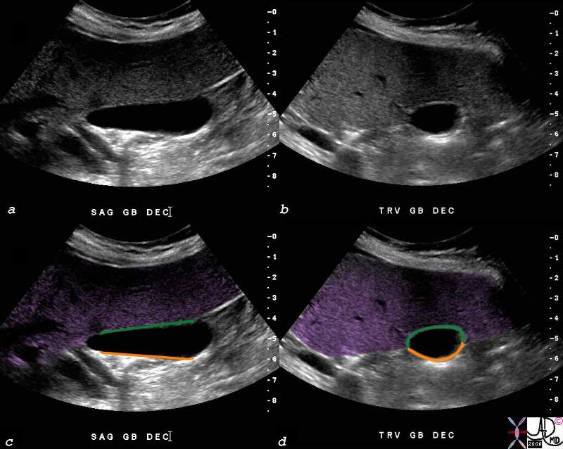
| The body of the gallbladder is attached to the liver.(purple) The US in sagittal section (a,c) and transverse section (b,d) shows the fixed portion (green) of the body attached to the gallbladder fossa and liver, and the free wall (orange) lying open to the peritoneal cavity. The image also reflects the megaphone or flask like shape to the body with a decreasing diameter as the gallbladder proceeds from fundus to the neck.shows th82428c03.8s gallbladder small normal transverse oval shape normal anatomy body of the gallbladder green is wall attached to the liver orange is free wall USscan ultrasound copyright 2008 Courtesy Ashley DAvidoff MD |
Applied Biology
When the gallbladder becomes inflammed the dominant location of fluid accumulation is in the gallbladdder fossa, where the body of the gallbladder is attached to the liver.

Fluid and Thickening in the Gallbladder Fossa
|
|
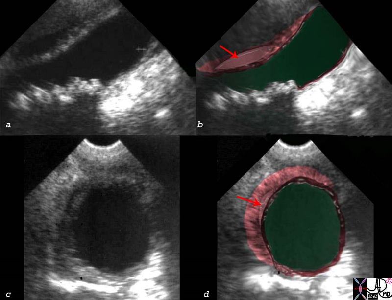
This US is from a patient who presented with classical signs of right upper quadrant pain and ther edema of the gallbladder is pedominantly in the gallbladder fossa since this is the location of the lymphatics and other congested vessels.
00543c03s.8 right upper quadrant pain RUQ pain positive Murphy?s sign gallbladder gallbladder fossa thickened linear lacy thickening gall stones calculi calculous multiple small stones stones dependant position shadowing multiple small stones cholelithiasis cholecystitis acute cholecystitis acute calculous cholecystitis USscan ultrasound Courtesy Ashley Davidoff MD copyright 2008 |
DOMElement Object
(
    [schemaTypeInfo] =>
    [tagName] => table
    [firstElementChild] => (object value omitted)
    [lastElementChild] => (object value omitted)
    [childElementCount] => 1
    [previousElementSibling] => (object value omitted)
    [nextElementSibling] =>
    [nodeName] => table
    [nodeValue] =>


Fluid and Thickening in the Gallbladder Fossa


This US is from a patient who presented with classical signs of right upper quadrant pain and ther edema of the gallbladder is pedominantly in the gallbladder fossa since this is the location of the lymphatics and other congested vessels.
00543c03s.8 right upper quadrant pain RUQ pain positive Murphy?s sign gallbladder gallbladder fossa thickened linear lacy thickening gall stones calculi calculous multiple small stones stones dependant position shadowing multiple small stones cholelithiasis cholecystitis acute cholecystitis acute calculous cholecystitis USscan ultrasound Courtesy Ashley Davidoff MD copyright 2008


    [nodeType] => 1
    [parentNode] => (object value omitted)
    [childNodes] => (object value omitted)
    [firstChild] => (object value omitted)
    [lastChild] => (object value omitted)
    [previousSibling] => (object value omitted)
    [nextSibling] => (object value omitted)
    [attributes] => (object value omitted)
    [ownerDocument] => (object value omitted)
    [namespaceURI] =>
    [prefix] =>
    [localName] => table
    [baseURI] =>
    [textContent] =>


Fluid and Thickening in the Gallbladder Fossa


This US is from a patient who presented with classical signs of right upper quadrant pain and ther edema of the gallbladder is pedominantly in the gallbladder fossa since this is the location of the lymphatics and other congested vessels.
00543c03s.8 right upper quadrant pain RUQ pain positive Murphy?s sign gallbladder gallbladder fossa thickened linear lacy thickening gall stones calculi calculous multiple small stones stones dependant position shadowing multiple small stones cholelithiasis cholecystitis acute cholecystitis acute calculous cholecystitis USscan ultrasound Courtesy Ashley Davidoff MD copyright 2008


)
DOMElement Object
(
    [schemaTypeInfo] =>
    [tagName] => td
    [firstElementChild] => (object value omitted)
    [lastElementChild] => (object value omitted)
    [childElementCount] => 2
    [previousElementSibling] =>
    [nextElementSibling] =>
    [nodeName] => td
    [nodeValue] =>
This US is from a patient who presented with classical signs of right upper quadrant pain and ther edema of the gallbladder is pedominantly in the gallbladder fossa since this is the location of the lymphatics and other congested vessels.
00543c03s.8 right upper quadrant pain RUQ pain positive Murphy?s sign gallbladder gallbladder fossa thickened linear lacy thickening gall stones calculi calculous multiple small stones stones dependant position shadowing multiple small stones cholelithiasis cholecystitis acute cholecystitis acute calculous cholecystitis USscan ultrasound Courtesy Ashley Davidoff MD copyright 2008
    [nodeType] => 1
    [parentNode] => (object value omitted)
    [childNodes] => (object value omitted)
    [firstChild] => (object value omitted)
    [lastChild] => (object value omitted)
    [previousSibling] => (object value omitted)
    [nextSibling] => (object value omitted)
    [attributes] => (object value omitted)
    [ownerDocument] => (object value omitted)
    [namespaceURI] =>
    [prefix] =>
    [localName] => td
    [baseURI] =>
    [textContent] =>
This US is from a patient who presented with classical signs of right upper quadrant pain and ther edema of the gallbladder is pedominantly in the gallbladder fossa since this is the location of the lymphatics and other congested vessels.
00543c03s.8 right upper quadrant pain RUQ pain positive Murphy?s sign gallbladder gallbladder fossa thickened linear lacy thickening gall stones calculi calculous multiple small stones stones dependant position shadowing multiple small stones cholelithiasis cholecystitis acute cholecystitis acute calculous cholecystitis USscan ultrasound Courtesy Ashley Davidoff MD copyright 2008
)
DOMElement Object
(
    [schemaTypeInfo] =>
    [tagName] => td
    [firstElementChild] => (object value omitted)
    [lastElementChild] => (object value omitted)
    [childElementCount] => 2
    [previousElementSibling] =>
    [nextElementSibling] =>
    [nodeName] => td
    [nodeValue] =>
Fluid and Thickening in the Gallbladder Fossa

    [nodeType] => 1
    [parentNode] => (object value omitted)
    [childNodes] => (object value omitted)
    [firstChild] => (object value omitted)
    [lastChild] => (object value omitted)
    [previousSibling] => (object value omitted)
    [nextSibling] => (object value omitted)
    [attributes] => (object value omitted)
    [ownerDocument] => (object value omitted)
    [namespaceURI] =>
    [prefix] =>
    [localName] => td
    [baseURI] =>
    [textContent] =>
Fluid and Thickening in the Gallbladder Fossa

)
DOMElement Object
(
    [schemaTypeInfo] =>
    [tagName] => table
    [firstElementChild] => (object value omitted)
    [lastElementChild] => (object value omitted)
    [childElementCount] => 1
    [previousElementSibling] => (object value omitted)
    [nextElementSibling] => (object value omitted)
    [nodeName] => table
    [nodeValue] =>


Body of the Gallbladder


The body of the gallbladder is attached to the liver.(purple)  The US in sagittal section (a,c) and transverse section (b,d) shows the fixed portion (green) of the body attached to the gallbladder fossa and liver, and the free wall (orange) lying open to the peritoneal cavity. The image also reflects the megaphone or flask like shape  to the body with a decreasing diameter as the gallbladder proceeds from fundus to the neck.shows th82428c03.8s gallbladder small normal transverse oval shape normal anatomy body of the gallbladder green is wall attached to the liver orange is free wall USscan ultrasound copyright 2008 Courtesy Ashley DAvidoff MD


    [nodeType] => 1
    [parentNode] => (object value omitted)
    [childNodes] => (object value omitted)
    [firstChild] => (object value omitted)
    [lastChild] => (object value omitted)
    [previousSibling] => (object value omitted)
    [nextSibling] => (object value omitted)
    [attributes] => (object value omitted)
    [ownerDocument] => (object value omitted)
    [namespaceURI] =>
    [prefix] =>
    [localName] => table
    [baseURI] =>
    [textContent] =>


Body of the Gallbladder


The body of the gallbladder is attached to the liver.(purple)  The US in sagittal section (a,c) and transverse section (b,d) shows the fixed portion (green) of the body attached to the gallbladder fossa and liver, and the free wall (orange) lying open to the peritoneal cavity. The image also reflects the megaphone or flask like shape  to the body with a decreasing diameter as the gallbladder proceeds from fundus to the neck.shows th82428c03.8s gallbladder small normal transverse oval shape normal anatomy body of the gallbladder green is wall attached to the liver orange is free wall USscan ultrasound copyright 2008 Courtesy Ashley DAvidoff MD


)
DOMElement Object
(
    [schemaTypeInfo] =>
    [tagName] => td
    [firstElementChild] =>
    [lastElementChild] =>
    [childElementCount] => 0
    [previousElementSibling] =>
    [nextElementSibling] =>
    [nodeName] => td
    [nodeValue] => The body of the gallbladder is attached to the liver.(purple)  The US in sagittal section (a,c) and transverse section (b,d) shows the fixed portion (green) of the body attached to the gallbladder fossa and liver, and the free wall (orange) lying open to the peritoneal cavity. The image also reflects the megaphone or flask like shape  to the body with a decreasing diameter as the gallbladder proceeds from fundus to the neck.shows th82428c03.8s gallbladder small normal transverse oval shape normal anatomy body of the gallbladder green is wall attached to the liver orange is free wall USscan ultrasound copyright 2008 Courtesy Ashley DAvidoff MD
    [nodeType] => 1
    [parentNode] => (object value omitted)
    [childNodes] => (object value omitted)
    [firstChild] => (object value omitted)
    [lastChild] => (object value omitted)
    [previousSibling] => (object value omitted)
    [nextSibling] => (object value omitted)
    [attributes] => (object value omitted)
    [ownerDocument] => (object value omitted)
    [namespaceURI] =>
    [prefix] =>
    [localName] => td
    [baseURI] =>
    [textContent] => The body of the gallbladder is attached to the liver.(purple)  The US in sagittal section (a,c) and transverse section (b,d) shows the fixed portion (green) of the body attached to the gallbladder fossa and liver, and the free wall (orange) lying open to the peritoneal cavity. The image also reflects the megaphone or flask like shape  to the body with a decreasing diameter as the gallbladder proceeds from fundus to the neck.shows th82428c03.8s gallbladder small normal transverse oval shape normal anatomy body of the gallbladder green is wall attached to the liver orange is free wall USscan ultrasound copyright 2008 Courtesy Ashley DAvidoff MD
)
DOMElement Object
(
    [schemaTypeInfo] =>
    [tagName] => td
    [firstElementChild] => (object value omitted)
    [lastElementChild] => (object value omitted)
    [childElementCount] => 2
    [previousElementSibling] =>
    [nextElementSibling] =>
    [nodeName] => td
    [nodeValue] =>
Body of the Gallbladder

    [nodeType] => 1
    [parentNode] => (object value omitted)
    [childNodes] => (object value omitted)
    [firstChild] => (object value omitted)
    [lastChild] => (object value omitted)
    [previousSibling] => (object value omitted)
    [nextSibling] => (object value omitted)
    [attributes] => (object value omitted)
    [ownerDocument] => (object value omitted)
    [namespaceURI] =>
    [prefix] =>
    [localName] => td
    [baseURI] =>
    [textContent] =>
Body of the Gallbladder

)


