The Common Vein Copyright 2008
Alok Anand Ashley Davidoff MD
Definition
Percutaneous cholecystostomy is a procedure for decompressing an obstructed, inflamed gallbladder. This is accomplished through the placement of tubes passing through the abdomen, directly into the gallbladder.
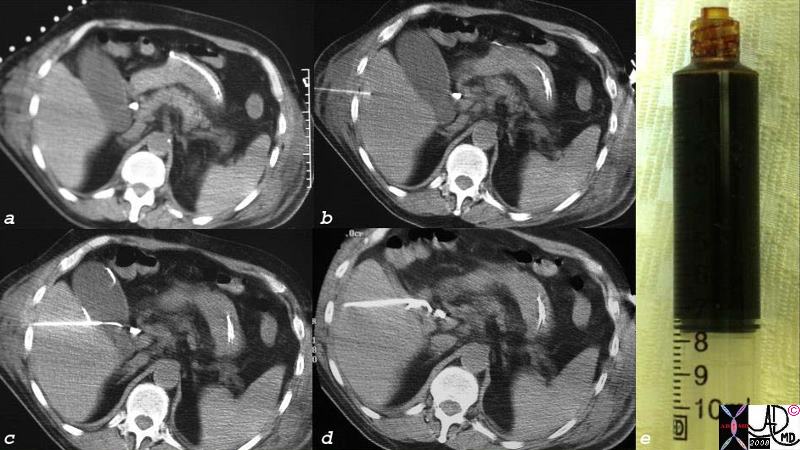
CT Guided Cholecystostomy with
Crankcase Oil Type Bile Aspirated
|
| CT guided percutaneous cholecystostomy shows external markers on the skin (a) followed by advancing guiding nededle (b) and guidewire in gallbladder (c) and fimnally the catheter with decompression of the gallbladder (d) with catheter in place. The aspirate was very black due to the dehdration of the bile in the gallbladder and is described as having the color of crankcase oil.
26067c01.8cs gallbladder acalculous cholecystitis percutaneous cholecystostomy crankcase oil black bile CTscan Courtesy Ashley Davidoff MD copyright 2008 |
Principles
The gallbladder that cannot empty due to long standing stasis, requires surgical or minimally invasive intervention to help decompress the prssure and prevent rupture
Indications
This procedure is the procedure of choice for patients with acalculous cholecystitis. Thus a patient with a fever of unknown origin who presents with acalculous right upper quadrant pain, and a distended sludge filled gallbladdder requires decompression by percutatneous cholecystostomy. For patients who present with calculous cholecystitis who would not be able to tolerate the hemodynamic stress of abdominal surgery, due to comorbid factors such as advanced age, CHF, sepsis, or end-stage liver disease, percutaneous drainage is indicated .
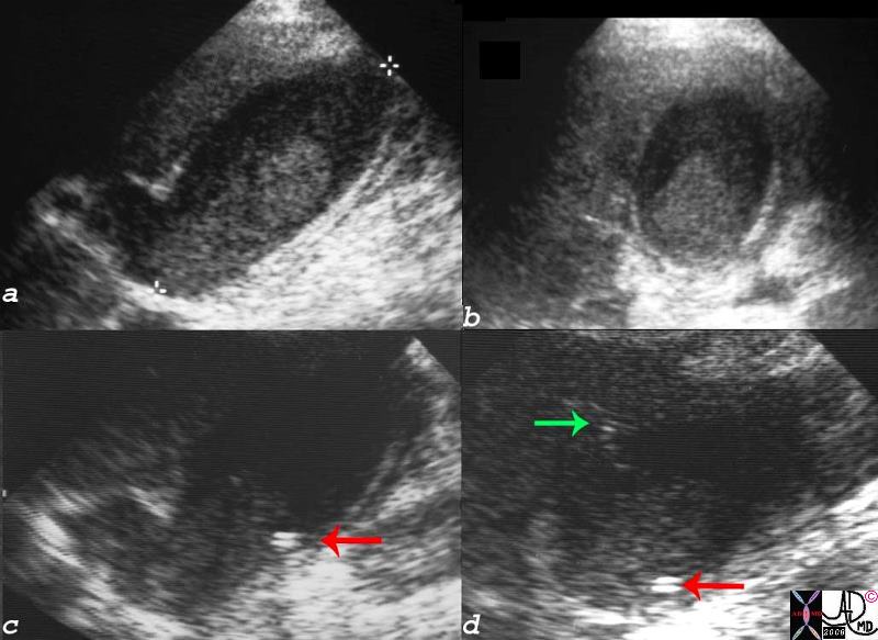 Ultrasound Guided Cholecystostomy Ultrasound Guided Cholecystostomy |
| The cholecystostomy was performed under ultrasound guidance. The thickened bile manifests as tumefactive sludge in this case (a,b,c,d) and the catheter can be seen in c and d (red arrows).
16136c01.8s gallbladder prolonged ICU stay right upper quadrant pain right upper quadrant tenderness cholestasis enlarged tumefactive bile sludge thick walled acalculous cholecystitis percutaneous cholecystostomy ultrasound USscan Courtesy Ashley Davidoff MD copyright 2008 |
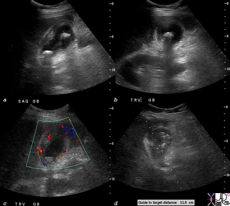 Cholecystostomy for Acute Calculous Cholecystitis Cholecystostomy for Acute Calculous Cholecystitis |
| The ultrasound from this elderly and frail patient shoews evidence of acute calculous cholecystitis characterized by shadowing stones (a,b) fluid in gallbladder fossa ((a,b) hyperemia in the wall by doppler interrogation (c) and planned route for cholecystostomy tube. The indication in this instance was due to the comorbid conditions existing in this elderly man for whom surgery was relatively contraindicated.
78304c01.8s elderly lady with multiple comorbid ilnnesses presents with RUQ pain fever, positive Murphy’s sign gallbladder distended cholelithiasis shadowing hyperemic wall complex fluid in the gallbladder fossa GBF stones acute cholelcystitis percutaneous cholecystostomy pigtail minimally invasive therapy MIT USscan ultrasound doppler color flow Courtesy Ashley Davidoff MD copyright 2008 |
Contraindications
There are relatively few contraindications to this procedure, as it is considered a safe alternative to emergent cholecystectomy. Septic shock, peritonitis, and gallbladder carcinoma. Coagulopathy would be a relative contraindication and should be reversed before attempting this interventional procedure.
Advantages
The advantage of this procedure is that it allows for decompression of the gallbladder to prevent further spread of infection and prevent chemical peritonitis caused by gallbladder rupture. It is also relatively safe to perform on patients with several underlying co-morbidities or who can not tolerate general anesthesia.
Disadvantages
The major disadvantage of cholecystostomy is that it is not a curative procedure, and patients undergoing this procedure will likely still need definitive treatment at a later point. Although the catheter is able to decompress the gallbladder, draining pus and bile, it is usually too small to pass gallstones. As gallstone disease is likely to recur patients will eventually require a cholecystectomy.
Aim
The aim of this procedure is to provide temporary decompression and drainage of purulent material. In conjunction with IV resuscitation and antibiotics, the use of percutaneous drainage gives physicians time to treat other medical instability that would make surgery risky.
Method
Patient Preparation
Patients are generally given local anesthesia. No specific preparation is required.
Equipment
The equipment used included various catheters, guide-wires, and the imaging equipment used. Although ultrasound is popular, CT is also often used for guidance.
Technique
The patient is placed on the operating table (supine, or right side up). Ultrasound or CT guidance is used to insert a catheter over guide-wire through the abdomen, liver, and into the gallbladder. By passing through the liver, the catheter remains outside the peritoneum and risk of bile leak is minimized. A pigtail catheter is generally used, as its tip can be ?kinked? to ensure it stays in place. The catheter is then connected to a drainage bag, so that output can be monitored and will remain in place until inflammation has resolved. Laparoscopic cholecystectomy may then be performed at a later point.
Result
Drainage through cholecystostomy helps control the spread of local infection. Although morbidity increases with progressive delay of definitive treatment, cholecystostomy may still provide temporary control of infection and inflammation while other more emergent pathological processes can be addressed.
Examples of Various Diseases
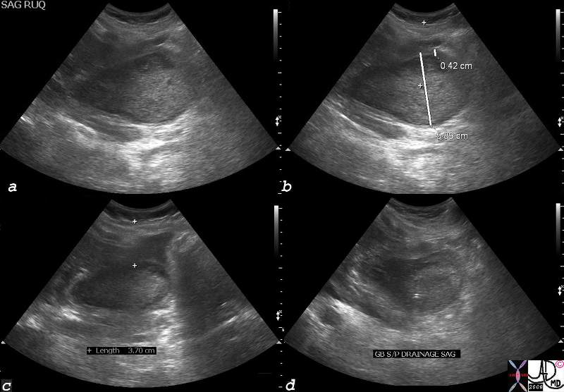 Hyperemesis Gravidarum and Acalculous Cholecystitis Hyperemesis Gravidarum and Acalculous Cholecystitis |
| The ultrasound of a 32 year old pregnant female with hyperemesis gravidarum and right upper quadrant pain. The ultrasound shows a distended gallbladder with tumefactive bile. Percutaneous cholecystostomy was performed and the catheter is seen as a double white line in the gallbladder in image d.
77695c04.8s 32 female pregnant with hyperemesis gravidarum gallbladder distended painful sludge tumefactive bile percutaneous cholecystostomy USscan Courtesy Ashley Davidoff MD copyright 2008 |
Complications
Percutaneous cholecystostomy is a procedure that is useful for temporary control of symptomatic gallbladder disease, particularly in patients who are poor surgical candidates. Complications include bleeding, organ injury, gallbladder perforation and peritonitis.
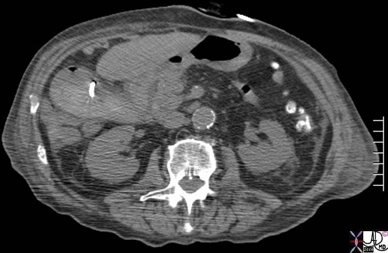 Hemorrhagic Cholecystitis Hemorrhagic Cholecystitis |
| The CT scan from an eledrerly man with RUQ pain and shows a catheter in a gallbladder that contains relatively dense bile consistent with hemorrhage. Hemorrhagic cholecystitis was present prior to the procedure, but the procedure also induced further hemorrhage into the lumen. The patient subsequently recovered.
81901.8s gallbladder hemorrhagic cholecystitis cholecystostomy further hemorrhage air iatrogenic catheter CTscan Courtsesy Ashley Davidoff MD copyright 2008 |
Conclusion
Percutaneous cholecystostomy is a highly effective and safe procedure, though complications can occur particularly if the gallbladder wall is necrotic, or technical factors or patient factors preclude optimal visualization of the gallbladder.
References
NDoherty, G. M. & Way, L. W. Current Surgical Diagnosis & Treatment Lange Medical Books/McGraw-Hill, 2006, 1453
Brunicardi, F. C. & Schwartz, S. I. Schwartz’s Principles of Surgery McGraw-Hill, Health Pub. Division, 2005, 1950
DOMElement Object
(
    [schemaTypeInfo] =>
    [tagName] => table
    [firstElementChild] => (object value omitted)
    [lastElementChild] => (object value omitted)
    [childElementCount] => 1
    [previousElementSibling] => (object value omitted)
    [nextElementSibling] => (object value omitted)
    [nodeName] => table
    [nodeValue] =>


Hemorrhagic Cholecystitis


The CT scan from an eledrerly man with RUQ pain and shows a catheter in a gallbladder that contains relatively dense bile consistent with hemorrhage.  Hemorrhagic cholecystitis was present prior to the procedure, but the procedure also induced further hemorrhage into the lumen.  The patient subsequently recovered.
81901.8s gallbladder hemorrhagic cholecystitis cholecystostomy further hemorrhage air iatrogenic catheter CTscan Courtsesy Ashley Davidoff MD copyright 2008


    [nodeType] => 1
    [parentNode] => (object value omitted)
    [childNodes] => (object value omitted)
    [firstChild] => (object value omitted)
    [lastChild] => (object value omitted)
    [previousSibling] => (object value omitted)
    [nextSibling] => (object value omitted)
    [attributes] => (object value omitted)
    [ownerDocument] => (object value omitted)
    [namespaceURI] =>
    [prefix] =>
    [localName] => table
    [baseURI] =>
    [textContent] =>


Hemorrhagic Cholecystitis


The CT scan from an eledrerly man with RUQ pain and shows a catheter in a gallbladder that contains relatively dense bile consistent with hemorrhage.  Hemorrhagic cholecystitis was present prior to the procedure, but the procedure also induced further hemorrhage into the lumen.  The patient subsequently recovered.
81901.8s gallbladder hemorrhagic cholecystitis cholecystostomy further hemorrhage air iatrogenic catheter CTscan Courtsesy Ashley Davidoff MD copyright 2008


)
DOMElement Object
(
    [schemaTypeInfo] =>
    [tagName] => td
    [firstElementChild] => (object value omitted)
    [lastElementChild] => (object value omitted)
    [childElementCount] => 1
    [previousElementSibling] =>
    [nextElementSibling] =>
    [nodeName] => td
    [nodeValue] => The CT scan from an eledrerly man with RUQ pain and shows a catheter in a gallbladder that contains relatively dense bile consistent with hemorrhage.  Hemorrhagic cholecystitis was present prior to the procedure, but the procedure also induced further hemorrhage into the lumen.  The patient subsequently recovered.
81901.8s gallbladder hemorrhagic cholecystitis cholecystostomy further hemorrhage air iatrogenic catheter CTscan Courtsesy Ashley Davidoff MD copyright 2008
    [nodeType] => 1
    [parentNode] => (object value omitted)
    [childNodes] => (object value omitted)
    [firstChild] => (object value omitted)
    [lastChild] => (object value omitted)
    [previousSibling] => (object value omitted)
    [nextSibling] => (object value omitted)
    [attributes] => (object value omitted)
    [ownerDocument] => (object value omitted)
    [namespaceURI] =>
    [prefix] =>
    [localName] => td
    [baseURI] =>
    [textContent] => The CT scan from an eledrerly man with RUQ pain and shows a catheter in a gallbladder that contains relatively dense bile consistent with hemorrhage.  Hemorrhagic cholecystitis was present prior to the procedure, but the procedure also induced further hemorrhage into the lumen.  The patient subsequently recovered.
81901.8s gallbladder hemorrhagic cholecystitis cholecystostomy further hemorrhage air iatrogenic catheter CTscan Courtsesy Ashley Davidoff MD copyright 2008
)
DOMElement Object
(
    [schemaTypeInfo] =>
    [tagName] => td
    [firstElementChild] => (object value omitted)
    [lastElementChild] => (object value omitted)
    [childElementCount] => 1
    [previousElementSibling] =>
    [nextElementSibling] =>
    [nodeName] => td
    [nodeValue] => Hemorrhagic Cholecystitis
    [nodeType] => 1
    [parentNode] => (object value omitted)
    [childNodes] => (object value omitted)
    [firstChild] => (object value omitted)
    [lastChild] => (object value omitted)
    [previousSibling] => (object value omitted)
    [nextSibling] => (object value omitted)
    [attributes] => (object value omitted)
    [ownerDocument] => (object value omitted)
    [namespaceURI] =>
    [prefix] =>
    [localName] => td
    [baseURI] =>
    [textContent] => Hemorrhagic Cholecystitis
)
DOMElement Object
(
    [schemaTypeInfo] =>
    [tagName] => table
    [firstElementChild] => (object value omitted)
    [lastElementChild] => (object value omitted)
    [childElementCount] => 1
    [previousElementSibling] => (object value omitted)
    [nextElementSibling] => (object value omitted)
    [nodeName] => table
    [nodeValue] =>


Hyperemesis Gravidarum and Acalculous Cholecystitis


The ultrasound of a 32 year old pregnant female with hyperemesis gravidarum and right upper quadrant pain.  The ultrasound shows a distended gallbladder with tumefactive bile.  Percutaneous cholecystostomy was performed and the catheter is seen as a double white line in the gallbladder in image d.
77695c04.8s 32 female pregnant with hyperemesis gravidarum gallbladder distended painful sludge tumefactive bile percutaneous cholecystostomy USscan Courtesy Ashley Davidoff MD copyright 2008


    [nodeType] => 1
    [parentNode] => (object value omitted)
    [childNodes] => (object value omitted)
    [firstChild] => (object value omitted)
    [lastChild] => (object value omitted)
    [previousSibling] => (object value omitted)
    [nextSibling] => (object value omitted)
    [attributes] => (object value omitted)
    [ownerDocument] => (object value omitted)
    [namespaceURI] =>
    [prefix] =>
    [localName] => table
    [baseURI] =>
    [textContent] =>


Hyperemesis Gravidarum and Acalculous Cholecystitis


The ultrasound of a 32 year old pregnant female with hyperemesis gravidarum and right upper quadrant pain.  The ultrasound shows a distended gallbladder with tumefactive bile.  Percutaneous cholecystostomy was performed and the catheter is seen as a double white line in the gallbladder in image d.
77695c04.8s 32 female pregnant with hyperemesis gravidarum gallbladder distended painful sludge tumefactive bile percutaneous cholecystostomy USscan Courtesy Ashley Davidoff MD copyright 2008


)
DOMElement Object
(
    [schemaTypeInfo] =>
    [tagName] => td
    [firstElementChild] => (object value omitted)
    [lastElementChild] => (object value omitted)
    [childElementCount] => 1
    [previousElementSibling] =>
    [nextElementSibling] =>
    [nodeName] => td
    [nodeValue] => The ultrasound of a 32 year old pregnant female with hyperemesis gravidarum and right upper quadrant pain.  The ultrasound shows a distended gallbladder with tumefactive bile.  Percutaneous cholecystostomy was performed and the catheter is seen as a double white line in the gallbladder in image d.
77695c04.8s 32 female pregnant with hyperemesis gravidarum gallbladder distended painful sludge tumefactive bile percutaneous cholecystostomy USscan Courtesy Ashley Davidoff MD copyright 2008
    [nodeType] => 1
    [parentNode] => (object value omitted)
    [childNodes] => (object value omitted)
    [firstChild] => (object value omitted)
    [lastChild] => (object value omitted)
    [previousSibling] => (object value omitted)
    [nextSibling] => (object value omitted)
    [attributes] => (object value omitted)
    [ownerDocument] => (object value omitted)
    [namespaceURI] =>
    [prefix] =>
    [localName] => td
    [baseURI] =>
    [textContent] => The ultrasound of a 32 year old pregnant female with hyperemesis gravidarum and right upper quadrant pain.  The ultrasound shows a distended gallbladder with tumefactive bile.  Percutaneous cholecystostomy was performed and the catheter is seen as a double white line in the gallbladder in image d.
77695c04.8s 32 female pregnant with hyperemesis gravidarum gallbladder distended painful sludge tumefactive bile percutaneous cholecystostomy USscan Courtesy Ashley Davidoff MD copyright 2008
)
DOMElement Object
(
    [schemaTypeInfo] =>
    [tagName] => td
    [firstElementChild] => (object value omitted)
    [lastElementChild] => (object value omitted)
    [childElementCount] => 1
    [previousElementSibling] =>
    [nextElementSibling] =>
    [nodeName] => td
    [nodeValue] => Hyperemesis Gravidarum and Acalculous Cholecystitis
    [nodeType] => 1
    [parentNode] => (object value omitted)
    [childNodes] => (object value omitted)
    [firstChild] => (object value omitted)
    [lastChild] => (object value omitted)
    [previousSibling] => (object value omitted)
    [nextSibling] => (object value omitted)
    [attributes] => (object value omitted)
    [ownerDocument] => (object value omitted)
    [namespaceURI] =>
    [prefix] =>
    [localName] => td
    [baseURI] =>
    [textContent] => Hyperemesis Gravidarum and Acalculous Cholecystitis
)
DOMElement Object
(
    [schemaTypeInfo] =>
    [tagName] => table
    [firstElementChild] => (object value omitted)
    [lastElementChild] => (object value omitted)
    [childElementCount] => 1
    [previousElementSibling] => (object value omitted)
    [nextElementSibling] => (object value omitted)
    [nodeName] => table
    [nodeValue] =>


Cholecystostomy for Acute Calculous Cholecystitis


The ultrasound from this elderly and frail patient shoews evidence of acute calculous cholecystitis characterized by shadowing stones (a,b) fluid in gallbladder fossa ((a,b) hyperemia in the wall by doppler interrogation (c) and planned route for cholecystostomy tube.  The indication in this instance was due to the comorbid conditions existing in this elderly man for whom surgery was relatively contraindicated.
78304c01.8s elderly lady with multiple comorbid ilnnesses presents with RUQ pain fever, positive Murphy’s sign gallbladder distended cholelithiasis shadowing hyperemic wall complex fluid in the gallbladder fossa GBF stones acute cholelcystitis percutaneous cholecystostomy pigtail minimally invasive therapy MIT USscan ultrasound doppler color flow Courtesy Ashley Davidoff MD copyright 2008


    [nodeType] => 1
    [parentNode] => (object value omitted)
    [childNodes] => (object value omitted)
    [firstChild] => (object value omitted)
    [lastChild] => (object value omitted)
    [previousSibling] => (object value omitted)
    [nextSibling] => (object value omitted)
    [attributes] => (object value omitted)
    [ownerDocument] => (object value omitted)
    [namespaceURI] =>
    [prefix] =>
    [localName] => table
    [baseURI] =>
    [textContent] =>


Cholecystostomy for Acute Calculous Cholecystitis


The ultrasound from this elderly and frail patient shoews evidence of acute calculous cholecystitis characterized by shadowing stones (a,b) fluid in gallbladder fossa ((a,b) hyperemia in the wall by doppler interrogation (c) and planned route for cholecystostomy tube.  The indication in this instance was due to the comorbid conditions existing in this elderly man for whom surgery was relatively contraindicated.
78304c01.8s elderly lady with multiple comorbid ilnnesses presents with RUQ pain fever, positive Murphy’s sign gallbladder distended cholelithiasis shadowing hyperemic wall complex fluid in the gallbladder fossa GBF stones acute cholelcystitis percutaneous cholecystostomy pigtail minimally invasive therapy MIT USscan ultrasound doppler color flow Courtesy Ashley Davidoff MD copyright 2008


)
DOMElement Object
(
    [schemaTypeInfo] =>
    [tagName] => td
    [firstElementChild] => (object value omitted)
    [lastElementChild] => (object value omitted)
    [childElementCount] => 1
    [previousElementSibling] =>
    [nextElementSibling] =>
    [nodeName] => td
    [nodeValue] => The ultrasound from this elderly and frail patient shoews evidence of acute calculous cholecystitis characterized by shadowing stones (a,b) fluid in gallbladder fossa ((a,b) hyperemia in the wall by doppler interrogation (c) and planned route for cholecystostomy tube.  The indication in this instance was due to the comorbid conditions existing in this elderly man for whom surgery was relatively contraindicated.
78304c01.8s elderly lady with multiple comorbid ilnnesses presents with RUQ pain fever, positive Murphy’s sign gallbladder distended cholelithiasis shadowing hyperemic wall complex fluid in the gallbladder fossa GBF stones acute cholelcystitis percutaneous cholecystostomy pigtail minimally invasive therapy MIT USscan ultrasound doppler color flow Courtesy Ashley Davidoff MD copyright 2008
    [nodeType] => 1
    [parentNode] => (object value omitted)
    [childNodes] => (object value omitted)
    [firstChild] => (object value omitted)
    [lastChild] => (object value omitted)
    [previousSibling] => (object value omitted)
    [nextSibling] => (object value omitted)
    [attributes] => (object value omitted)
    [ownerDocument] => (object value omitted)
    [namespaceURI] =>
    [prefix] =>
    [localName] => td
    [baseURI] =>
    [textContent] => The ultrasound from this elderly and frail patient shoews evidence of acute calculous cholecystitis characterized by shadowing stones (a,b) fluid in gallbladder fossa ((a,b) hyperemia in the wall by doppler interrogation (c) and planned route for cholecystostomy tube.  The indication in this instance was due to the comorbid conditions existing in this elderly man for whom surgery was relatively contraindicated.
78304c01.8s elderly lady with multiple comorbid ilnnesses presents with RUQ pain fever, positive Murphy’s sign gallbladder distended cholelithiasis shadowing hyperemic wall complex fluid in the gallbladder fossa GBF stones acute cholelcystitis percutaneous cholecystostomy pigtail minimally invasive therapy MIT USscan ultrasound doppler color flow Courtesy Ashley Davidoff MD copyright 2008
)
DOMElement Object
(
    [schemaTypeInfo] =>
    [tagName] => td
    [firstElementChild] => (object value omitted)
    [lastElementChild] => (object value omitted)
    [childElementCount] => 1
    [previousElementSibling] =>
    [nextElementSibling] =>
    [nodeName] => td
    [nodeValue] => Cholecystostomy for Acute Calculous Cholecystitis
    [nodeType] => 1
    [parentNode] => (object value omitted)
    [childNodes] => (object value omitted)
    [firstChild] => (object value omitted)
    [lastChild] => (object value omitted)
    [previousSibling] => (object value omitted)
    [nextSibling] => (object value omitted)
    [attributes] => (object value omitted)
    [ownerDocument] => (object value omitted)
    [namespaceURI] =>
    [prefix] =>
    [localName] => td
    [baseURI] =>
    [textContent] => Cholecystostomy for Acute Calculous Cholecystitis
)
DOMElement Object
(
    [schemaTypeInfo] =>
    [tagName] => table
    [firstElementChild] => (object value omitted)
    [lastElementChild] => (object value omitted)
    [childElementCount] => 1
    [previousElementSibling] => (object value omitted)
    [nextElementSibling] => (object value omitted)
    [nodeName] => table
    [nodeValue] =>


Ultrasound Guided Cholecystostomy


The cholecystostomy was performed under ultrasound guidance.  The thickened bile manifests as tumefactive sludge in this case (a,b,c,d) and the catheter can be seen in c and d (red arrows).
16136c01.8s gallbladder prolonged ICU stay right upper quadrant pain right upper quadrant tenderness cholestasis enlarged tumefactive bile sludge thick walled acalculous cholecystitis percutaneous cholecystostomy ultrasound USscan Courtesy Ashley Davidoff MD copyright 2008


    [nodeType] => 1
    [parentNode] => (object value omitted)
    [childNodes] => (object value omitted)
    [firstChild] => (object value omitted)
    [lastChild] => (object value omitted)
    [previousSibling] => (object value omitted)
    [nextSibling] => (object value omitted)
    [attributes] => (object value omitted)
    [ownerDocument] => (object value omitted)
    [namespaceURI] =>
    [prefix] =>
    [localName] => table
    [baseURI] =>
    [textContent] =>


Ultrasound Guided Cholecystostomy


The cholecystostomy was performed under ultrasound guidance.  The thickened bile manifests as tumefactive sludge in this case (a,b,c,d) and the catheter can be seen in c and d (red arrows).
16136c01.8s gallbladder prolonged ICU stay right upper quadrant pain right upper quadrant tenderness cholestasis enlarged tumefactive bile sludge thick walled acalculous cholecystitis percutaneous cholecystostomy ultrasound USscan Courtesy Ashley Davidoff MD copyright 2008


)
DOMElement Object
(
    [schemaTypeInfo] =>
    [tagName] => td
    [firstElementChild] => (object value omitted)
    [lastElementChild] => (object value omitted)
    [childElementCount] => 1
    [previousElementSibling] =>
    [nextElementSibling] =>
    [nodeName] => td
    [nodeValue] => The cholecystostomy was performed under ultrasound guidance.  The thickened bile manifests as tumefactive sludge in this case (a,b,c,d) and the catheter can be seen in c and d (red arrows).
16136c01.8s gallbladder prolonged ICU stay right upper quadrant pain right upper quadrant tenderness cholestasis enlarged tumefactive bile sludge thick walled acalculous cholecystitis percutaneous cholecystostomy ultrasound USscan Courtesy Ashley Davidoff MD copyright 2008
    [nodeType] => 1
    [parentNode] => (object value omitted)
    [childNodes] => (object value omitted)
    [firstChild] => (object value omitted)
    [lastChild] => (object value omitted)
    [previousSibling] => (object value omitted)
    [nextSibling] => (object value omitted)
    [attributes] => (object value omitted)
    [ownerDocument] => (object value omitted)
    [namespaceURI] =>
    [prefix] =>
    [localName] => td
    [baseURI] =>
    [textContent] => The cholecystostomy was performed under ultrasound guidance.  The thickened bile manifests as tumefactive sludge in this case (a,b,c,d) and the catheter can be seen in c and d (red arrows).
16136c01.8s gallbladder prolonged ICU stay right upper quadrant pain right upper quadrant tenderness cholestasis enlarged tumefactive bile sludge thick walled acalculous cholecystitis percutaneous cholecystostomy ultrasound USscan Courtesy Ashley Davidoff MD copyright 2008
)
DOMElement Object
(
    [schemaTypeInfo] =>
    [tagName] => td
    [firstElementChild] => (object value omitted)
    [lastElementChild] => (object value omitted)
    [childElementCount] => 1
    [previousElementSibling] =>
    [nextElementSibling] =>
    [nodeName] => td
    [nodeValue] => Ultrasound Guided Cholecystostomy
    [nodeType] => 1
    [parentNode] => (object value omitted)
    [childNodes] => (object value omitted)
    [firstChild] => (object value omitted)
    [lastChild] => (object value omitted)
    [previousSibling] => (object value omitted)
    [nextSibling] => (object value omitted)
    [attributes] => (object value omitted)
    [ownerDocument] => (object value omitted)
    [namespaceURI] =>
    [prefix] =>
    [localName] => td
    [baseURI] =>
    [textContent] => Ultrasound Guided Cholecystostomy
)
DOMElement Object
(
    [schemaTypeInfo] =>
    [tagName] => table
    [firstElementChild] => (object value omitted)
    [lastElementChild] => (object value omitted)
    [childElementCount] => 1
    [previousElementSibling] => (object value omitted)
    [nextElementSibling] => (object value omitted)
    [nodeName] => table
    [nodeValue] =>


CT Guided Cholecystostomy with
Crankcase Oil Type Bile Aspirated


CT guided percutaneous cholecystostomy shows external markers on the skin (a) followed by advancing guiding nededle (b) and guidewire in gallbladder (c) and fimnally the catheter with decompression of the gallbladder (d) with catheter in place.  The aspirate was very black due to the dehdration of the bile in the gallbladder and is described as having the color of crankcase oil.
26067c01.8cs gallbladder acalculous cholecystitis percutaneous cholecystostomy crankcase oil black bile CTscan Courtesy Ashley Davidoff MD copyright 2008


    [nodeType] => 1
    [parentNode] => (object value omitted)
    [childNodes] => (object value omitted)
    [firstChild] => (object value omitted)
    [lastChild] => (object value omitted)
    [previousSibling] => (object value omitted)
    [nextSibling] => (object value omitted)
    [attributes] => (object value omitted)
    [ownerDocument] => (object value omitted)
    [namespaceURI] =>
    [prefix] =>
    [localName] => table
    [baseURI] =>
    [textContent] =>


CT Guided Cholecystostomy with
Crankcase Oil Type Bile Aspirated


CT guided percutaneous cholecystostomy shows external markers on the skin (a) followed by advancing guiding nededle (b) and guidewire in gallbladder (c) and fimnally the catheter with decompression of the gallbladder (d) with catheter in place.  The aspirate was very black due to the dehdration of the bile in the gallbladder and is described as having the color of crankcase oil.
26067c01.8cs gallbladder acalculous cholecystitis percutaneous cholecystostomy crankcase oil black bile CTscan Courtesy Ashley Davidoff MD copyright 2008


)
DOMElement Object
(
    [schemaTypeInfo] =>
    [tagName] => td
    [firstElementChild] => (object value omitted)
    [lastElementChild] => (object value omitted)
    [childElementCount] => 2
    [previousElementSibling] =>
    [nextElementSibling] =>
    [nodeName] => td
    [nodeValue] => CT guided percutaneous cholecystostomy shows external markers on the skin (a) followed by advancing guiding nededle (b) and guidewire in gallbladder (c) and fimnally the catheter with decompression of the gallbladder (d) with catheter in place.  The aspirate was very black due to the dehdration of the bile in the gallbladder and is described as having the color of crankcase oil.
26067c01.8cs gallbladder acalculous cholecystitis percutaneous cholecystostomy crankcase oil black bile CTscan Courtesy Ashley Davidoff MD copyright 2008
    [nodeType] => 1
    [parentNode] => (object value omitted)
    [childNodes] => (object value omitted)
    [firstChild] => (object value omitted)
    [lastChild] => (object value omitted)
    [previousSibling] => (object value omitted)
    [nextSibling] => (object value omitted)
    [attributes] => (object value omitted)
    [ownerDocument] => (object value omitted)
    [namespaceURI] =>
    [prefix] =>
    [localName] => td
    [baseURI] =>
    [textContent] => CT guided percutaneous cholecystostomy shows external markers on the skin (a) followed by advancing guiding nededle (b) and guidewire in gallbladder (c) and fimnally the catheter with decompression of the gallbladder (d) with catheter in place.  The aspirate was very black due to the dehdration of the bile in the gallbladder and is described as having the color of crankcase oil.
26067c01.8cs gallbladder acalculous cholecystitis percutaneous cholecystostomy crankcase oil black bile CTscan Courtesy Ashley Davidoff MD copyright 2008
)
DOMElement Object
(
    [schemaTypeInfo] =>
    [tagName] => td
    [firstElementChild] => (object value omitted)
    [lastElementChild] => (object value omitted)
    [childElementCount] => 3
    [previousElementSibling] =>
    [nextElementSibling] =>
    [nodeName] => td
    [nodeValue] =>
CT Guided Cholecystostomy with
Crankcase Oil Type Bile Aspirated

    [nodeType] => 1
    [parentNode] => (object value omitted)
    [childNodes] => (object value omitted)
    [firstChild] => (object value omitted)
    [lastChild] => (object value omitted)
    [previousSibling] => (object value omitted)
    [nextSibling] => (object value omitted)
    [attributes] => (object value omitted)
    [ownerDocument] => (object value omitted)
    [namespaceURI] =>
    [prefix] =>
    [localName] => td
    [baseURI] =>
    [textContent] =>
CT Guided Cholecystostomy with
Crankcase Oil Type Bile Aspirated

)


