|
The transverse colon is the longest and most mobile part of the large bowel. It extends from the right upper quadrant to the LUQ usually dipping down in its mid portion, sometimes as far as the pelvis and then ascending to its highest position of the colon in the abdomen in the LUQ at the level of the splenic flexure.
The transverse colon ranges from 30-60cms.in length and has a diameter that should measure less than 5cms.
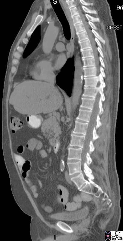
It is the most anterior portion of the large bowel, which is an important fact to remember and apply in the execution of a double contrast barium enema.
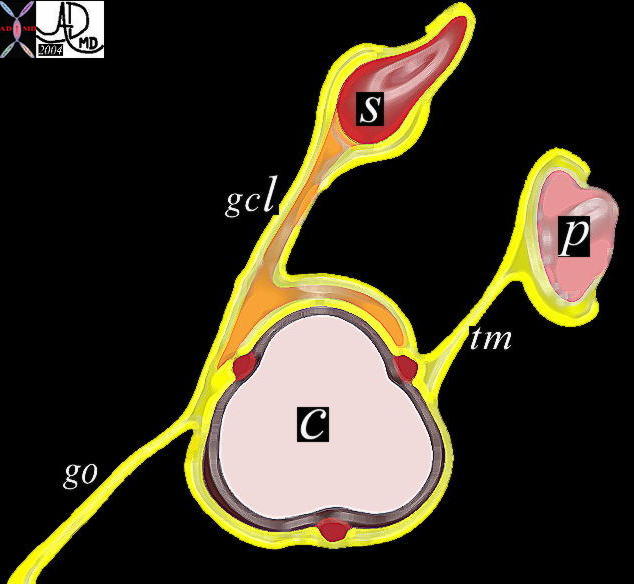
The first phase of the enema is to get the contrast into the cecum. The radiologist does not want to overfill the bowel with barium since for the double contrast study the barium is just needed to coat the wall and not to fill the lumen. It is the object of the air to fill the lumen and for the barium to outline the mucosa. Less barium is better in the performance and optimization of the barium enema examination. Therefore once the barium column reaches the splenic flexure the radiologist really wants to just roll the barium down the transverse colon up to the hepatic flexure, and then down the ascending colon. ? a slow roller coaster ride. As the barium column reaches the splenic flexure, the patient is placed in the prone position so that the barium will be aided by gravity as it moves anteriorly in the transverse colon. In this position the barium should traverse most of the transverse colon after which a right decubitus should take the barium to the hepatic flexure. Getting it over the hepatic flexure hump is sometimes difficult but once accomplished it is downhill for the barium. The patient is placed supine as the technologists uncoils the enema tubes from the patients legs and the table tipped up in the grand hope of bringing the barium home safely into the cecum. Not always so easy. Once it is reached ? huge relief for the radiologist. Double contrast enemas are one of the most challenging examinations for the radiologist and knowledge of the position of the parts of the colon is essential to enlist gravity as an aide to move the barium and air. The transverse colon is suspended by two ligaments and acts as a support for a third. The two supporting ligaments are the gastrocolic ligament and the transverse mesocolon. The transverse mesocolon ™ runs between the pancreas and the posterior and superior aspect of the colon. The gastrocolic ligament (gcl) also known as the lesser omentum runs between the inferior aspect of the stomach and the antero-superior aspect of the transverse colon. The greater omentum is a thin apron of fat that hangs off the inferior aspect of the transverse colon.
The transverse colon receives blood mainly from the middle colic artery, but has crossover supply from both the right and left colic arteries. Venous drainage is via the corresponding veins and into the SMV and portal vein Applied AnatomyDisease can spread along these ligaments so that in the case of diseases of the stomach, they can spread along the gastrocolic ligament to the superior aspect of the colon, while diseases such as pancreatitis can spread to the posterior and sometimes superior aspect of the colon. The greater omentum is notorious for collecting ovarian cancer deposits and when identified the complex of the fat of the omentum and the soft tissue elements of the cancer has been described as ?omental cake? It is therefore very important for the radiologist to examine the superior, posterior and inferior borders of the transverse colon which may hold clues to important diseases in the abdomen.
Since the transverse colon is anterior and the greater omentum forms an apron of protection just below the anterior abdominal wall, they are both subject to traumatic injury. The following case represents the abdominal CTscan of a football player.
As stated above the greater omentum is a thin double layer of mesentery consisting of fat and connective tissue, that hangs off the transverse colon and seems to collect metastases particularly from the ovary, but also from gastric, pancreatic and colonic carcinoma as well. The nodules on the omentum are described as omental cake.
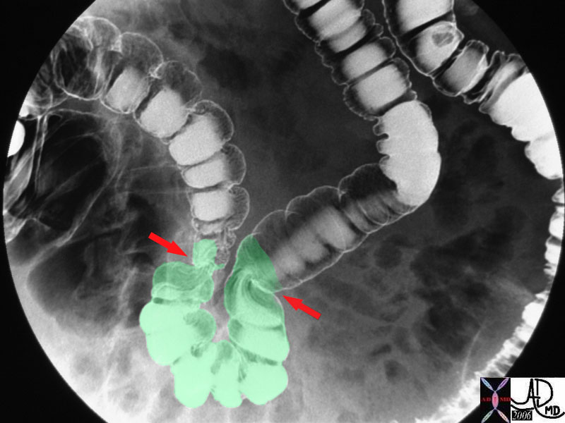
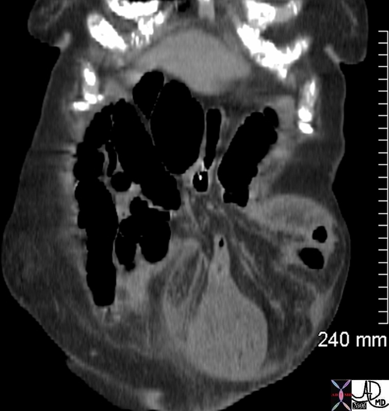
The transverse colon due to its anterior position as well as its redundancy is sometimes trapped in abdominal wall defects resulting in herniation.. When the defect is large the colon can be reduced either spontaneously or manually. However if the orifice is small strangulation and incarceration of the colon can occur, resulting in a surgical emergency.
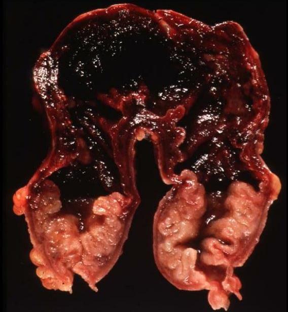
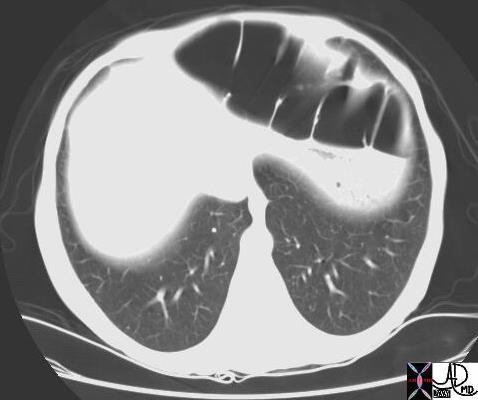
When the hernial orifice, the hole through which the bowel protrudes is small, and the structural components in the hernia become more bulky for one of many reasons, the bowel in the hernial sac gets stuck, and cannot be reduced. This entity may be complicated by venous and then arterial compromise with resulting ischemia and infarction ? an entity known as an incarcerated hernia. 5 129852985 Another more uncommon hernia (3% of abdominal hernias) is the Morgagni hernia, where the transverse colon or stomach protrudes through an anterior, retrosternal defect of the diaphragm. Although this hernia is a result of a congenital defect, it may not become apparent until adulthood. Sometimes it presents as a mass in the chest on a CXR and hence it is one of the causes of a pseudotumor of the lung. Obstruction of the colon can occur with this hernia.
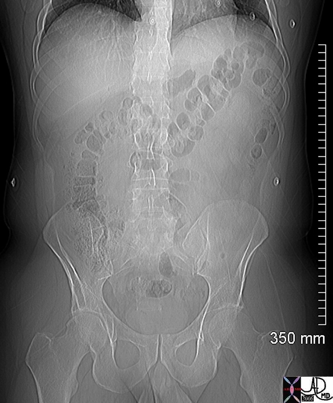
Toxic megacolon (eMedicine) is a life threatening condition where an underlying colonic disease suddenly accelerates and the patient presents with a severe clinical syndrome. The colon is not always dilated in this syndrome and hence a better term is toxic colitis. The dilatation, when it occurs is caused by an ileus thought to originate as a result of damage to the nerves in the submucosa by the disease. In the absence of normal function of these nerves, motility of the bowel is limited and hence an ileus results. When toxic colitis presents with megacolon it is usually the transverse colon that is affected and the theory for its involvement relates to the position of the transverse colon. The ill patient usually being confined to bed is lies predominantly in the supine position. Air in any part of the colon will rise to the most anterior part of the colon and therefore accumulates in the transverse colon. The transverse colon is therefore stretched and since the air cannot be transported due to the ileus, there is progressive dilatation of the transverse colon and hence progressive ischemia and worsening of the clinical situation. There are multiple causes of the toxic colitis or toxic megacolon including ulcerative colitis, ischemic colitis, and pseudomembranous colitis.
|
DOMElement Object
(
    [schemaTypeInfo] =>
    [tagName] => table
    [firstElementChild] => (object value omitted)
    [lastElementChild] => (object value omitted)
    [childElementCount] => 1
    [previousElementSibling] => (object value omitted)
    [nextElementSibling] =>
    [nodeName] => table
    [nodeValue] =>


Toxic megacolon in ulcerative colitis


The plain film of the abdomen shows a transverse colon that is greater than 6cms in a patient who has known ulcerative colitis with sudden clinical deterioration and marked systemic changes.  Note that the distal descending colon has evidence of pinky printing (fine undulations of the air column) suggesting associated involvement with the colitis.
Courtesy Ashley Davidoff MD
00306


    [nodeType] => 1
    [parentNode] => (object value omitted)
    [childNodes] => (object value omitted)
    [firstChild] => (object value omitted)
    [lastChild] => (object value omitted)
    [previousSibling] => (object value omitted)
    [nextSibling] => (object value omitted)
    [attributes] => (object value omitted)
    [ownerDocument] => (object value omitted)
    [namespaceURI] =>
    [prefix] =>
    [localName] => table
    [baseURI] =>
    [textContent] =>


Toxic megacolon in ulcerative colitis


The plain film of the abdomen shows a transverse colon that is greater than 6cms in a patient who has known ulcerative colitis with sudden clinical deterioration and marked systemic changes.  Note that the distal descending colon has evidence of pinky printing (fine undulations of the air column) suggesting associated involvement with the colitis.
Courtesy Ashley Davidoff MD
00306


)
DOMElement Object
(
    [schemaTypeInfo] =>
    [tagName] => td
    [firstElementChild] => (object value omitted)
    [lastElementChild] => (object value omitted)
    [childElementCount] => 2
    [previousElementSibling] =>
    [nextElementSibling] =>
    [nodeName] => td
    [nodeValue] => The plain film of the abdomen shows a transverse colon that is greater than 6cms in a patient who has known ulcerative colitis with sudden clinical deterioration and marked systemic changes.  Note that the distal descending colon has evidence of pinky printing (fine undulations of the air column) suggesting associated involvement with the colitis.
Courtesy Ashley Davidoff MD
00306
    [nodeType] => 1
    [parentNode] => (object value omitted)
    [childNodes] => (object value omitted)
    [firstChild] => (object value omitted)
    [lastChild] => (object value omitted)
    [previousSibling] => (object value omitted)
    [nextSibling] => (object value omitted)
    [attributes] => (object value omitted)
    [ownerDocument] => (object value omitted)
    [namespaceURI] =>
    [prefix] =>
    [localName] => td
    [baseURI] =>
    [textContent] => The plain film of the abdomen shows a transverse colon that is greater than 6cms in a patient who has known ulcerative colitis with sudden clinical deterioration and marked systemic changes.  Note that the distal descending colon has evidence of pinky printing (fine undulations of the air column) suggesting associated involvement with the colitis.
Courtesy Ashley Davidoff MD
00306
)
DOMElement Object
(
    [schemaTypeInfo] =>
    [tagName] => td
    [firstElementChild] => (object value omitted)
    [lastElementChild] => (object value omitted)
    [childElementCount] => 1
    [previousElementSibling] =>
    [nextElementSibling] =>
    [nodeName] => td
    [nodeValue] => Toxic megacolon in ulcerative colitis
    [nodeType] => 1
    [parentNode] => (object value omitted)
    [childNodes] => (object value omitted)
    [firstChild] => (object value omitted)
    [lastChild] => (object value omitted)
    [previousSibling] => (object value omitted)
    [nextSibling] => (object value omitted)
    [attributes] => (object value omitted)
    [ownerDocument] => (object value omitted)
    [namespaceURI] =>
    [prefix] =>
    [localName] => td
    [baseURI] =>
    [textContent] => Toxic megacolon in ulcerative colitis
)
DOMElement Object
(
    [schemaTypeInfo] =>
    [tagName] => table
    [firstElementChild] => (object value omitted)
    [lastElementChild] => (object value omitted)
    [childElementCount] => 1
    [previousElementSibling] => (object value omitted)
    [nextElementSibling] => (object value omitted)
    [nodeName] => table
    [nodeValue] =>


Morgagni hernia


The transverse colon is recognized by its large diameter and haustral folds and is noted to be too high and intrathoracic.  The Morgagni hernia   is caused by a congenital defect of the diaphragm.
Courtesy Ashley Davidoff MD
17226


    [nodeType] => 1
    [parentNode] => (object value omitted)
    [childNodes] => (object value omitted)
    [firstChild] => (object value omitted)
    [lastChild] => (object value omitted)
    [previousSibling] => (object value omitted)
    [nextSibling] => (object value omitted)
    [attributes] => (object value omitted)
    [ownerDocument] => (object value omitted)
    [namespaceURI] =>
    [prefix] =>
    [localName] => table
    [baseURI] =>
    [textContent] =>


Morgagni hernia


The transverse colon is recognized by its large diameter and haustral folds and is noted to be too high and intrathoracic.  The Morgagni hernia   is caused by a congenital defect of the diaphragm.
Courtesy Ashley Davidoff MD
17226


)
DOMElement Object
(
    [schemaTypeInfo] =>
    [tagName] => td
    [firstElementChild] => (object value omitted)
    [lastElementChild] => (object value omitted)
    [childElementCount] => 2
    [previousElementSibling] =>
    [nextElementSibling] =>
    [nodeName] => td
    [nodeValue] => The transverse colon is recognized by its large diameter and haustral folds and is noted to be too high and intrathoracic.  The Morgagni hernia   is caused by a congenital defect of the diaphragm.
Courtesy Ashley Davidoff MD
17226
    [nodeType] => 1
    [parentNode] => (object value omitted)
    [childNodes] => (object value omitted)
    [firstChild] => (object value omitted)
    [lastChild] => (object value omitted)
    [previousSibling] => (object value omitted)
    [nextSibling] => (object value omitted)
    [attributes] => (object value omitted)
    [ownerDocument] => (object value omitted)
    [namespaceURI] =>
    [prefix] =>
    [localName] => td
    [baseURI] =>
    [textContent] => The transverse colon is recognized by its large diameter and haustral folds and is noted to be too high and intrathoracic.  The Morgagni hernia   is caused by a congenital defect of the diaphragm.
Courtesy Ashley Davidoff MD
17226
)
DOMElement Object
(
    [schemaTypeInfo] =>
    [tagName] => td
    [firstElementChild] => (object value omitted)
    [lastElementChild] => (object value omitted)
    [childElementCount] => 1
    [previousElementSibling] =>
    [nextElementSibling] =>
    [nodeName] => td
    [nodeValue] => Morgagni hernia
    [nodeType] => 1
    [parentNode] => (object value omitted)
    [childNodes] => (object value omitted)
    [firstChild] => (object value omitted)
    [lastChild] => (object value omitted)
    [previousSibling] => (object value omitted)
    [nextSibling] => (object value omitted)
    [attributes] => (object value omitted)
    [ownerDocument] => (object value omitted)
    [namespaceURI] =>
    [prefix] =>
    [localName] => td
    [baseURI] =>
    [textContent] => Morgagni hernia
)
DOMElement Object
(
    [schemaTypeInfo] =>
    [tagName] => table
    [firstElementChild] => (object value omitted)
    [lastElementChild] => (object value omitted)
    [childElementCount] => 1
    [previousElementSibling] => (object value omitted)
    [nextElementSibling] => (object value omitted)
    [nodeName] => table
    [nodeValue] =>


Incarcerated hernia of the transverse colon


The CT images from a coronal reformat just off the anterior abdominal wall show a loop of transverse colon (overlaid in red associated with surrounding edema of the subcutaneous fat.  At surgery this was shown to be incarcerated.  The pathology specimen is not from the same case but shows hemorrhagic infarction of small bowel.
Courtesy Ashley Davidoff MD grosspathology Courtesy Barbara Banner MD
45488b01 45488b01 12985


    [nodeType] => 1
    [parentNode] => (object value omitted)
    [childNodes] => (object value omitted)
    [firstChild] => (object value omitted)
    [lastChild] => (object value omitted)
    [previousSibling] => (object value omitted)
    [nextSibling] => (object value omitted)
    [attributes] => (object value omitted)
    [ownerDocument] => (object value omitted)
    [namespaceURI] =>
    [prefix] =>
    [localName] => table
    [baseURI] =>
    [textContent] =>


Incarcerated hernia of the transverse colon


The CT images from a coronal reformat just off the anterior abdominal wall show a loop of transverse colon (overlaid in red associated with surrounding edema of the subcutaneous fat.  At surgery this was shown to be incarcerated.  The pathology specimen is not from the same case but shows hemorrhagic infarction of small bowel.
Courtesy Ashley Davidoff MD grosspathology Courtesy Barbara Banner MD
45488b01 45488b01 12985


)
DOMElement Object
(
    [schemaTypeInfo] =>
    [tagName] => td
    [firstElementChild] => (object value omitted)
    [lastElementChild] => (object value omitted)
    [childElementCount] => 2
    [previousElementSibling] =>
    [nextElementSibling] =>
    [nodeName] => td
    [nodeValue] => The CT images from a coronal reformat just off the anterior abdominal wall show a loop of transverse colon (overlaid in red associated with surrounding edema of the subcutaneous fat.  At surgery this was shown to be incarcerated.  The pathology specimen is not from the same case but shows hemorrhagic infarction of small bowel.
Courtesy Ashley Davidoff MD grosspathology Courtesy Barbara Banner MD
45488b01 45488b01 12985
    [nodeType] => 1
    [parentNode] => (object value omitted)
    [childNodes] => (object value omitted)
    [firstChild] => (object value omitted)
    [lastChild] => (object value omitted)
    [previousSibling] => (object value omitted)
    [nextSibling] => (object value omitted)
    [attributes] => (object value omitted)
    [ownerDocument] => (object value omitted)
    [namespaceURI] =>
    [prefix] =>
    [localName] => td
    [baseURI] =>
    [textContent] => The CT images from a coronal reformat just off the anterior abdominal wall show a loop of transverse colon (overlaid in red associated with surrounding edema of the subcutaneous fat.  At surgery this was shown to be incarcerated.  The pathology specimen is not from the same case but shows hemorrhagic infarction of small bowel.
Courtesy Ashley Davidoff MD grosspathology Courtesy Barbara Banner MD
45488b01 45488b01 12985
)
DOMElement Object
(
    [schemaTypeInfo] =>
    [tagName] => td
    [firstElementChild] => (object value omitted)
    [lastElementChild] => (object value omitted)
    [childElementCount] => 1
    [previousElementSibling] =>
    [nextElementSibling] =>
    [nodeName] => td
    [nodeValue] => Incarcerated hernia of the transverse colon
    [nodeType] => 1
    [parentNode] => (object value omitted)
    [childNodes] => (object value omitted)
    [firstChild] => (object value omitted)
    [lastChild] => (object value omitted)
    [previousSibling] => (object value omitted)
    [nextSibling] => (object value omitted)
    [attributes] => (object value omitted)
    [ownerDocument] => (object value omitted)
    [namespaceURI] =>
    [prefix] =>
    [localName] => td
    [baseURI] =>
    [textContent] => Incarcerated hernia of the transverse colon
)
DOMElement Object
(
    [schemaTypeInfo] =>
    [tagName] => table
    [firstElementChild] => (object value omitted)
    [lastElementChild] => (object value omitted)
    [childElementCount] => 1
    [previousElementSibling] => (object value omitted)
    [nextElementSibling] => (object value omitted)
    [nodeName] => table
    [nodeValue] =>


Anterior abdominal wall hernia


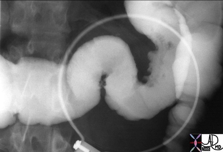
The double contrast barium enema demonstrates a napkin ring like constriction across the transverse colon which is trapped in an anterior abdominal wall defect.  The overlay in green is within the hernial sac, while the neck depicted at the arrows reflects the hernial orifice through which the bowel penetrates the anterior abdominal wall.  Since the caliber of the colon in the sac is normal it implies that there is no obstruction, but the potential for future obstruction is present.
Courtesy Ashley Davidoff MD
33647 33647b02


    [nodeType] => 1
    [parentNode] => (object value omitted)
    [childNodes] => (object value omitted)
    [firstChild] => (object value omitted)
    [lastChild] => (object value omitted)
    [previousSibling] => (object value omitted)
    [nextSibling] => (object value omitted)
    [attributes] => (object value omitted)
    [ownerDocument] => (object value omitted)
    [namespaceURI] =>
    [prefix] =>
    [localName] => table
    [baseURI] =>
    [textContent] =>


Anterior abdominal wall hernia


The double contrast barium enema demonstrates a napkin ring like constriction across the transverse colon which is trapped in an anterior abdominal wall defect.  The overlay in green is within the hernial sac, while the neck depicted at the arrows reflects the hernial orifice through which the bowel penetrates the anterior abdominal wall.  Since the caliber of the colon in the sac is normal it implies that there is no obstruction, but the potential for future obstruction is present.
Courtesy Ashley Davidoff MD
33647 33647b02


)
DOMElement Object
(
    [schemaTypeInfo] =>
    [tagName] => td
    [firstElementChild] => (object value omitted)
    [lastElementChild] => (object value omitted)
    [childElementCount] => 2
    [previousElementSibling] =>
    [nextElementSibling] =>
    [nodeName] => td
    [nodeValue] => The double contrast barium enema demonstrates a napkin ring like constriction across the transverse colon which is trapped in an anterior abdominal wall defect.  The overlay in green is within the hernial sac, while the neck depicted at the arrows reflects the hernial orifice through which the bowel penetrates the anterior abdominal wall.  Since the caliber of the colon in the sac is normal it implies that there is no obstruction, but the potential for future obstruction is present.
Courtesy Ashley Davidoff MD
33647 33647b02
    [nodeType] => 1
    [parentNode] => (object value omitted)
    [childNodes] => (object value omitted)
    [firstChild] => (object value omitted)
    [lastChild] => (object value omitted)
    [previousSibling] => (object value omitted)
    [nextSibling] => (object value omitted)
    [attributes] => (object value omitted)
    [ownerDocument] => (object value omitted)
    [namespaceURI] =>
    [prefix] =>
    [localName] => td
    [baseURI] =>
    [textContent] => The double contrast barium enema demonstrates a napkin ring like constriction across the transverse colon which is trapped in an anterior abdominal wall defect.  The overlay in green is within the hernial sac, while the neck depicted at the arrows reflects the hernial orifice through which the bowel penetrates the anterior abdominal wall.  Since the caliber of the colon in the sac is normal it implies that there is no obstruction, but the potential for future obstruction is present.
Courtesy Ashley Davidoff MD
33647 33647b02
)
DOMElement Object
(
    [schemaTypeInfo] =>
    [tagName] => td
    [firstElementChild] => (object value omitted)
    [lastElementChild] => (object value omitted)
    [childElementCount] => 1
    [previousElementSibling] =>
    [nextElementSibling] =>
    [nodeName] => td
    [nodeValue] => Anterior abdominal wall hernia
    [nodeType] => 1
    [parentNode] => (object value omitted)
    [childNodes] => (object value omitted)
    [firstChild] => (object value omitted)
    [lastChild] => (object value omitted)
    [previousSibling] => (object value omitted)
    [nextSibling] => (object value omitted)
    [attributes] => (object value omitted)
    [ownerDocument] => (object value omitted)
    [namespaceURI] =>
    [prefix] =>
    [localName] => td
    [baseURI] =>
    [textContent] => Anterior abdominal wall hernia
)
DOMElement Object
(
    [schemaTypeInfo] =>
    [tagName] => table
    [firstElementChild] => (object value omitted)
    [lastElementChild] => (object value omitted)
    [childElementCount] => 1
    [previousElementSibling] => (object value omitted)
    [nextElementSibling] => (object value omitted)
    [nodeName] => table
    [nodeValue] =>


Omental cake


The CT is from an 81 year old patient with metastatic pancreatic carcinoma with spread to the peritoneal cavity causing ascites (light yellow).  In this instance the metastases can be identified as nodules on the greater omentum (orange) described by radiologists as omental cake.  Image c is the overlay for a and image d is the overlay for b.
Courtesy Ashley Davidoff MD
41324c02


    [nodeType] => 1
    [parentNode] => (object value omitted)
    [childNodes] => (object value omitted)
    [firstChild] => (object value omitted)
    [lastChild] => (object value omitted)
    [previousSibling] => (object value omitted)
    [nextSibling] => (object value omitted)
    [attributes] => (object value omitted)
    [ownerDocument] => (object value omitted)
    [namespaceURI] =>
    [prefix] =>
    [localName] => table
    [baseURI] =>
    [textContent] =>


Omental cake


The CT is from an 81 year old patient with metastatic pancreatic carcinoma with spread to the peritoneal cavity causing ascites (light yellow).  In this instance the metastases can be identified as nodules on the greater omentum (orange) described by radiologists as omental cake.  Image c is the overlay for a and image d is the overlay for b.
Courtesy Ashley Davidoff MD
41324c02


)
DOMElement Object
(
    [schemaTypeInfo] =>
    [tagName] => td
    [firstElementChild] => (object value omitted)
    [lastElementChild] => (object value omitted)
    [childElementCount] => 2
    [previousElementSibling] =>
    [nextElementSibling] =>
    [nodeName] => td
    [nodeValue] => The CT is from an 81 year old patient with metastatic pancreatic carcinoma with spread to the peritoneal cavity causing ascites (light yellow).  In this instance the metastases can be identified as nodules on the greater omentum (orange) described by radiologists as omental cake.  Image c is the overlay for a and image d is the overlay for b.
Courtesy Ashley Davidoff MD
41324c02
    [nodeType] => 1
    [parentNode] => (object value omitted)
    [childNodes] => (object value omitted)
    [firstChild] => (object value omitted)
    [lastChild] => (object value omitted)
    [previousSibling] => (object value omitted)
    [nextSibling] => (object value omitted)
    [attributes] => (object value omitted)
    [ownerDocument] => (object value omitted)
    [namespaceURI] =>
    [prefix] =>
    [localName] => td
    [baseURI] =>
    [textContent] => The CT is from an 81 year old patient with metastatic pancreatic carcinoma with spread to the peritoneal cavity causing ascites (light yellow).  In this instance the metastases can be identified as nodules on the greater omentum (orange) described by radiologists as omental cake.  Image c is the overlay for a and image d is the overlay for b.
Courtesy Ashley Davidoff MD
41324c02
)
DOMElement Object
(
    [schemaTypeInfo] =>
    [tagName] => td
    [firstElementChild] => (object value omitted)
    [lastElementChild] => (object value omitted)
    [childElementCount] => 1
    [previousElementSibling] =>
    [nextElementSibling] =>
    [nodeName] => td
    [nodeValue] => Omental cake
    [nodeType] => 1
    [parentNode] => (object value omitted)
    [childNodes] => (object value omitted)
    [firstChild] => (object value omitted)
    [lastChild] => (object value omitted)
    [previousSibling] => (object value omitted)
    [nextSibling] => (object value omitted)
    [attributes] => (object value omitted)
    [ownerDocument] => (object value omitted)
    [namespaceURI] =>
    [prefix] =>
    [localName] => td
    [baseURI] =>
    [textContent] => Omental cake
)
DOMElement Object
(
    [schemaTypeInfo] =>
    [tagName] => table
    [firstElementChild] => (object value omitted)
    [lastElementChild] => (object value omitted)
    [childElementCount] => 1
    [previousElementSibling] => (object value omitted)
    [nextElementSibling] => (object value omitted)
    [nodeName] => table
    [nodeValue] =>


Traumatic injury to the greater omentum


This coronal reformat is from a young college football player who presented with RUQ pain and fever.  There is a region of homogeneous increase in attenuation of the greater omentum in the right upper quadrant (bright orange) which was shown to be hemorrhage in the omentum.  The patient subsequently recalled that he had previously been hit in the abdomen ? an event which he had forgotten when questioned prior to the surgery.  Note also the gastrocolic ligament (in light yellow aka lesser omentum) which connects the stomach above with the transverse colon below.
Courtesy Ashley Davidoff MD
44692 44692b01


    [nodeType] => 1
    [parentNode] => (object value omitted)
    [childNodes] => (object value omitted)
    [firstChild] => (object value omitted)
    [lastChild] => (object value omitted)
    [previousSibling] => (object value omitted)
    [nextSibling] => (object value omitted)
    [attributes] => (object value omitted)
    [ownerDocument] => (object value omitted)
    [namespaceURI] =>
    [prefix] =>
    [localName] => table
    [baseURI] =>
    [textContent] =>


Traumatic injury to the greater omentum


This coronal reformat is from a young college football player who presented with RUQ pain and fever.  There is a region of homogeneous increase in attenuation of the greater omentum in the right upper quadrant (bright orange) which was shown to be hemorrhage in the omentum.  The patient subsequently recalled that he had previously been hit in the abdomen ? an event which he had forgotten when questioned prior to the surgery.  Note also the gastrocolic ligament (in light yellow aka lesser omentum) which connects the stomach above with the transverse colon below.
Courtesy Ashley Davidoff MD
44692 44692b01


)
DOMElement Object
(
    [schemaTypeInfo] =>
    [tagName] => td
    [firstElementChild] => (object value omitted)
    [lastElementChild] => (object value omitted)
    [childElementCount] => 2
    [previousElementSibling] =>
    [nextElementSibling] =>
    [nodeName] => td
    [nodeValue] => This coronal reformat is from a young college football player who presented with RUQ pain and fever.  There is a region of homogeneous increase in attenuation of the greater omentum in the right upper quadrant (bright orange) which was shown to be hemorrhage in the omentum.  The patient subsequently recalled that he had previously been hit in the abdomen ? an event which he had forgotten when questioned prior to the surgery.  Note also the gastrocolic ligament (in light yellow aka lesser omentum) which connects the stomach above with the transverse colon below.
Courtesy Ashley Davidoff MD
44692 44692b01
    [nodeType] => 1
    [parentNode] => (object value omitted)
    [childNodes] => (object value omitted)
    [firstChild] => (object value omitted)
    [lastChild] => (object value omitted)
    [previousSibling] => (object value omitted)
    [nextSibling] => (object value omitted)
    [attributes] => (object value omitted)
    [ownerDocument] => (object value omitted)
    [namespaceURI] =>
    [prefix] =>
    [localName] => td
    [baseURI] =>
    [textContent] => This coronal reformat is from a young college football player who presented with RUQ pain and fever.  There is a region of homogeneous increase in attenuation of the greater omentum in the right upper quadrant (bright orange) which was shown to be hemorrhage in the omentum.  The patient subsequently recalled that he had previously been hit in the abdomen ? an event which he had forgotten when questioned prior to the surgery.  Note also the gastrocolic ligament (in light yellow aka lesser omentum) which connects the stomach above with the transverse colon below.
Courtesy Ashley Davidoff MD
44692 44692b01
)
DOMElement Object
(
    [schemaTypeInfo] =>
    [tagName] => td
    [firstElementChild] => (object value omitted)
    [lastElementChild] => (object value omitted)
    [childElementCount] => 1
    [previousElementSibling] =>
    [nextElementSibling] =>
    [nodeName] => td
    [nodeValue] => Traumatic injury to the greater omentum
    [nodeType] => 1
    [parentNode] => (object value omitted)
    [childNodes] => (object value omitted)
    [firstChild] => (object value omitted)
    [lastChild] => (object value omitted)
    [previousSibling] => (object value omitted)
    [nextSibling] => (object value omitted)
    [attributes] => (object value omitted)
    [ownerDocument] => (object value omitted)
    [namespaceURI] =>
    [prefix] =>
    [localName] => td
    [baseURI] =>
    [textContent] => Traumatic injury to the greater omentum
)
DOMElement Object
(
    [schemaTypeInfo] =>
    [tagName] => table
    [firstElementChild] => (object value omitted)
    [lastElementChild] => (object value omitted)
    [childElementCount] => 1
    [previousElementSibling] => (object value omitted)
    [nextElementSibling] => (object value omitted)
    [nodeName] => table
    [nodeValue] =>


Spread of disease via the transverse mesocolon


This patient has pancreatitis.  Note the irregular and nodular changes of the superior border of the distal transverse colon just prior to the splenic flexure.  These findings are caused by spread of the inflammatory process from the pancreas via the transverse mesocolon to the postero-superior border of the colon.
Courtesy Ashley Davidoff MD
16530


    [nodeType] => 1
    [parentNode] => (object value omitted)
    [childNodes] => (object value omitted)
    [firstChild] => (object value omitted)
    [lastChild] => (object value omitted)
    [previousSibling] => (object value omitted)
    [nextSibling] => (object value omitted)
    [attributes] => (object value omitted)
    [ownerDocument] => (object value omitted)
    [namespaceURI] =>
    [prefix] =>
    [localName] => table
    [baseURI] =>
    [textContent] =>


Spread of disease via the transverse mesocolon


This patient has pancreatitis.  Note the irregular and nodular changes of the superior border of the distal transverse colon just prior to the splenic flexure.  These findings are caused by spread of the inflammatory process from the pancreas via the transverse mesocolon to the postero-superior border of the colon.
Courtesy Ashley Davidoff MD
16530


)
DOMElement Object
(
    [schemaTypeInfo] =>
    [tagName] => td
    [firstElementChild] => (object value omitted)
    [lastElementChild] => (object value omitted)
    [childElementCount] => 2
    [previousElementSibling] =>
    [nextElementSibling] =>
    [nodeName] => td
    [nodeValue] => This patient has pancreatitis.  Note the irregular and nodular changes of the superior border of the distal transverse colon just prior to the splenic flexure.  These findings are caused by spread of the inflammatory process from the pancreas via the transverse mesocolon to the postero-superior border of the colon.
Courtesy Ashley Davidoff MD
16530
    [nodeType] => 1
    [parentNode] => (object value omitted)
    [childNodes] => (object value omitted)
    [firstChild] => (object value omitted)
    [lastChild] => (object value omitted)
    [previousSibling] => (object value omitted)
    [nextSibling] => (object value omitted)
    [attributes] => (object value omitted)
    [ownerDocument] => (object value omitted)
    [namespaceURI] =>
    [prefix] =>
    [localName] => td
    [baseURI] =>
    [textContent] => This patient has pancreatitis.  Note the irregular and nodular changes of the superior border of the distal transverse colon just prior to the splenic flexure.  These findings are caused by spread of the inflammatory process from the pancreas via the transverse mesocolon to the postero-superior border of the colon.
Courtesy Ashley Davidoff MD
16530
)
DOMElement Object
(
    [schemaTypeInfo] =>
    [tagName] => td
    [firstElementChild] => (object value omitted)
    [lastElementChild] => (object value omitted)
    [childElementCount] => 1
    [previousElementSibling] =>
    [nextElementSibling] =>
    [nodeName] => td
    [nodeValue] => Spread of disease via the transverse mesocolon
    [nodeType] => 1
    [parentNode] => (object value omitted)
    [childNodes] => (object value omitted)
    [firstChild] => (object value omitted)
    [lastChild] => (object value omitted)
    [previousSibling] => (object value omitted)
    [nextSibling] => (object value omitted)
    [attributes] => (object value omitted)
    [ownerDocument] => (object value omitted)
    [namespaceURI] =>
    [prefix] =>
    [localName] => td
    [baseURI] =>
    [textContent] => Spread of disease via the transverse mesocolon
)
DOMElement Object
(
    [schemaTypeInfo] =>
    [tagName] => table
    [firstElementChild] => (object value omitted)
    [lastElementChild] => (object value omitted)
    [childElementCount] => 1
    [previousElementSibling] => (object value omitted)
    [nextElementSibling] => (object value omitted)
    [nodeName] => table
    [nodeValue] =>


Ligaments of the transverse colon


The transverse colon is suspended by two ligaments and acts as a support for a third.  The gastrocolic ligament (gcl) extends from the stomach (S) to the antero-superior aspect of the transverse colon, while the transverse mesocolon ™ extends from the pancreas to the postero-superior aspect.  The greater omentum extends as an apron of fat from the anterior aspect of the transverse colon for a variable distance sometimes reaching all the way to the pelvis.
Courtesy Ashley Davidoff MD
01443b05


    [nodeType] => 1
    [parentNode] => (object value omitted)
    [childNodes] => (object value omitted)
    [firstChild] => (object value omitted)
    [lastChild] => (object value omitted)
    [previousSibling] => (object value omitted)
    [nextSibling] => (object value omitted)
    [attributes] => (object value omitted)
    [ownerDocument] => (object value omitted)
    [namespaceURI] =>
    [prefix] =>
    [localName] => table
    [baseURI] =>
    [textContent] =>


Ligaments of the transverse colon


The transverse colon is suspended by two ligaments and acts as a support for a third.  The gastrocolic ligament (gcl) extends from the stomach (S) to the antero-superior aspect of the transverse colon, while the transverse mesocolon ™ extends from the pancreas to the postero-superior aspect.  The greater omentum extends as an apron of fat from the anterior aspect of the transverse colon for a variable distance sometimes reaching all the way to the pelvis.
Courtesy Ashley Davidoff MD
01443b05


)
DOMElement Object
(
    [schemaTypeInfo] =>
    [tagName] => td
    [firstElementChild] => (object value omitted)
    [lastElementChild] => (object value omitted)
    [childElementCount] => 2
    [previousElementSibling] =>
    [nextElementSibling] =>
    [nodeName] => td
    [nodeValue] => The transverse colon is suspended by two ligaments and acts as a support for a third.  The gastrocolic ligament (gcl) extends from the stomach (S) to the antero-superior aspect of the transverse colon, while the transverse mesocolon ™ extends from the pancreas to the postero-superior aspect.  The greater omentum extends as an apron of fat from the anterior aspect of the transverse colon for a variable distance sometimes reaching all the way to the pelvis.
Courtesy Ashley Davidoff MD
01443b05
    [nodeType] => 1
    [parentNode] => (object value omitted)
    [childNodes] => (object value omitted)
    [firstChild] => (object value omitted)
    [lastChild] => (object value omitted)
    [previousSibling] => (object value omitted)
    [nextSibling] => (object value omitted)
    [attributes] => (object value omitted)
    [ownerDocument] => (object value omitted)
    [namespaceURI] =>
    [prefix] =>
    [localName] => td
    [baseURI] =>
    [textContent] => The transverse colon is suspended by two ligaments and acts as a support for a third.  The gastrocolic ligament (gcl) extends from the stomach (S) to the antero-superior aspect of the transverse colon, while the transverse mesocolon ™ extends from the pancreas to the postero-superior aspect.  The greater omentum extends as an apron of fat from the anterior aspect of the transverse colon for a variable distance sometimes reaching all the way to the pelvis.
Courtesy Ashley Davidoff MD
01443b05
)
DOMElement Object
(
    [schemaTypeInfo] =>
    [tagName] => td
    [firstElementChild] => (object value omitted)
    [lastElementChild] => (object value omitted)
    [childElementCount] => 1
    [previousElementSibling] =>
    [nextElementSibling] =>
    [nodeName] => td
    [nodeValue] => Ligaments of the transverse colon
    [nodeType] => 1
    [parentNode] => (object value omitted)
    [childNodes] => (object value omitted)
    [firstChild] => (object value omitted)
    [lastChild] => (object value omitted)
    [previousSibling] => (object value omitted)
    [nextSibling] => (object value omitted)
    [attributes] => (object value omitted)
    [ownerDocument] => (object value omitted)
    [namespaceURI] =>
    [prefix] =>
    [localName] => td
    [baseURI] =>
    [textContent] => Ligaments of the transverse colon
)
DOMElement Object
(
    [schemaTypeInfo] =>
    [tagName] => table
    [firstElementChild] => (object value omitted)
    [lastElementChild] => (object value omitted)
    [childElementCount] => 1
    [previousElementSibling] => (object value omitted)
    [nextElementSibling] => (object value omitted)
    [nodeName] => table
    [nodeValue] =>


Transverse colon – anterior position


The sagittal MPR of this abdominal CTscan shows the anterior position of the transverse colon.
Courtesy Ashley Davidoff MD
45482 45482b01


    [nodeType] => 1
    [parentNode] => (object value omitted)
    [childNodes] => (object value omitted)
    [firstChild] => (object value omitted)
    [lastChild] => (object value omitted)
    [previousSibling] => (object value omitted)
    [nextSibling] => (object value omitted)
    [attributes] => (object value omitted)
    [ownerDocument] => (object value omitted)
    [namespaceURI] =>
    [prefix] =>
    [localName] => table
    [baseURI] =>
    [textContent] =>


Transverse colon – anterior position


The sagittal MPR of this abdominal CTscan shows the anterior position of the transverse colon.
Courtesy Ashley Davidoff MD
45482 45482b01


)
DOMElement Object
(
    [schemaTypeInfo] =>
    [tagName] => td
    [firstElementChild] => (object value omitted)
    [lastElementChild] => (object value omitted)
    [childElementCount] => 2
    [previousElementSibling] =>
    [nextElementSibling] =>
    [nodeName] => td
    [nodeValue] => The sagittal MPR of this abdominal CTscan shows the anterior position of the transverse colon.
Courtesy Ashley Davidoff MD
45482 45482b01
    [nodeType] => 1
    [parentNode] => (object value omitted)
    [childNodes] => (object value omitted)
    [firstChild] => (object value omitted)
    [lastChild] => (object value omitted)
    [previousSibling] => (object value omitted)
    [nextSibling] => (object value omitted)
    [attributes] => (object value omitted)
    [ownerDocument] => (object value omitted)
    [namespaceURI] =>
    [prefix] =>
    [localName] => td
    [baseURI] =>
    [textContent] => The sagittal MPR of this abdominal CTscan shows the anterior position of the transverse colon.
Courtesy Ashley Davidoff MD
45482 45482b01
)
DOMElement Object
(
    [schemaTypeInfo] =>
    [tagName] => td
    [firstElementChild] => (object value omitted)
    [lastElementChild] => (object value omitted)
    [childElementCount] => 1
    [previousElementSibling] =>
    [nextElementSibling] =>
    [nodeName] => td
    [nodeValue] => Transverse colon – anterior position
    [nodeType] => 1
    [parentNode] => (object value omitted)
    [childNodes] => (object value omitted)
    [firstChild] => (object value omitted)
    [lastChild] => (object value omitted)
    [previousSibling] => (object value omitted)
    [nextSibling] => (object value omitted)
    [attributes] => (object value omitted)
    [ownerDocument] => (object value omitted)
    [namespaceURI] =>
    [prefix] =>
    [localName] => td
    [baseURI] =>
    [textContent] => Transverse colon – anterior position
)
DOMElement Object
(
    [schemaTypeInfo] =>
    [tagName] => table
    [firstElementChild] => (object value omitted)
    [lastElementChild] => (object value omitted)
    [childElementCount] => 1
    [previousElementSibling] => (object value omitted)
    [nextElementSibling] => (object value omitted)
    [nodeName] => table
    [nodeValue] =>


MRI of the normal transverse colon – a big dipper


T1 and T2 weighted MRI images of the normal and relatively decompressed images of the transverse colon demonstrating its location and shape.  The low intensity of content on the T1 combined with high intensity seen on the second T2 weighted image reflects fluid in the colon.  In this case the transverse colon dips down deep into the pelvis.
Courtesy Ashley Davidoff MD
38900 38901


    [nodeType] => 1
    [parentNode] => (object value omitted)
    [childNodes] => (object value omitted)
    [firstChild] => (object value omitted)
    [lastChild] => (object value omitted)
    [previousSibling] => (object value omitted)
    [nextSibling] => (object value omitted)
    [attributes] => (object value omitted)
    [ownerDocument] => (object value omitted)
    [namespaceURI] =>
    [prefix] =>
    [localName] => table
    [baseURI] =>
    [textContent] =>


MRI of the normal transverse colon – a big dipper


T1 and T2 weighted MRI images of the normal and relatively decompressed images of the transverse colon demonstrating its location and shape.  The low intensity of content on the T1 combined with high intensity seen on the second T2 weighted image reflects fluid in the colon.  In this case the transverse colon dips down deep into the pelvis.
Courtesy Ashley Davidoff MD
38900 38901


)
DOMElement Object
(
    [schemaTypeInfo] =>
    [tagName] => td
    [firstElementChild] => (object value omitted)
    [lastElementChild] => (object value omitted)
    [childElementCount] => 2
    [previousElementSibling] =>
    [nextElementSibling] =>
    [nodeName] => td
    [nodeValue] => T1 and T2 weighted MRI images of the normal and relatively decompressed images of the transverse colon demonstrating its location and shape.  The low intensity of content on the T1 combined with high intensity seen on the second T2 weighted image reflects fluid in the colon.  In this case the transverse colon dips down deep into the pelvis.
Courtesy Ashley Davidoff MD
38900 38901
    [nodeType] => 1
    [parentNode] => (object value omitted)
    [childNodes] => (object value omitted)
    [firstChild] => (object value omitted)
    [lastChild] => (object value omitted)
    [previousSibling] => (object value omitted)
    [nextSibling] => (object value omitted)
    [attributes] => (object value omitted)
    [ownerDocument] => (object value omitted)
    [namespaceURI] =>
    [prefix] =>
    [localName] => td
    [baseURI] =>
    [textContent] => T1 and T2 weighted MRI images of the normal and relatively decompressed images of the transverse colon demonstrating its location and shape.  The low intensity of content on the T1 combined with high intensity seen on the second T2 weighted image reflects fluid in the colon.  In this case the transverse colon dips down deep into the pelvis.
Courtesy Ashley Davidoff MD
38900 38901
)
DOMElement Object
(
    [schemaTypeInfo] =>
    [tagName] => td
    [firstElementChild] => (object value omitted)
    [lastElementChild] => (object value omitted)
    [childElementCount] => 1
    [previousElementSibling] =>
    [nextElementSibling] =>
    [nodeName] => td
    [nodeValue] => MRI of the normal transverse colon – a big dipper
    [nodeType] => 1
    [parentNode] => (object value omitted)
    [childNodes] => (object value omitted)
    [firstChild] => (object value omitted)
    [lastChild] => (object value omitted)
    [previousSibling] => (object value omitted)
    [nextSibling] => (object value omitted)
    [attributes] => (object value omitted)
    [ownerDocument] => (object value omitted)
    [namespaceURI] =>
    [prefix] =>
    [localName] => td
    [baseURI] =>
    [textContent] => MRI of the normal transverse colon – a big dipper
)
DOMElement Object
(
    [schemaTypeInfo] =>
    [tagName] => table
    [firstElementChild] => (object value omitted)
    [lastElementChild] => (object value omitted)
    [childElementCount] => 1
    [previousElementSibling] => (object value omitted)
    [nextElementSibling] => (object value omitted)
    [nodeName] => table
    [nodeValue] =>


Normal transverse colon


In this patient the transverse colon is relatively short and only dips down a short distance before it ascends to the splenic flexure.  The haustra are well demonstrated in this image. The right colon sigmoid and rectum are relatively empty suggesting recent evacuation.  Note also that when the descending colon is empty it approximates the size of the small bowel.  It has the smallest diameter of the colon.
Courtesy Ashley Davidoff MD
45442


    [nodeType] => 1
    [parentNode] => (object value omitted)
    [childNodes] => (object value omitted)
    [firstChild] => (object value omitted)
    [lastChild] => (object value omitted)
    [previousSibling] => (object value omitted)
    [nextSibling] => (object value omitted)
    [attributes] => (object value omitted)
    [ownerDocument] => (object value omitted)
    [namespaceURI] =>
    [prefix] =>
    [localName] => table
    [baseURI] =>
    [textContent] =>


Normal transverse colon


In this patient the transverse colon is relatively short and only dips down a short distance before it ascends to the splenic flexure.  The haustra are well demonstrated in this image. The right colon sigmoid and rectum are relatively empty suggesting recent evacuation.  Note also that when the descending colon is empty it approximates the size of the small bowel.  It has the smallest diameter of the colon.
Courtesy Ashley Davidoff MD
45442


)
DOMElement Object
(
    [schemaTypeInfo] =>
    [tagName] => td
    [firstElementChild] => (object value omitted)
    [lastElementChild] => (object value omitted)
    [childElementCount] => 2
    [previousElementSibling] =>
    [nextElementSibling] =>
    [nodeName] => td
    [nodeValue] => In this patient the transverse colon is relatively short and only dips down a short distance before it ascends to the splenic flexure.  The haustra are well demonstrated in this image. The right colon sigmoid and rectum are relatively empty suggesting recent evacuation.  Note also that when the descending colon is empty it approximates the size of the small bowel.  It has the smallest diameter of the colon.
Courtesy Ashley Davidoff MD
45442
    [nodeType] => 1
    [parentNode] => (object value omitted)
    [childNodes] => (object value omitted)
    [firstChild] => (object value omitted)
    [lastChild] => (object value omitted)
    [previousSibling] => (object value omitted)
    [nextSibling] => (object value omitted)
    [attributes] => (object value omitted)
    [ownerDocument] => (object value omitted)
    [namespaceURI] =>
    [prefix] =>
    [localName] => td
    [baseURI] =>
    [textContent] => In this patient the transverse colon is relatively short and only dips down a short distance before it ascends to the splenic flexure.  The haustra are well demonstrated in this image. The right colon sigmoid and rectum are relatively empty suggesting recent evacuation.  Note also that when the descending colon is empty it approximates the size of the small bowel.  It has the smallest diameter of the colon.
Courtesy Ashley Davidoff MD
45442
)
DOMElement Object
(
    [schemaTypeInfo] =>
    [tagName] => td
    [firstElementChild] => (object value omitted)
    [lastElementChild] => (object value omitted)
    [childElementCount] => 1
    [previousElementSibling] =>
    [nextElementSibling] =>
    [nodeName] => td
    [nodeValue] => Normal transverse colon
    [nodeType] => 1
    [parentNode] => (object value omitted)
    [childNodes] => (object value omitted)
    [firstChild] => (object value omitted)
    [lastChild] => (object value omitted)
    [previousSibling] => (object value omitted)
    [nextSibling] => (object value omitted)
    [attributes] => (object value omitted)
    [ownerDocument] => (object value omitted)
    [namespaceURI] =>
    [prefix] =>
    [localName] => td
    [baseURI] =>
    [textContent] => Normal transverse colon
)
DOMElement Object
(
    [schemaTypeInfo] =>
    [tagName] => table
    [firstElementChild] => (object value omitted)
    [lastElementChild] => (object value omitted)
    [childElementCount] => 1
    [previousElementSibling] =>
    [nextElementSibling] =>
    [nodeName] => table
    [nodeValue] =>


The transverse colon is the longest and most mobile part of the large bowel. It extends from the right upper quadrant to the LUQ usually dipping down in its mid portion, sometimes as far as the pelvis and then ascending to its highest position of the colon in the abdomen in the LUQ at the level of the splenic flexure.


Normal transverse colon


In this patient the transverse colon is relatively short and only dips down a short distance before it ascends to the splenic flexure.  The haustra are well demonstrated in this image. The right colon sigmoid and rectum are relatively empty suggesting recent evacuation.  Note also that when the descending colon is empty it approximates the size of the small bowel.  It has the smallest diameter of the colon.
Courtesy Ashley Davidoff MD
45442


The transverse colon ranges from 30-60cms.in length and has a diameter that should measure less than 5cms.


MRI of the normal transverse colon – a big dipper


T1 and T2 weighted MRI images of the normal and relatively decompressed images of the transverse colon demonstrating its location and shape.  The low intensity of content on the T1 combined with high intensity seen on the second T2 weighted image reflects fluid in the colon.  In this case the transverse colon dips down deep into the pelvis.
Courtesy Ashley Davidoff MD
38900 38901


It is the most anterior portion of the large bowel, which is an important fact to remember and apply in the execution of a double contrast barium enema.


Transverse colon – anterior position


The sagittal MPR of this abdominal CTscan shows the anterior position of the transverse colon.
Courtesy Ashley Davidoff MD
45482 45482b01


The first phase of the enema is to get the contrast into the cecum.  The radiologist does not want to overfill the bowel with barium since for the double contrast study the barium is just needed to coat the wall and not to fill the lumen.  It is the object of the air to fill the lumen and for the barium to outline the mucosa. Less barium is better in the performance and optimization of the barium enema examination.  Therefore once the barium column reaches the splenic flexure the radiologist  really wants to just roll the barium down the transverse colon  up to the hepatic flexure, and then down the ascending colon.  ? a slow roller coaster ride.  As the barium column reaches the splenic flexure, the patient is placed in the prone position so that the barium will be aided by gravity as it moves anteriorly in the transverse colon.  In this position the barium should traverse most of the transverse colon after which a right decubitus should take the barium to the hepatic flexure.  Getting it over the hepatic flexure hump is sometimes difficult but once accomplished it is downhill for the barium.  The patient is placed supine as the technologists uncoils the enema tubes from the patients legs and the table tipped up in the grand hope of bringing the barium home safely into the cecum.  Not always so easy.  Once it is reached ? huge relief for the radiologist.  Double contrast enemas are one of the most challenging examinations for the radiologist and knowledge of the position of the parts of the colon is essential to enlist gravity as an aide to move the barium and air.
The transverse colon is suspended by two ligaments and acts as a support for a third.  The two supporting ligaments are the gastrocolic ligament and the transverse mesocolon.  The transverse mesocolon ™ runs between the pancreas and the posterior and superior aspect of the colon.  The gastrocolic ligament (gcl) also known as the lesser omentum runs between the inferior aspect of the stomach and the antero-superior aspect of the transverse colon.  The greater omentum is a thin apron of fat that hangs off the inferior aspect of the transverse colon.


Ligaments of the transverse colon


The transverse colon is suspended by two ligaments and acts as a support for a third.  The gastrocolic ligament (gcl) extends from the stomach (S) to the antero-superior aspect of the transverse colon, while the transverse mesocolon ™ extends from the pancreas to the postero-superior aspect.  The greater omentum extends as an apron of fat from the anterior aspect of the transverse colon for a variable distance sometimes reaching all the way to the pelvis.
Courtesy Ashley Davidoff MD
01443b05


The transverse colon receives blood mainly from the middle colic artery, but has crossover supply from both the  right and left colic arteries.  Venous drainage is via the corresponding veins and into the SMV and portal vein
Applied Anatomy
Disease can spread along these ligaments so that in the case of diseases of the stomach, they can spread along the gastrocolic ligament to the superior aspect of the colon, while diseases such as pancreatitis can spread to the posterior and sometimes superior aspect of the colon.  The greater omentum is notorious for collecting ovarian cancer deposits and when identified the complex of the fat of the omentum and the soft tissue elements of the cancer has been described as ?omental cake? It is therefore very important for the radiologist to examine the superior, posterior and inferior borders of the transverse colon which may hold clues to important diseases in the abdomen.


Spread of disease via the transverse mesocolon


This patient has pancreatitis.  Note the irregular and nodular changes of the superior border of the distal transverse colon just prior to the splenic flexure.  These findings are caused by spread of the inflammatory process from the pancreas via the transverse mesocolon to the postero-superior border of the colon.
Courtesy Ashley Davidoff MD
16530


Since the transverse colon is anterior and the greater omentum forms an apron of protection just below the anterior abdominal wall, they are both subject to traumatic injury.  The following case represents the abdominal CTscan of a football player.


Traumatic injury to the greater omentum


This coronal reformat is from a young college football player who presented with RUQ pain and fever.  There is a region of homogeneous increase in attenuation of the greater omentum in the right upper quadrant (bright orange) which was shown to be hemorrhage in the omentum.  The patient subsequently recalled that he had previously been hit in the abdomen ? an event which he had forgotten when questioned prior to the surgery.  Note also the gastrocolic ligament (in light yellow aka lesser omentum) which connects the stomach above with the transverse colon below.
Courtesy Ashley Davidoff MD
44692 44692b01


As stated above the greater omentum is a thin double layer of mesentery consisting of fat and connective tissue, that hangs off the transverse colon and seems to collect metastases particularly from the ovary, but also from gastric, pancreatic and colonic carcinoma as well.  The nodules on the omentum are described as omental cake.


Omental cake


The CT is from an 81 year old patient with metastatic pancreatic carcinoma with spread to the peritoneal cavity causing ascites (light yellow).  In this instance the metastases can be identified as nodules on the greater omentum (orange) described by radiologists as omental cake.  Image c is the overlay for a and image d is the overlay for b.
Courtesy Ashley Davidoff MD
41324c02


The transverse colon due to its anterior position as well as its redundancy is sometimes trapped in abdominal wall defects resulting in herniation..  When the defect is large the colon can be reduced either spontaneously or manually. However if the orifice is small strangulation and incarceration of the colon can occur, resulting in a surgical emergency.


Anterior abdominal wall hernia


The double contrast barium enema demonstrates a napkin ring like constriction across the transverse colon which is trapped in an anterior abdominal wall defect.  The overlay in green is within the hernial sac, while the neck depicted at the arrows reflects the hernial orifice through which the bowel penetrates the anterior abdominal wall.  Since the caliber of the colon in the sac is normal it implies that there is no obstruction, but the potential for future obstruction is present.
Courtesy Ashley Davidoff MD
33647 33647b02


When the hernial orifice, the hole through which the bowel protrudes is small, and the structural components in the hernia become more bulky for one of many reasons, the bowel in the hernial sac gets stuck, and cannot be reduced.  This entity may be complicated by venous and then arterial compromise with resulting ischemia and infarction ? an entity known as an incarcerated hernia.
5


Incarcerated hernia of the transverse colon


The CT images from a coronal reformat just off the anterior abdominal wall show a loop of transverse colon (overlaid in red associated with surrounding edema of the subcutaneous fat.  At surgery this was shown to be incarcerated.  The pathology specimen is not from the same case but shows hemorrhagic infarction of small bowel.
Courtesy Ashley Davidoff MD grosspathology Courtesy Barbara Banner MD
45488b01 45488b01 12985


129852985
Another more uncommon hernia (3% of abdominal hernias) is the Morgagni hernia, where the transverse colon or stomach protrudes through an anterior, retrosternal defect of the diaphragm.  Although this hernia is a result of a congenital defect, it may not become apparent until adulthood.  Sometimes it presents as a mass in the chest on a CXR and hence it is one of the causes of a pseudotumor of the lung.  Obstruction of the colon can occur with this hernia.


Morgagni hernia


The transverse colon is recognized by its large diameter and haustral folds and is noted to be too high and intrathoracic.  The Morgagni hernia   is caused by a congenital defect of the diaphragm.
Courtesy Ashley Davidoff MD
17226


Toxic megacolon (eMedicine) is a life threatening condition where an underlying colonic disease suddenly accelerates and the patient presents with a severe clinical syndrome.  The colon is not always dilated in this syndrome and hence a better term is toxic colitis.  The dilatation, when it occurs is caused by an ileus thought to originate as a result of damage to the nerves in the submucosa by the disease.  In the absence of normal function of these nerves, motility of the bowel is limited and hence an ileus results.
When toxic colitis presents with megacolon it is usually the transverse colon that is affected and the theory for its involvement relates to the position of the transverse colon.  The ill patient usually being confined to bed is lies predominantly in the supine position.  Air in any part of the colon will rise to the most anterior part of the colon and therefore accumulates in the transverse colon.  The transverse colon is therefore stretched and since the air cannot be transported due to the ileus, there is progressive dilatation of the transverse colon and hence progressive ischemia and worsening of the clinical situation.
There are multiple causes of the toxic colitis or toxic megacolon including ulcerative colitis, ischemic colitis, and pseudomembranous colitis.


Toxic megacolon in ulcerative colitis


The plain film of the abdomen shows a transverse colon that is greater than 6cms in a patient who has known ulcerative colitis with sudden clinical deterioration and marked systemic changes.  Note that the distal descending colon has evidence of pinky printing (fine undulations of the air column) suggesting associated involvement with the colitis.
Courtesy Ashley Davidoff MD
00306


    [nodeType] => 1
    [parentNode] => (object value omitted)
    [childNodes] => (object value omitted)
    [firstChild] => (object value omitted)
    [lastChild] => (object value omitted)
    [previousSibling] =>
    [nextSibling] => (object value omitted)
    [attributes] => (object value omitted)
    [ownerDocument] => (object value omitted)
    [namespaceURI] =>
    [prefix] =>
    [localName] => table
    [baseURI] =>
    [textContent] =>


The transverse colon is the longest and most mobile part of the large bowel. It extends from the right upper quadrant to the LUQ usually dipping down in its mid portion, sometimes as far as the pelvis and then ascending to its highest position of the colon in the abdomen in the LUQ at the level of the splenic flexure.


Normal transverse colon


In this patient the transverse colon is relatively short and only dips down a short distance before it ascends to the splenic flexure.  The haustra are well demonstrated in this image. The right colon sigmoid and rectum are relatively empty suggesting recent evacuation.  Note also that when the descending colon is empty it approximates the size of the small bowel.  It has the smallest diameter of the colon.
Courtesy Ashley Davidoff MD
45442


The transverse colon ranges from 30-60cms.in length and has a diameter that should measure less than 5cms.


MRI of the normal transverse colon – a big dipper


T1 and T2 weighted MRI images of the normal and relatively decompressed images of the transverse colon demonstrating its location and shape.  The low intensity of content on the T1 combined with high intensity seen on the second T2 weighted image reflects fluid in the colon.  In this case the transverse colon dips down deep into the pelvis.
Courtesy Ashley Davidoff MD
38900 38901


It is the most anterior portion of the large bowel, which is an important fact to remember and apply in the execution of a double contrast barium enema.


Transverse colon – anterior position


The sagittal MPR of this abdominal CTscan shows the anterior position of the transverse colon.
Courtesy Ashley Davidoff MD
45482 45482b01


The first phase of the enema is to get the contrast into the cecum.  The radiologist does not want to overfill the bowel with barium since for the double contrast study the barium is just needed to coat the wall and not to fill the lumen.  It is the object of the air to fill the lumen and for the barium to outline the mucosa. Less barium is better in the performance and optimization of the barium enema examination.  Therefore once the barium column reaches the splenic flexure the radiologist  really wants to just roll the barium down the transverse colon  up to the hepatic flexure, and then down the ascending colon.  ? a slow roller coaster ride.  As the barium column reaches the splenic flexure, the patient is placed in the prone position so that the barium will be aided by gravity as it moves anteriorly in the transverse colon.  In this position the barium should traverse most of the transverse colon after which a right decubitus should take the barium to the hepatic flexure.  Getting it over the hepatic flexure hump is sometimes difficult but once accomplished it is downhill for the barium.  The patient is placed supine as the technologists uncoils the enema tubes from the patients legs and the table tipped up in the grand hope of bringing the barium home safely into the cecum.  Not always so easy.  Once it is reached ? huge relief for the radiologist.  Double contrast enemas are one of the most challenging examinations for the radiologist and knowledge of the position of the parts of the colon is essential to enlist gravity as an aide to move the barium and air.
The transverse colon is suspended by two ligaments and acts as a support for a third.  The two supporting ligaments are the gastrocolic ligament and the transverse mesocolon.  The transverse mesocolon ™ runs between the pancreas and the posterior and superior aspect of the colon.  The gastrocolic ligament (gcl) also known as the lesser omentum runs between the inferior aspect of the stomach and the antero-superior aspect of the transverse colon.  The greater omentum is a thin apron of fat that hangs off the inferior aspect of the transverse colon.


Ligaments of the transverse colon


The transverse colon is suspended by two ligaments and acts as a support for a third.  The gastrocolic ligament (gcl) extends from the stomach (S) to the antero-superior aspect of the transverse colon, while the transverse mesocolon ™ extends from the pancreas to the postero-superior aspect.  The greater omentum extends as an apron of fat from the anterior aspect of the transverse colon for a variable distance sometimes reaching all the way to the pelvis.
Courtesy Ashley Davidoff MD
01443b05


The transverse colon receives blood mainly from the middle colic artery, but has crossover supply from both the  right and left colic arteries.  Venous drainage is via the corresponding veins and into the SMV and portal vein
Applied Anatomy
Disease can spread along these ligaments so that in the case of diseases of the stomach, they can spread along the gastrocolic ligament to the superior aspect of the colon, while diseases such as pancreatitis can spread to the posterior and sometimes superior aspect of the colon.  The greater omentum is notorious for collecting ovarian cancer deposits and when identified the complex of the fat of the omentum and the soft tissue elements of the cancer has been described as ?omental cake? It is therefore very important for the radiologist to examine the superior, posterior and inferior borders of the transverse colon which may hold clues to important diseases in the abdomen.


Spread of disease via the transverse mesocolon


This patient has pancreatitis.  Note the irregular and nodular changes of the superior border of the distal transverse colon just prior to the splenic flexure.  These findings are caused by spread of the inflammatory process from the pancreas via the transverse mesocolon to the postero-superior border of the colon.
Courtesy Ashley Davidoff MD
16530


Since the transverse colon is anterior and the greater omentum forms an apron of protection just below the anterior abdominal wall, they are both subject to traumatic injury.  The following case represents the abdominal CTscan of a football player.


Traumatic injury to the greater omentum


This coronal reformat is from a young college football player who presented with RUQ pain and fever.  There is a region of homogeneous increase in attenuation of the greater omentum in the right upper quadrant (bright orange) which was shown to be hemorrhage in the omentum.  The patient subsequently recalled that he had previously been hit in the abdomen ? an event which he had forgotten when questioned prior to the surgery.  Note also the gastrocolic ligament (in light yellow aka lesser omentum) which connects the stomach above with the transverse colon below.
Courtesy Ashley Davidoff MD
44692 44692b01


As stated above the greater omentum is a thin double layer of mesentery consisting of fat and connective tissue, that hangs off the transverse colon and seems to collect metastases particularly from the ovary, but also from gastric, pancreatic and colonic carcinoma as well.  The nodules on the omentum are described as omental cake.


Omental cake


The CT is from an 81 year old patient with metastatic pancreatic carcinoma with spread to the peritoneal cavity causing ascites (light yellow).  In this instance the metastases can be identified as nodules on the greater omentum (orange) described by radiologists as omental cake.  Image c is the overlay for a and image d is the overlay for b.
Courtesy Ashley Davidoff MD
41324c02


The transverse colon due to its anterior position as well as its redundancy is sometimes trapped in abdominal wall defects resulting in herniation..  When the defect is large the colon can be reduced either spontaneously or manually. However if the orifice is small strangulation and incarceration of the colon can occur, resulting in a surgical emergency.


Anterior abdominal wall hernia


The double contrast barium enema demonstrates a napkin ring like constriction across the transverse colon which is trapped in an anterior abdominal wall defect.  The overlay in green is within the hernial sac, while the neck depicted at the arrows reflects the hernial orifice through which the bowel penetrates the anterior abdominal wall.  Since the caliber of the colon in the sac is normal it implies that there is no obstruction, but the potential for future obstruction is present.
Courtesy Ashley Davidoff MD
33647 33647b02


When the hernial orifice, the hole through which the bowel protrudes is small, and the structural components in the hernia become more bulky for one of many reasons, the bowel in the hernial sac gets stuck, and cannot be reduced.  This entity may be complicated by venous and then arterial compromise with resulting ischemia and infarction ? an entity known as an incarcerated hernia.
5


Incarcerated hernia of the transverse colon


The CT images from a coronal reformat just off the anterior abdominal wall show a loop of transverse colon (overlaid in red associated with surrounding edema of the subcutaneous fat.  At surgery this was shown to be incarcerated.  The pathology specimen is not from the same case but shows hemorrhagic infarction of small bowel.
Courtesy Ashley Davidoff MD grosspathology Courtesy Barbara Banner MD
45488b01 45488b01 12985


129852985
Another more uncommon hernia (3% of abdominal hernias) is the Morgagni hernia, where the transverse colon or stomach protrudes through an anterior, retrosternal defect of the diaphragm.  Although this hernia is a result of a congenital defect, it may not become apparent until adulthood.  Sometimes it presents as a mass in the chest on a CXR and hence it is one of the causes of a pseudotumor of the lung.  Obstruction of the colon can occur with this hernia.


Morgagni hernia


The transverse colon is recognized by its large diameter and haustral folds and is noted to be too high and intrathoracic.  The Morgagni hernia   is caused by a congenital defect of the diaphragm.
Courtesy Ashley Davidoff MD
17226


Toxic megacolon (eMedicine) is a life threatening condition where an underlying colonic disease suddenly accelerates and the patient presents with a severe clinical syndrome.  The colon is not always dilated in this syndrome and hence a better term is toxic colitis.  The dilatation, when it occurs is caused by an ileus thought to originate as a result of damage to the nerves in the submucosa by the disease.  In the absence of normal function of these nerves, motility of the bowel is limited and hence an ileus results.
When toxic colitis presents with megacolon it is usually the transverse colon that is affected and the theory for its involvement relates to the position of the transverse colon.  The ill patient usually being confined to bed is lies predominantly in the supine position.  Air in any part of the colon will rise to the most anterior part of the colon and therefore accumulates in the transverse colon.  The transverse colon is therefore stretched and since the air cannot be transported due to the ileus, there is progressive dilatation of the transverse colon and hence progressive ischemia and worsening of the clinical situation.
There are multiple causes of the toxic colitis or toxic megacolon including ulcerative colitis, ischemic colitis, and pseudomembranous colitis.


Toxic megacolon in ulcerative colitis


The plain film of the abdomen shows a transverse colon that is greater than 6cms in a patient who has known ulcerative colitis with sudden clinical deterioration and marked systemic changes.  Note that the distal descending colon has evidence of pinky printing (fine undulations of the air column) suggesting associated involvement with the colitis.
Courtesy Ashley Davidoff MD
00306


)
DOMElement Object
(
    [schemaTypeInfo] =>
    [tagName] => td
    [firstElementChild] => (object value omitted)
    [lastElementChild] => (object value omitted)
    [childElementCount] => 2
    [previousElementSibling] =>
    [nextElementSibling] =>
    [nodeName] => td
    [nodeValue] => The plain film of the abdomen shows a transverse colon that is greater than 6cms in a patient who has known ulcerative colitis with sudden clinical deterioration and marked systemic changes.  Note that the distal descending colon has evidence of pinky printing (fine undulations of the air column) suggesting associated involvement with the colitis.
Courtesy Ashley Davidoff MD
00306
    [nodeType] => 1
    [parentNode] => (object value omitted)
    [childNodes] => (object value omitted)
    [firstChild] => (object value omitted)
    [lastChild] => (object value omitted)
    [previousSibling] => (object value omitted)
    [nextSibling] => (object value omitted)
    [attributes] => (object value omitted)
    [ownerDocument] => (object value omitted)
    [namespaceURI] =>
    [prefix] =>
    [localName] => td
    [baseURI] =>
    [textContent] => The plain film of the abdomen shows a transverse colon that is greater than 6cms in a patient who has known ulcerative colitis with sudden clinical deterioration and marked systemic changes.  Note that the distal descending colon has evidence of pinky printing (fine undulations of the air column) suggesting associated involvement with the colitis.
Courtesy Ashley Davidoff MD
00306
)
DOMElement Object
(
    [schemaTypeInfo] =>
    [tagName] => td
    [firstElementChild] => (object value omitted)
    [lastElementChild] => (object value omitted)
    [childElementCount] => 2
    [previousElementSibling] =>
    [nextElementSibling] =>
    [nodeName] => td
    [nodeValue] => Toxic megacolon in ulcerative colitis
    [nodeType] => 1
    [parentNode] => (object value omitted)
    [childNodes] => (object value omitted)
    [firstChild] => (object value omitted)
    [lastChild] => (object value omitted)
    [previousSibling] => (object value omitted)
    [nextSibling] => (object value omitted)
    [attributes] => (object value omitted)
    [ownerDocument] => (object value omitted)
    [namespaceURI] =>
    [prefix] =>
    [localName] => td
    [baseURI] =>
    [textContent] => Toxic megacolon in ulcerative colitis
)
DOMElement Object
(
    [schemaTypeInfo] =>
    [tagName] => td
    [firstElementChild] => (object value omitted)
    [lastElementChild] => (object value omitted)
    [childElementCount] => 2
    [previousElementSibling] =>
    [nextElementSibling] =>
    [nodeName] => td
    [nodeValue] => The transverse colon is recognized by its large diameter and haustral folds and is noted to be too high and intrathoracic.  The Morgagni hernia   is caused by a congenital defect of the diaphragm.
Courtesy Ashley Davidoff MD
17226
    [nodeType] => 1
    [parentNode] => (object value omitted)
    [childNodes] => (object value omitted)
    [firstChild] => (object value omitted)
    [lastChild] => (object value omitted)
    [previousSibling] => (object value omitted)
    [nextSibling] => (object value omitted)
    [attributes] => (object value omitted)
    [ownerDocument] => (object value omitted)
    [namespaceURI] =>
    [prefix] =>
    [localName] => td
    [baseURI] =>
    [textContent] => The transverse colon is recognized by its large diameter and haustral folds and is noted to be too high and intrathoracic.  The Morgagni hernia   is caused by a congenital defect of the diaphragm.
Courtesy Ashley Davidoff MD
17226
)
DOMElement Object
(
    [schemaTypeInfo] =>
    [tagName] => td
    [firstElementChild] => (object value omitted)
    [lastElementChild] => (object value omitted)
    [childElementCount] => 2
    [previousElementSibling] =>
    [nextElementSibling] =>
    [nodeName] => td
    [nodeValue] => Morgagni hernia
    [nodeType] => 1
    [parentNode] => (object value omitted)
    [childNodes] => (object value omitted)
    [firstChild] => (object value omitted)
    [lastChild] => (object value omitted)
    [previousSibling] => (object value omitted)
    [nextSibling] => (object value omitted)
    [attributes] => (object value omitted)
    [ownerDocument] => (object value omitted)
    [namespaceURI] =>
    [prefix] =>
    [localName] => td
    [baseURI] =>
    [textContent] => Morgagni hernia
)
DOMElement Object
(
    [schemaTypeInfo] =>
    [tagName] => td
    [firstElementChild] => (object value omitted)
    [lastElementChild] => (object value omitted)
    [childElementCount] => 2
    [previousElementSibling] =>
    [nextElementSibling] =>
    [nodeName] => td
    [nodeValue] => The CT images from a coronal reformat just off the anterior abdominal wall show a loop of transverse colon (overlaid in red associated with surrounding edema of the subcutaneous fat.  At surgery this was shown to be incarcerated.  The pathology specimen is not from the same case but shows hemorrhagic infarction of small bowel.
Courtesy Ashley Davidoff MD grosspathology Courtesy Barbara Banner MD
45488b01 45488b01 12985
    [nodeType] => 1
    [parentNode] => (object value omitted)
    [childNodes] => (object value omitted)
    [firstChild] => (object value omitted)
    [lastChild] => (object value omitted)
    [previousSibling] => (object value omitted)
    [nextSibling] => (object value omitted)
    [attributes] => (object value omitted)
    [ownerDocument] => (object value omitted)
    [namespaceURI] =>
    [prefix] =>
    [localName] => td
    [baseURI] =>
    [textContent] => The CT images from a coronal reformat just off the anterior abdominal wall show a loop of transverse colon (overlaid in red associated with surrounding edema of the subcutaneous fat.  At surgery this was shown to be incarcerated.  The pathology specimen is not from the same case but shows hemorrhagic infarction of small bowel.
Courtesy Ashley Davidoff MD grosspathology Courtesy Barbara Banner MD
45488b01 45488b01 12985
)
DOMElement Object
(
    [schemaTypeInfo] =>
    [tagName] => td
    [firstElementChild] => (object value omitted)
    [lastElementChild] => (object value omitted)
    [childElementCount] => 3
    [previousElementSibling] =>
    [nextElementSibling] =>
    [nodeName] => td
    [nodeValue] => Incarcerated hernia of the transverse colon
    [nodeType] => 1
    [parentNode] => (object value omitted)
    [childNodes] => (object value omitted)
    [firstChild] => (object value omitted)
    [lastChild] => (object value omitted)
    [previousSibling] => (object value omitted)
    [nextSibling] => (object value omitted)
    [attributes] => (object value omitted)
    [ownerDocument] => (object value omitted)
    [namespaceURI] =>
    [prefix] =>
    [localName] => td
    [baseURI] =>
    [textContent] => Incarcerated hernia of the transverse colon
)
DOMElement Object
(
    [schemaTypeInfo] =>
    [tagName] => td
    [firstElementChild] => (object value omitted)
    [lastElementChild] => (object value omitted)
    [childElementCount] => 2
    [previousElementSibling] =>
    [nextElementSibling] =>
    [nodeName] => td
    [nodeValue] => The double contrast barium enema demonstrates a napkin ring like constriction across the transverse colon which is trapped in an anterior abdominal wall defect.  The overlay in green is within the hernial sac, while the neck depicted at the arrows reflects the hernial orifice through which the bowel penetrates the anterior abdominal wall.  Since the caliber of the colon in the sac is normal it implies that there is no obstruction, but the potential for future obstruction is present.
Courtesy Ashley Davidoff MD
33647 33647b02
    [nodeType] => 1
    [parentNode] => (object value omitted)
    [childNodes] => (object value omitted)
    [firstChild] => (object value omitted)
    [lastChild] => (object value omitted)
    [previousSibling] => (object value omitted)
    [nextSibling] => (object value omitted)
    [attributes] => (object value omitted)
    [ownerDocument] => (object value omitted)
    [namespaceURI] =>
    [prefix] =>
    [localName] => td
    [baseURI] =>
    [textContent] => The double contrast barium enema demonstrates a napkin ring like constriction across the transverse colon which is trapped in an anterior abdominal wall defect.  The overlay in green is within the hernial sac, while the neck depicted at the arrows reflects the hernial orifice through which the bowel penetrates the anterior abdominal wall.  Since the caliber of the colon in the sac is normal it implies that there is no obstruction, but the potential for future obstruction is present.
Courtesy Ashley Davidoff MD
33647 33647b02
)
DOMElement Object
(
    [schemaTypeInfo] =>
    [tagName] => td
    [firstElementChild] => (object value omitted)
    [lastElementChild] => (object value omitted)
    [childElementCount] => 2
    [previousElementSibling] =>
    [nextElementSibling] =>
    [nodeName] => td
    [nodeValue] => Anterior abdominal wall hernia
    [nodeType] => 1
    [parentNode] => (object value omitted)
    [childNodes] => (object value omitted)
    [firstChild] => (object value omitted)
    [lastChild] => (object value omitted)
    [previousSibling] => (object value omitted)
    [nextSibling] => (object value omitted)
    [attributes] => (object value omitted)
    [ownerDocument] => (object value omitted)
    [namespaceURI] =>
    [prefix] =>
    [localName] => td
    [baseURI] =>
    [textContent] => Anterior abdominal wall hernia
)
DOMElement Object
(
    [schemaTypeInfo] =>
    [tagName] => td
    [firstElementChild] => (object value omitted)
    [lastElementChild] => (object value omitted)
    [childElementCount] => 2
    [previousElementSibling] =>
    [nextElementSibling] =>
    [nodeName] => td
    [nodeValue] => The CT is from an 81 year old patient with metastatic pancreatic carcinoma with spread to the peritoneal cavity causing ascites (light yellow).  In this instance the metastases can be identified as nodules on the greater omentum (orange) described by radiologists as omental cake.  Image c is the overlay for a and image d is the overlay for b.
Courtesy Ashley Davidoff MD
41324c02
    [nodeType] => 1
    [parentNode] => (object value omitted)
    [childNodes] => (object value omitted)
    [firstChild] => (object value omitted)
    [lastChild] => (object value omitted)
    [previousSibling] => (object value omitted)
    [nextSibling] => (object value omitted)
    [attributes] => (object value omitted)
    [ownerDocument] => (object value omitted)
    [namespaceURI] =>
    [prefix] =>
    [localName] => td
    [baseURI] =>
    [textContent] => The CT is from an 81 year old patient with metastatic pancreatic carcinoma with spread to the peritoneal cavity causing ascites (light yellow).  In this instance the metastases can be identified as nodules on the greater omentum (orange) described by radiologists as omental cake.  Image c is the overlay for a and image d is the overlay for b.
Courtesy Ashley Davidoff MD
41324c02
)
DOMElement Object
(
    [schemaTypeInfo] =>
    [tagName] => td
    [firstElementChild] => (object value omitted)
    [lastElementChild] => (object value omitted)
    [childElementCount] => 2
    [previousElementSibling] =>
    [nextElementSibling] =>
    [nodeName] => td
    [nodeValue] => Omental cake
    [nodeType] => 1
    [parentNode] => (object value omitted)
    [childNodes] => (object value omitted)
    [firstChild] => (object value omitted)
    [lastChild] => (object value omitted)
    [previousSibling] => (object value omitted)
    [nextSibling] => (object value omitted)
    [attributes] => (object value omitted)
    [ownerDocument] => (object value omitted)
    [namespaceURI] =>
    [prefix] =>
    [localName] => td
    [baseURI] =>
    [textContent] => Omental cake
)
DOMElement Object
(
    [schemaTypeInfo] =>
    [tagName] => td
    [firstElementChild] => (object value omitted)
    [lastElementChild] => (object value omitted)
    [childElementCount] => 2
    [previousElementSibling] =>
    [nextElementSibling] =>
    [nodeName] => td
    [nodeValue] => This coronal reformat is from a young college football player who presented with RUQ pain and fever.  There is a region of homogeneous increase in attenuation of the greater omentum in the right upper quadrant (bright orange) which was shown to be hemorrhage in the omentum.  The patient subsequently recalled that he had previously been hit in the abdomen ? an event which he had forgotten when questioned prior to the surgery.  Note also the gastrocolic ligament (in light yellow aka lesser omentum) which connects the stomach above with the transverse colon below.
Courtesy Ashley Davidoff MD
44692 44692b01
    [nodeType] => 1
    [parentNode] => (object value omitted)
    [childNodes] => (object value omitted)
    [firstChild] => (object value omitted)
    [lastChild] => (object value omitted)
    [previousSibling] => (object value omitted)
    [nextSibling] => (object value omitted)
    [attributes] => (object value omitted)
    [ownerDocument] => (object value omitted)
    [namespaceURI] =>
    [prefix] =>
    [localName] => td
    [baseURI] =>
    [textContent] => This coronal reformat is from a young college football player who presented with RUQ pain and fever.  There is a region of homogeneous increase in attenuation of the greater omentum in the right upper quadrant (bright orange) which was shown to be hemorrhage in the omentum.  The patient subsequently recalled that he had previously been hit in the abdomen ? an event which he had forgotten when questioned prior to the surgery.  Note also the gastrocolic ligament (in light yellow aka lesser omentum) which connects the stomach above with the transverse colon below.
Courtesy Ashley Davidoff MD
44692 44692b01
)
DOMElement Object
(
    [schemaTypeInfo] =>
    [tagName] => td
    [firstElementChild] => (object value omitted)
    [lastElementChild] => (object value omitted)
    [childElementCount] => 3
    [previousElementSibling] =>
    [nextElementSibling] =>
    [nodeName] => td
    [nodeValue] => Traumatic injury to the greater omentum
    [nodeType] => 1
    [parentNode] => (object value omitted)
    [childNodes] => (object value omitted)
    [firstChild] => (object value omitted)
    [lastChild] => (object value omitted)
    [previousSibling] => (object value omitted)
    [nextSibling] => (object value omitted)
    [attributes] => (object value omitted)
    [ownerDocument] => (object value omitted)
    [namespaceURI] =>
    [prefix] =>
    [localName] => td
    [baseURI] =>
    [textContent] => Traumatic injury to the greater omentum
)
DOMElement Object
(
    [schemaTypeInfo] =>
    [tagName] => td
    [firstElementChild] => (object value omitted)
    [lastElementChild] => (object value omitted)
    [childElementCount] => 2
    [previousElementSibling] =>
    [nextElementSibling] =>
    [nodeName] => td
    [nodeValue] => This patient has pancreatitis.  Note the irregular and nodular changes of the superior border of the distal transverse colon just prior to the splenic flexure.  These findings are caused by spread of the inflammatory process from the pancreas via the transverse mesocolon to the postero-superior border of the colon.
Courtesy Ashley Davidoff MD
16530
    [nodeType] => 1
    [parentNode] => (object value omitted)
    [childNodes] => (object value omitted)
    [firstChild] => (object value omitted)
    [lastChild] => (object value omitted)
    [previousSibling] => (object value omitted)
    [nextSibling] => (object value omitted)
    [attributes] => (object value omitted)
    [ownerDocument] => (object value omitted)
    [namespaceURI] =>
    [prefix] =>
    [localName] => td
    [baseURI] =>
    [textContent] => This patient has pancreatitis.  Note the irregular and nodular changes of the superior border of the distal transverse colon just prior to the splenic flexure.  These findings are caused by spread of the inflammatory process from the pancreas via the transverse mesocolon to the postero-superior border of the colon.
Courtesy Ashley Davidoff MD
16530
)
DOMElement Object
(
    [schemaTypeInfo] =>
    [tagName] => td
    [firstElementChild] => (object value omitted)
    [lastElementChild] => (object value omitted)
    [childElementCount] => 2
    [previousElementSibling] =>
    [nextElementSibling] =>
    [nodeName] => td
    [nodeValue] => Spread of disease via the transverse mesocolon
    [nodeType] => 1
    [parentNode] => (object value omitted)
    [childNodes] => (object value omitted)
    [firstChild] => (object value omitted)
    [lastChild] => (object value omitted)
    [previousSibling] => (object value omitted)
    [nextSibling] => (object value omitted)
    [attributes] => (object value omitted)
    [ownerDocument] => (object value omitted)
    [namespaceURI] =>
    [prefix] =>
    [localName] => td
    [baseURI] =>
    [textContent] => Spread of disease via the transverse mesocolon
)
DOMElement Object
(
    [schemaTypeInfo] =>
    [tagName] => td
    [firstElementChild] => (object value omitted)
    [lastElementChild] => (object value omitted)
    [childElementCount] => 2
    [previousElementSibling] =>
    [nextElementSibling] =>
    [nodeName] => td
    [nodeValue] => The transverse colon is suspended by two ligaments and acts as a support for a third.  The gastrocolic ligament (gcl) extends from the stomach (S) to the antero-superior aspect of the transverse colon, while the transverse mesocolon ™ extends from the pancreas to the postero-superior aspect.  The greater omentum extends as an apron of fat from the anterior aspect of the transverse colon for a variable distance sometimes reaching all the way to the pelvis.
Courtesy Ashley Davidoff MD
01443b05
    [nodeType] => 1
    [parentNode] => (object value omitted)
    [childNodes] => (object value omitted)
    [firstChild] => (object value omitted)
    [lastChild] => (object value omitted)
    [previousSibling] => (object value omitted)
    [nextSibling] => (object value omitted)
    [attributes] => (object value omitted)
    [ownerDocument] => (object value omitted)
    [namespaceURI] =>
    [prefix] =>
    [localName] => td
    [baseURI] =>
    [textContent] => The transverse colon is suspended by two ligaments and acts as a support for a third.  The gastrocolic ligament (gcl) extends from the stomach (S) to the antero-superior aspect of the transverse colon, while the transverse mesocolon ™ extends from the pancreas to the postero-superior aspect.  The greater omentum extends as an apron of fat from the anterior aspect of the transverse colon for a variable distance sometimes reaching all the way to the pelvis.
Courtesy Ashley Davidoff MD
01443b05
)
DOMElement Object
(
    [schemaTypeInfo] =>
    [tagName] => td
    [firstElementChild] => (object value omitted)
    [lastElementChild] => (object value omitted)
    [childElementCount] => 2
    [previousElementSibling] =>
    [nextElementSibling] =>
    [nodeName] => td
    [nodeValue] => Ligaments of the transverse colon
    [nodeType] => 1
    [parentNode] => (object value omitted)
    [childNodes] => (object value omitted)
    [firstChild] => (object value omitted)
    [lastChild] => (object value omitted)
    [previousSibling] => (object value omitted)
    [nextSibling] => (object value omitted)
    [attributes] => (object value omitted)
    [ownerDocument] => (object value omitted)
    [namespaceURI] =>
    [prefix] =>
    [localName] => td
    [baseURI] =>
    [textContent] => Ligaments of the transverse colon
)
DOMElement Object
(
    [schemaTypeInfo] =>
    [tagName] => td
    [firstElementChild] => (object value omitted)
    [lastElementChild] => (object value omitted)
    [childElementCount] => 2
    [previousElementSibling] =>
    [nextElementSibling] =>
    [nodeName] => td
    [nodeValue] => The sagittal MPR of this abdominal CTscan shows the anterior position of the transverse colon.
Courtesy Ashley Davidoff MD
45482 45482b01
    [nodeType] => 1
    [parentNode] => (object value omitted)
    [childNodes] => (object value omitted)
    [firstChild] => (object value omitted)
    [lastChild] => (object value omitted)
    [previousSibling] => (object value omitted)
    [nextSibling] => (object value omitted)
    [attributes] => (object value omitted)
    [ownerDocument] => (object value omitted)
    [namespaceURI] =>
    [prefix] =>
    [localName] => td
    [baseURI] =>
    [textContent] => The sagittal MPR of this abdominal CTscan shows the anterior position of the transverse colon.
Courtesy Ashley Davidoff MD
45482 45482b01
)
DOMElement Object
(
    [schemaTypeInfo] =>
    [tagName] => td
    [firstElementChild] => (object value omitted)
    [lastElementChild] => (object value omitted)
    [childElementCount] => 2
    [previousElementSibling] =>
    [nextElementSibling] =>
    [nodeName] => td
    [nodeValue] => Transverse colon – anterior position
    [nodeType] => 1
    [parentNode] => (object value omitted)
    [childNodes] => (object value omitted)
    [firstChild] => (object value omitted)
    [lastChild] => (object value omitted)
    [previousSibling] => (object value omitted)
    [nextSibling] => (object value omitted)
    [attributes] => (object value omitted)
    [ownerDocument] => (object value omitted)
    [namespaceURI] =>
    [prefix] =>
    [localName] => td
    [baseURI] =>
    [textContent] => Transverse colon – anterior position
)
DOMElement Object
(
    [schemaTypeInfo] =>
    [tagName] => td
    [firstElementChild] => (object value omitted)
    [lastElementChild] => (object value omitted)
    [childElementCount] => 2
    [previousElementSibling] =>
    [nextElementSibling] =>
    [nodeName] => td
    [nodeValue] => T1 and T2 weighted MRI images of the normal and relatively decompressed images of the transverse colon demonstrating its location and shape.  The low intensity of content on the T1 combined with high intensity seen on the second T2 weighted image reflects fluid in the colon.  In this case the transverse colon dips down deep into the pelvis.
Courtesy Ashley Davidoff MD
38900 38901
    [nodeType] => 1
    [parentNode] => (object value omitted)
    [childNodes] => (object value omitted)
    [firstChild] => (object value omitted)
    [lastChild] => (object value omitted)
    [previousSibling] => (object value omitted)
    [nextSibling] => (object value omitted)
    [attributes] => (object value omitted)
    [ownerDocument] => (object value omitted)
    [namespaceURI] =>
    [prefix] =>
    [localName] => td
    [baseURI] =>
    [textContent] => T1 and T2 weighted MRI images of the normal and relatively decompressed images of the transverse colon demonstrating its location and shape.  The low intensity of content on the T1 combined with high intensity seen on the second T2 weighted image reflects fluid in the colon.  In this case the transverse colon dips down deep into the pelvis.
Courtesy Ashley Davidoff MD
38900 38901
)
DOMElement Object
(
    [schemaTypeInfo] =>
    [tagName] => td
    [firstElementChild] => (object value omitted)
    [lastElementChild] => (object value omitted)
    [childElementCount] => 3
    [previousElementSibling] =>
    [nextElementSibling] =>
    [nodeName] => td
    [nodeValue] => MRI of the normal transverse colon – a big dipper
    [nodeType] => 1
    [parentNode] => (object value omitted)
    [childNodes] => (object value omitted)
    [firstChild] => (object value omitted)
    [lastChild] => (object value omitted)
    [previousSibling] => (object value omitted)
    [nextSibling] => (object value omitted)
    [attributes] => (object value omitted)
    [ownerDocument] => (object value omitted)
    [namespaceURI] =>
    [prefix] =>
    [localName] => td
    [baseURI] =>
    [textContent] => MRI of the normal transverse colon – a big dipper
)
DOMElement Object
(
    [schemaTypeInfo] =>
    [tagName] => td
    [firstElementChild] => (object value omitted)
    [lastElementChild] => (object value omitted)
    [childElementCount] => 2
    [previousElementSibling] =>
    [nextElementSibling] =>
    [nodeName] => td
    [nodeValue] => In this patient the transverse colon is relatively short and only dips down a short distance before it ascends to the splenic flexure.  The haustra are well demonstrated in this image. The right colon sigmoid and rectum are relatively empty suggesting recent evacuation.  Note also that when the descending colon is empty it approximates the size of the small bowel.  It has the smallest diameter of the colon.
Courtesy Ashley Davidoff MD
45442
    [nodeType] => 1
    [parentNode] => (object value omitted)
    [childNodes] => (object value omitted)
    [firstChild] => (object value omitted)
    [lastChild] => (object value omitted)
    [previousSibling] => (object value omitted)
    [nextSibling] => (object value omitted)
    [attributes] => (object value omitted)
    [ownerDocument] => (object value omitted)
    [namespaceURI] =>
    [prefix] =>
    [localName] => td
    [baseURI] =>
    [textContent] => In this patient the transverse colon is relatively short and only dips down a short distance before it ascends to the splenic flexure.  The haustra are well demonstrated in this image. The right colon sigmoid and rectum are relatively empty suggesting recent evacuation.  Note also that when the descending colon is empty it approximates the size of the small bowel.  It has the smallest diameter of the colon.
Courtesy Ashley Davidoff MD
45442
)
DOMElement Object
(
    [schemaTypeInfo] =>
    [tagName] => td
    [firstElementChild] => (object value omitted)
    [lastElementChild] => (object value omitted)
    [childElementCount] => 2
    [previousElementSibling] =>
    [nextElementSibling] =>
    [nodeName] => td
    [nodeValue] => Normal transverse colon
    [nodeType] => 1
    [parentNode] => (object value omitted)
    [childNodes] => (object value omitted)
    [firstChild] => (object value omitted)
    [lastChild] => (object value omitted)
    [previousSibling] => (object value omitted)
    [nextSibling] => (object value omitted)
    [attributes] => (object value omitted)
    [ownerDocument] => (object value omitted)
    [namespaceURI] =>
    [prefix] =>
    [localName] => td
    [baseURI] =>
    [textContent] => Normal transverse colon
)
DOMElement Object
(
    [schemaTypeInfo] =>
    [tagName] => td
    [firstElementChild] => (object value omitted)
    [lastElementChild] => (object value omitted)
    [childElementCount] => 49
    [previousElementSibling] =>
    [nextElementSibling] =>
    [nodeName] => td
    [nodeValue] =>

The transverse colon is the longest and most mobile part of the large bowel. It extends from the right upper quadrant to the LUQ usually dipping down in its mid portion, sometimes as far as the pelvis and then ascending to its highest position of the colon in the abdomen in the LUQ at the level of the splenic flexure.


Normal transverse colon


In this patient the transverse colon is relatively short and only dips down a short distance before it ascends to the splenic flexure.  The haustra are well demonstrated in this image. The right colon sigmoid and rectum are relatively empty suggesting recent evacuation.  Note also that when the descending colon is empty it approximates the size of the small bowel.  It has the smallest diameter of the colon.
Courtesy Ashley Davidoff MD
45442


The transverse colon ranges from 30-60cms.in length and has a diameter that should measure less than 5cms.


MRI of the normal transverse colon – a big dipper


T1 and T2 weighted MRI images of the normal and relatively decompressed images of the transverse colon demonstrating its location and shape.  The low intensity of content on the T1 combined with high intensity seen on the second T2 weighted image reflects fluid in the colon.  In this case the transverse colon dips down deep into the pelvis.
Courtesy Ashley Davidoff MD
38900 38901


It is the most anterior portion of the large bowel, which is an important fact to remember and apply in the execution of a double contrast barium enema.


Transverse colon – anterior position


The sagittal MPR of this abdominal CTscan shows the anterior position of the transverse colon.
Courtesy Ashley Davidoff MD
45482 45482b01


The first phase of the enema is to get the contrast into the cecum.  The radiologist does not want to overfill the bowel with barium since for the double contrast study the barium is just needed to coat the wall and not to fill the lumen.  It is the object of the air to fill the lumen and for the barium to outline the mucosa. Less barium is better in the performance and optimization of the barium enema examination.  Therefore once the barium column reaches the splenic flexure the radiologist  really wants to just roll the barium down the transverse colon  up to the hepatic flexure, and then down the ascending colon.  ? a slow roller coaster ride.  As the barium column reaches the splenic flexure, the patient is placed in the prone position so that the barium will be aided by gravity as it moves anteriorly in the transverse colon.  In this position the barium should traverse most of the transverse colon after which a right decubitus should take the barium to the hepatic flexure.  Getting it over the hepatic flexure hump is sometimes difficult but once accomplished it is downhill for the barium.  The patient is placed supine as the technologists uncoils the enema tubes from the patients legs and the table tipped up in the grand hope of bringing the barium home safely into the cecum.  Not always so easy.  Once it is reached ? huge relief for the radiologist.  Double contrast enemas are one of the most challenging examinations for the radiologist and knowledge of the position of the parts of the colon is essential to enlist gravity as an aide to move the barium and air.
The transverse colon is suspended by two ligaments and acts as a support for a third.  The two supporting ligaments are the gastrocolic ligament and the transverse mesocolon.  The transverse mesocolon ™ runs between the pancreas and the posterior and superior aspect of the colon.  The gastrocolic ligament (gcl) also known as the lesser omentum runs between the inferior aspect of the stomach and the antero-superior aspect of the transverse colon.  The greater omentum is a thin apron of fat that hangs off the inferior aspect of the transverse colon.


Ligaments of the transverse colon


The transverse colon is suspended by two ligaments and acts as a support for a third.  The gastrocolic ligament (gcl) extends from the stomach (S) to the antero-superior aspect of the transverse colon, while the transverse mesocolon ™ extends from the pancreas to the postero-superior aspect.  The greater omentum extends as an apron of fat from the anterior aspect of the transverse colon for a variable distance sometimes reaching all the way to the pelvis.
Courtesy Ashley Davidoff MD
01443b05


The transverse colon receives blood mainly from the middle colic artery, but has crossover supply from both the  right and left colic arteries.  Venous drainage is via the corresponding veins and into the SMV and portal vein
Applied Anatomy
Disease can spread along these ligaments so that in the case of diseases of the stomach, they can spread along the gastrocolic ligament to the superior aspect of the colon, while diseases such as pancreatitis can spread to the posterior and sometimes superior aspect of the colon.  The greater omentum is notorious for collecting ovarian cancer deposits and when identified the complex of the fat of the omentum and the soft tissue elements of the cancer has been described as ?omental cake? It is therefore very important for the radiologist to examine the superior, posterior and inferior borders of the transverse colon which may hold clues to important diseases in the abdomen.


Spread of disease via the transverse mesocolon


This patient has pancreatitis.  Note the irregular and nodular changes of the superior border of the distal transverse colon just prior to the splenic flexure.  These findings are caused by spread of the inflammatory process from the pancreas via the transverse mesocolon to the postero-superior border of the colon.
Courtesy Ashley Davidoff MD
16530


Since the transverse colon is anterior and the greater omentum forms an apron of protection just below the anterior abdominal wall, they are both subject to traumatic injury.  The following case represents the abdominal CTscan of a football player.


Traumatic injury to the greater omentum


This coronal reformat is from a young college football player who presented with RUQ pain and fever.  There is a region of homogeneous increase in attenuation of the greater omentum in the right upper quadrant (bright orange) which was shown to be hemorrhage in the omentum.  The patient subsequently recalled that he had previously been hit in the abdomen ? an event which he had forgotten when questioned prior to the surgery.  Note also the gastrocolic ligament (in light yellow aka lesser omentum) which connects the stomach above with the transverse colon below.
Courtesy Ashley Davidoff MD
44692 44692b01


As stated above the greater omentum is a thin double layer of mesentery consisting of fat and connective tissue, that hangs off the transverse colon and seems to collect metastases particularly from the ovary, but also from gastric, pancreatic and colonic carcinoma as well.  The nodules on the omentum are described as omental cake.


Omental cake


The CT is from an 81 year old patient with metastatic pancreatic carcinoma with spread to the peritoneal cavity causing ascites (light yellow).  In this instance the metastases can be identified as nodules on the greater omentum (orange) described by radiologists as omental cake.  Image c is the overlay for a and image d is the overlay for b.
Courtesy Ashley Davidoff MD
41324c02


The transverse colon due to its anterior position as well as its redundancy is sometimes trapped in abdominal wall defects resulting in herniation..  When the defect is large the colon can be reduced either spontaneously or manually. However if the orifice is small strangulation and incarceration of the colon can occur, resulting in a surgical emergency.


Anterior abdominal wall hernia


The double contrast barium enema demonstrates a napkin ring like constriction across the transverse colon which is trapped in an anterior abdominal wall defect.  The overlay in green is within the hernial sac, while the neck depicted at the arrows reflects the hernial orifice through which the bowel penetrates the anterior abdominal wall.  Since the caliber of the colon in the sac is normal it implies that there is no obstruction, but the potential for future obstruction is present.
Courtesy Ashley Davidoff MD
33647 33647b02


When the hernial orifice, the hole through which the bowel protrudes is small, and the structural components in the hernia become more bulky for one of many reasons, the bowel in the hernial sac gets stuck, and cannot be reduced.  This entity may be complicated by venous and then arterial compromise with resulting ischemia and infarction ? an entity known as an incarcerated hernia.
5


Incarcerated hernia of the transverse colon


The CT images from a coronal reformat just off the anterior abdominal wall show a loop of transverse colon (overlaid in red associated with surrounding edema of the subcutaneous fat.  At surgery this was shown to be incarcerated.  The pathology specimen is not from the same case but shows hemorrhagic infarction of small bowel.
Courtesy Ashley Davidoff MD grosspathology Courtesy Barbara Banner MD
45488b01 45488b01 12985


129852985
Another more uncommon hernia (3% of abdominal hernias) is the Morgagni hernia, where the transverse colon or stomach protrudes through an anterior, retrosternal defect of the diaphragm.  Although this hernia is a result of a congenital defect, it may not become apparent until adulthood.  Sometimes it presents as a mass in the chest on a CXR and hence it is one of the causes of a pseudotumor of the lung.  Obstruction of the colon can occur with this hernia.


Morgagni hernia


The transverse colon is recognized by its large diameter and haustral folds and is noted to be too high and intrathoracic.  The Morgagni hernia   is caused by a congenital defect of the diaphragm.
Courtesy Ashley Davidoff MD
17226


Toxic megacolon (eMedicine) is a life threatening condition where an underlying colonic disease suddenly accelerates and the patient presents with a severe clinical syndrome.  The colon is not always dilated in this syndrome and hence a better term is toxic colitis.  The dilatation, when it occurs is caused by an ileus thought to originate as a result of damage to the nerves in the submucosa by the disease.  In the absence of normal function of these nerves, motility of the bowel is limited and hence an ileus results.
When toxic colitis presents with megacolon it is usually the transverse colon that is affected and the theory for its involvement relates to the position of the transverse colon.  The ill patient usually being confined to bed is lies predominantly in the supine position.  Air in any part of the colon will rise to the most anterior part of the colon and therefore accumulates in the transverse colon.  The transverse colon is therefore stretched and since the air cannot be transported due to the ileus, there is progressive dilatation of the transverse colon and hence progressive ischemia and worsening of the clinical situation.
There are multiple causes of the toxic colitis or toxic megacolon including ulcerative colitis, ischemic colitis, and pseudomembranous colitis.


Toxic megacolon in ulcerative colitis


The plain film of the abdomen shows a transverse colon that is greater than 6cms in a patient who has known ulcerative colitis with sudden clinical deterioration and marked systemic changes.  Note that the distal descending colon has evidence of pinky printing (fine undulations of the air column) suggesting associated involvement with the colitis.
Courtesy Ashley Davidoff MD
00306


    [nodeType] => 1
    [parentNode] => (object value omitted)
    [childNodes] => (object value omitted)
    [firstChild] => (object value omitted)
    [lastChild] => (object value omitted)
    [previousSibling] => (object value omitted)
    [nextSibling] => (object value omitted)
    [attributes] => (object value omitted)
    [ownerDocument] => (object value omitted)
    [namespaceURI] =>
    [prefix] =>
    [localName] => td
    [baseURI] =>
    [textContent] =>

The transverse colon is the longest and most mobile part of the large bowel. It extends from the right upper quadrant to the LUQ usually dipping down in its mid portion, sometimes as far as the pelvis and then ascending to its highest position of the colon in the abdomen in the LUQ at the level of the splenic flexure.


Normal transverse colon


In this patient the transverse colon is relatively short and only dips down a short distance before it ascends to the splenic flexure.  The haustra are well demonstrated in this image. The right colon sigmoid and rectum are relatively empty suggesting recent evacuation.  Note also that when the descending colon is empty it approximates the size of the small bowel.  It has the smallest diameter of the colon.
Courtesy Ashley Davidoff MD
45442


The transverse colon ranges from 30-60cms.in length and has a diameter that should measure less than 5cms.


MRI of the normal transverse colon – a big dipper


T1 and T2 weighted MRI images of the normal and relatively decompressed images of the transverse colon demonstrating its location and shape.  The low intensity of content on the T1 combined with high intensity seen on the second T2 weighted image reflects fluid in the colon.  In this case the transverse colon dips down deep into the pelvis.
Courtesy Ashley Davidoff MD
38900 38901


It is the most anterior portion of the large bowel, which is an important fact to remember and apply in the execution of a double contrast barium enema.


Transverse colon – anterior position


The sagittal MPR of this abdominal CTscan shows the anterior position of the transverse colon.
Courtesy Ashley Davidoff MD
45482 45482b01


The first phase of the enema is to get the contrast into the cecum.  The radiologist does not want to overfill the bowel with barium since for the double contrast study the barium is just needed to coat the wall and not to fill the lumen.  It is the object of the air to fill the lumen and for the barium to outline the mucosa. Less barium is better in the performance and optimization of the barium enema examination.  Therefore once the barium column reaches the splenic flexure the radiologist  really wants to just roll the barium down the transverse colon  up to the hepatic flexure, and then down the ascending colon.  ? a slow roller coaster ride.  As the barium column reaches the splenic flexure, the patient is placed in the prone position so that the barium will be aided by gravity as it moves anteriorly in the transverse colon.  In this position the barium should traverse most of the transverse colon after which a right decubitus should take the barium to the hepatic flexure.  Getting it over the hepatic flexure hump is sometimes difficult but once accomplished it is downhill for the barium.  The patient is placed supine as the technologists uncoils the enema tubes from the patients legs and the table tipped up in the grand hope of bringing the barium home safely into the cecum.  Not always so easy.  Once it is reached ? huge relief for the radiologist.  Double contrast enemas are one of the most challenging examinations for the radiologist and knowledge of the position of the parts of the colon is essential to enlist gravity as an aide to move the barium and air.
The transverse colon is suspended by two ligaments and acts as a support for a third.  The two supporting ligaments are the gastrocolic ligament and the transverse mesocolon.  The transverse mesocolon ™ runs between the pancreas and the posterior and superior aspect of the colon.  The gastrocolic ligament (gcl) also known as the lesser omentum runs between the inferior aspect of the stomach and the antero-superior aspect of the transverse colon.  The greater omentum is a thin apron of fat that hangs off the inferior aspect of the transverse colon.


Ligaments of the transverse colon


The transverse colon is suspended by two ligaments and acts as a support for a third.  The gastrocolic ligament (gcl) extends from the stomach (S) to the antero-superior aspect of the transverse colon, while the transverse mesocolon ™ extends from the pancreas to the postero-superior aspect.  The greater omentum extends as an apron of fat from the anterior aspect of the transverse colon for a variable distance sometimes reaching all the way to the pelvis.
Courtesy Ashley Davidoff MD
01443b05


The transverse colon receives blood mainly from the middle colic artery, but has crossover supply from both the  right and left colic arteries.  Venous drainage is via the corresponding veins and into the SMV and portal vein
Applied Anatomy
Disease can spread along these ligaments so that in the case of diseases of the stomach, they can spread along the gastrocolic ligament to the superior aspect of the colon, while diseases such as pancreatitis can spread to the posterior and sometimes superior aspect of the colon.  The greater omentum is notorious for collecting ovarian cancer deposits and when identified the complex of the fat of the omentum and the soft tissue elements of the cancer has been described as ?omental cake? It is therefore very important for the radiologist to examine the superior, posterior and inferior borders of the transverse colon which may hold clues to important diseases in the abdomen.


Spread of disease via the transverse mesocolon


This patient has pancreatitis.  Note the irregular and nodular changes of the superior border of the distal transverse colon just prior to the splenic flexure.  These findings are caused by spread of the inflammatory process from the pancreas via the transverse mesocolon to the postero-superior border of the colon.
Courtesy Ashley Davidoff MD
16530


Since the transverse colon is anterior and the greater omentum forms an apron of protection just below the anterior abdominal wall, they are both subject to traumatic injury.  The following case represents the abdominal CTscan of a football player.


Traumatic injury to the greater omentum


This coronal reformat is from a young college football player who presented with RUQ pain and fever.  There is a region of homogeneous increase in attenuation of the greater omentum in the right upper quadrant (bright orange) which was shown to be hemorrhage in the omentum.  The patient subsequently recalled that he had previously been hit in the abdomen ? an event which he had forgotten when questioned prior to the surgery.  Note also the gastrocolic ligament (in light yellow aka lesser omentum) which connects the stomach above with the transverse colon below.
Courtesy Ashley Davidoff MD
44692 44692b01


As stated above the greater omentum is a thin double layer of mesentery consisting of fat and connective tissue, that hangs off the transverse colon and seems to collect metastases particularly from the ovary, but also from gastric, pancreatic and colonic carcinoma as well.  The nodules on the omentum are described as omental cake.


Omental cake


The CT is from an 81 year old patient with metastatic pancreatic carcinoma with spread to the peritoneal cavity causing ascites (light yellow).  In this instance the metastases can be identified as nodules on the greater omentum (orange) described by radiologists as omental cake.  Image c is the overlay for a and image d is the overlay for b.
Courtesy Ashley Davidoff MD
41324c02


The transverse colon due to its anterior position as well as its redundancy is sometimes trapped in abdominal wall defects resulting in herniation..  When the defect is large the colon can be reduced either spontaneously or manually. However if the orifice is small strangulation and incarceration of the colon can occur, resulting in a surgical emergency.


Anterior abdominal wall hernia


The double contrast barium enema demonstrates a napkin ring like constriction across the transverse colon which is trapped in an anterior abdominal wall defect.  The overlay in green is within the hernial sac, while the neck depicted at the arrows reflects the hernial orifice through which the bowel penetrates the anterior abdominal wall.  Since the caliber of the colon in the sac is normal it implies that there is no obstruction, but the potential for future obstruction is present.
Courtesy Ashley Davidoff MD
33647 33647b02


When the hernial orifice, the hole through which the bowel protrudes is small, and the structural components in the hernia become more bulky for one of many reasons, the bowel in the hernial sac gets stuck, and cannot be reduced.  This entity may be complicated by venous and then arterial compromise with resulting ischemia and infarction ? an entity known as an incarcerated hernia.
5


Incarcerated hernia of the transverse colon


The CT images from a coronal reformat just off the anterior abdominal wall show a loop of transverse colon (overlaid in red associated with surrounding edema of the subcutaneous fat.  At surgery this was shown to be incarcerated.  The pathology specimen is not from the same case but shows hemorrhagic infarction of small bowel.
Courtesy Ashley Davidoff MD grosspathology Courtesy Barbara Banner MD
45488b01 45488b01 12985


129852985
Another more uncommon hernia (3% of abdominal hernias) is the Morgagni hernia, where the transverse colon or stomach protrudes through an anterior, retrosternal defect of the diaphragm.  Although this hernia is a result of a congenital defect, it may not become apparent until adulthood.  Sometimes it presents as a mass in the chest on a CXR and hence it is one of the causes of a pseudotumor of the lung.  Obstruction of the colon can occur with this hernia.


Morgagni hernia


The transverse colon is recognized by its large diameter and haustral folds and is noted to be too high and intrathoracic.  The Morgagni hernia   is caused by a congenital defect of the diaphragm.
Courtesy Ashley Davidoff MD
17226


Toxic megacolon (eMedicine) is a life threatening condition where an underlying colonic disease suddenly accelerates and the patient presents with a severe clinical syndrome.  The colon is not always dilated in this syndrome and hence a better term is toxic colitis.  The dilatation, when it occurs is caused by an ileus thought to originate as a result of damage to the nerves in the submucosa by the disease.  In the absence of normal function of these nerves, motility of the bowel is limited and hence an ileus results.
When toxic colitis presents with megacolon it is usually the transverse colon that is affected and the theory for its involvement relates to the position of the transverse colon.  The ill patient usually being confined to bed is lies predominantly in the supine position.  Air in any part of the colon will rise to the most anterior part of the colon and therefore accumulates in the transverse colon.  The transverse colon is therefore stretched and since the air cannot be transported due to the ileus, there is progressive dilatation of the transverse colon and hence progressive ischemia and worsening of the clinical situation.
There are multiple causes of the toxic colitis or toxic megacolon including ulcerative colitis, ischemic colitis, and pseudomembranous colitis.


Toxic megacolon in ulcerative colitis


The plain film of the abdomen shows a transverse colon that is greater than 6cms in a patient who has known ulcerative colitis with sudden clinical deterioration and marked systemic changes.  Note that the distal descending colon has evidence of pinky printing (fine undulations of the air column) suggesting associated involvement with the colitis.
Courtesy Ashley Davidoff MD
00306


)


