|
The sigmoid colon pounces out of the retroperitoneum like a horse out of the barn, as it gains its freedom from the confines of the retroperitoneum. It acquires a cloak of long mesentery ? the sigmoid mesocolon that fans out from the base to the apex of the sigmoid colon. The freedom does come with a price and it is at risk for torsion or volvulus in the same way as the cecum is predisposed, though sigmoid volvulus is far more common than cecal volvulus. In the perfect world the sigmoid colon holds on to the feces it receives from the straight and narrow descending colon. Its shape and mobility allows it to store fairly large volumes feces. Although it usually resides in the pelvis it has the freedom to move up into the abdomen because of its mobility. When opportunity knocks it passes its product onto the rectum which signals to its owner the need to move on and out. If this message is received at an inconvenient time, the stool is forced back into the sigmoid which holds onto the feces in storage until a more convenient time to evacuate is found.
The sigmoid is ?s? or sigmoid shaped and hence its name. It is very different in shape to its predecessor in the chain ? the descending colon which is straight and the rectum which comes after it which is also relatively straight. It measures about 40 cms. in length but can sometimes assume immense proportions both in diameter and length. (Bhatnagar) It is large in patients and cultures that have larger volumes of fiber in their diet and thus in Africa for example where dietary fiber is high the sigmoid colon is much larger. The incidence of sigmoid volvulus in these cultures is also much higher. The sigmoid colon transitions to the rectum at about the level of the 3rd sacral vertebra. At the transition point the longitudinal muscles ? taenia coli, spread and incorporate the entire rectum. The pleating effect therefore disappears and consequently the rectum does not have haustra.
The sigmoid colon is supplied by branches of the inferior mesenteric artery and the venous drainage is into the inferior mesenteric vein. Applied AnatomyMegacolon and megarectum
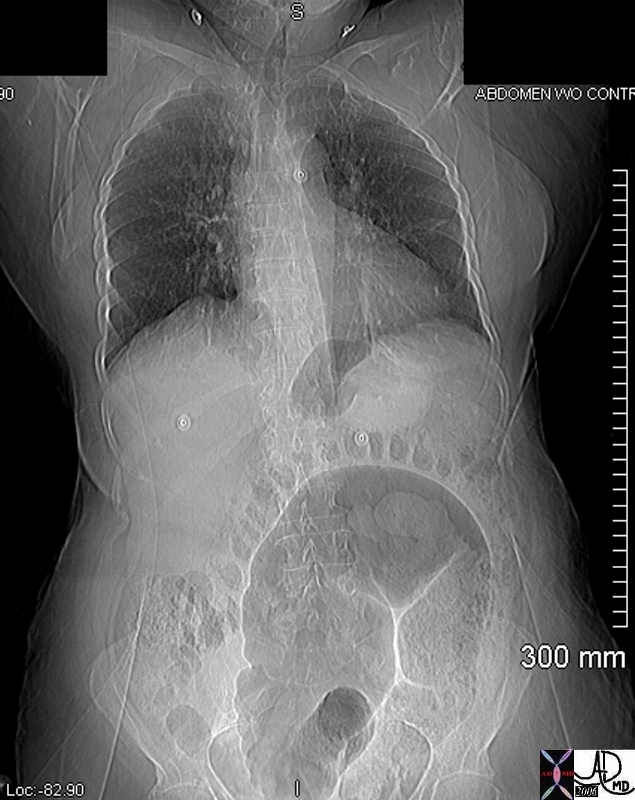
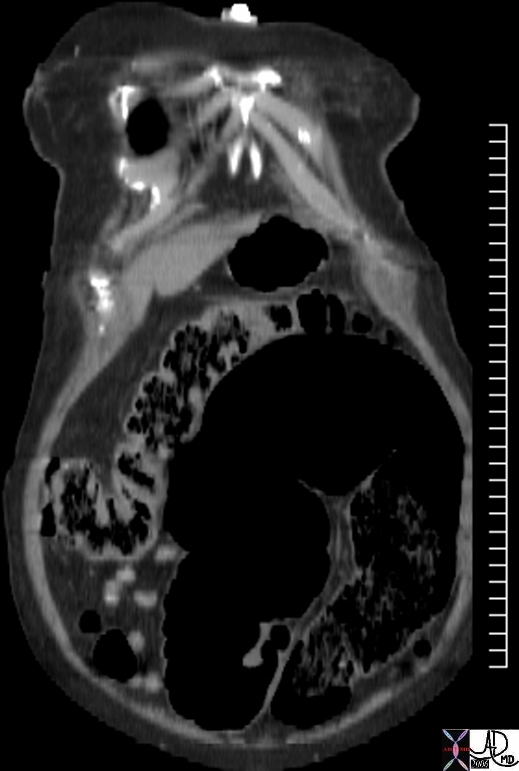
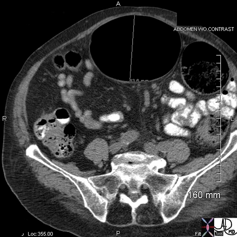
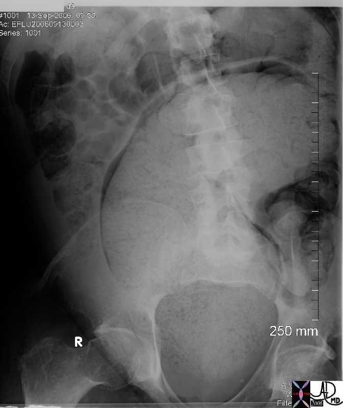
This entity describes an enlarged colon that is not related to mechanical obstruction and is characterized by a cecum that is larger than 12cms, an ascending colon larger than 8cms, or a rectosigmoid that is greater than 6.5cms. It has three forms. We have discussed toxic megacolon and the other two are acute megacolon (Ogilvie?s syndrome) and chronic megacolon. In the cases shown below the megacolon and megarectum are of the chronic form.
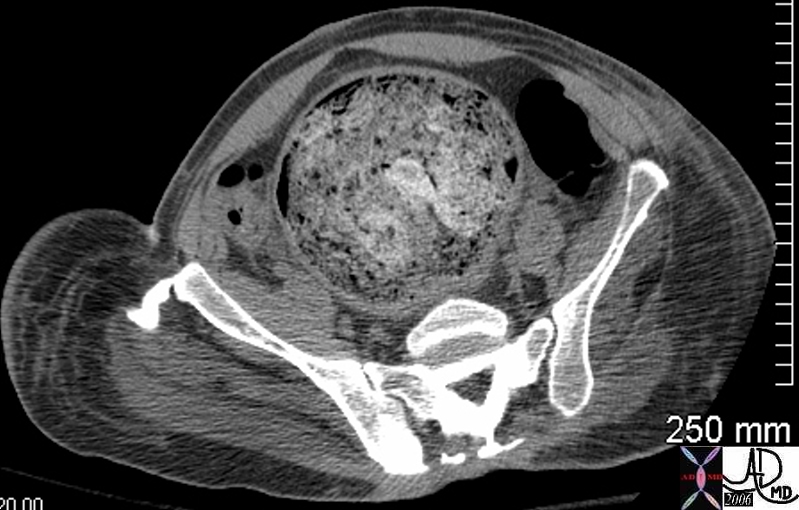
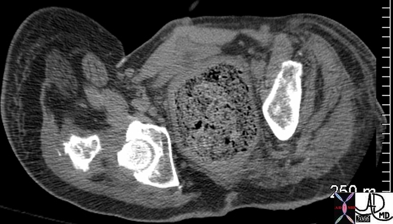
Although it is rather surprising that the patient above was not obstructed other complications such as stercoral ulcers due to chronic irritation of the feces on the mucosa, and bowel ischemia from vascular compromise must be considered in the appropriate clinical setting.
Diverticulosis
Diverticulosis is a very common entity in Western civilizations and is thought to be due to the lack of fiber bulk in our diets. In the USA approximately 50-60% of the elderly have evidence of diverticulosis and it is found predominantly on the left side of the colon and most commonly in the sigmoid colon. In the Asian population interestingly, diverticulosis occurs predominantly on the right side. The rectum is not affected presumably because of the extra layer of longitudinal muscle that completely encompasses the circumference. The pathogenesis of the entity is thought to relate to excessive pressure build up in the colon due to strong peristaltic action on the low bulk content, causing excessive intraluminal pressure, muscular hypertrophy, mucosal thickening, bowel shortening, and to some degree luminal narrowing. The increased luminal pressure is transferred to the walls of the bowel and blow out predominantly of the mucosa through weak regions in the bowel wall occurs. As stated in the histology section above, the regions of weakness occur where the blood vessels enter and leave the bowel wall. (Cleveland) (eMedicine)
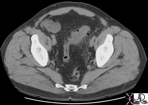
The diverticuli are easily identified on any of the diagnostic studies performed including endoscopy barium enema and CTscan.
Diverticulitis
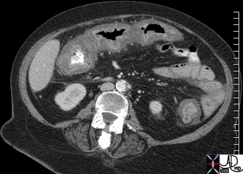
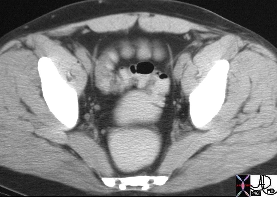
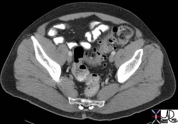
Feces and debris may get trapped in the narrow necked diverticulum and result in an inflammatory response. Mucus secretion and swelling of the mucosa result, which in turn cause further pressure build up in the diverticulum. The pressure on the walls may cause ischemia with microperforation with extension of the inflammatory process and infection to the surrounding fat. Complications include abscess formation, peritonitis, and fistulization to the bladder or skin. CTscan is the study of choice, and the diagnosis is made by finding inflammatory changes in the pericolic fat or of changes within the sigmoid mesocolon.
|
DOMElement Object
(
    [schemaTypeInfo] =>
    [tagName] => table
    [firstElementChild] => (object value omitted)
    [lastElementChild] => (object value omitted)
    [childElementCount] => 1
    [previousElementSibling] => (object value omitted)
    [nextElementSibling] =>
    [nodeName] => table
    [nodeValue] =>


Diverticulitis caused by c difficile colitis


This  72 female with intractable pseudomembranous colitis developed toxic megacolon and required surgical colectomy.  In the descending colon the enhancing mucosa and submucosa is seen herniating through the thickened muscularis and is associated with induration of the fat and the lateral portion of Gerota?s fascia.  This is another type of diverticulitis.
Courtesy Ashley Davidoff MD
45589


    [nodeType] => 1
    [parentNode] => (object value omitted)
    [childNodes] => (object value omitted)
    [firstChild] => (object value omitted)
    [lastChild] => (object value omitted)
    [previousSibling] => (object value omitted)
    [nextSibling] => (object value omitted)
    [attributes] => (object value omitted)
    [ownerDocument] => (object value omitted)
    [namespaceURI] =>
    [prefix] =>
    [localName] => table
    [baseURI] =>
    [textContent] =>


Diverticulitis caused by c difficile colitis


This  72 female with intractable pseudomembranous colitis developed toxic megacolon and required surgical colectomy.  In the descending colon the enhancing mucosa and submucosa is seen herniating through the thickened muscularis and is associated with induration of the fat and the lateral portion of Gerota?s fascia.  This is another type of diverticulitis.
Courtesy Ashley Davidoff MD
45589


)
DOMElement Object
(
    [schemaTypeInfo] =>
    [tagName] => td
    [firstElementChild] => (object value omitted)
    [lastElementChild] => (object value omitted)
    [childElementCount] => 2
    [previousElementSibling] =>
    [nextElementSibling] =>
    [nodeName] => td
    [nodeValue] => This  72 female with intractable pseudomembranous colitis developed toxic megacolon and required surgical colectomy.  In the descending colon the enhancing mucosa and submucosa is seen herniating through the thickened muscularis and is associated with induration of the fat and the lateral portion of Gerota?s fascia.  This is another type of diverticulitis.
Courtesy Ashley Davidoff MD
45589
    [nodeType] => 1
    [parentNode] => (object value omitted)
    [childNodes] => (object value omitted)
    [firstChild] => (object value omitted)
    [lastChild] => (object value omitted)
    [previousSibling] => (object value omitted)
    [nextSibling] => (object value omitted)
    [attributes] => (object value omitted)
    [ownerDocument] => (object value omitted)
    [namespaceURI] =>
    [prefix] =>
    [localName] => td
    [baseURI] =>
    [textContent] => This  72 female with intractable pseudomembranous colitis developed toxic megacolon and required surgical colectomy.  In the descending colon the enhancing mucosa and submucosa is seen herniating through the thickened muscularis and is associated with induration of the fat and the lateral portion of Gerota?s fascia.  This is another type of diverticulitis.
Courtesy Ashley Davidoff MD
45589
)
DOMElement Object
(
    [schemaTypeInfo] =>
    [tagName] => td
    [firstElementChild] => (object value omitted)
    [lastElementChild] => (object value omitted)
    [childElementCount] => 1
    [previousElementSibling] =>
    [nextElementSibling] =>
    [nodeName] => td
    [nodeValue] => Diverticulitis caused by c difficile colitis
    [nodeType] => 1
    [parentNode] => (object value omitted)
    [childNodes] => (object value omitted)
    [firstChild] => (object value omitted)
    [lastChild] => (object value omitted)
    [previousSibling] => (object value omitted)
    [nextSibling] => (object value omitted)
    [attributes] => (object value omitted)
    [ownerDocument] => (object value omitted)
    [namespaceURI] =>
    [prefix] =>
    [localName] => td
    [baseURI] =>
    [textContent] => Diverticulitis caused by c difficile colitis
)
DOMElement Object
(
    [schemaTypeInfo] =>
    [tagName] => table
    [firstElementChild] => (object value omitted)
    [lastElementChild] => (object value omitted)
    [childElementCount] => 1
    [previousElementSibling] => (object value omitted)
    [nextElementSibling] => (object value omitted)
    [nodeName] => table
    [nodeValue] =>


Mild diverticulitis


In this case a few small air filled diverticuli are noted in the sigmoid colon, but the key to the diagnosis is the mild induration (dirty fat) of the pericolic fat, and the thickening of the base of the sigmoid colon.  The sigmoid mesocolon attaches to the left side of the pelvis (orange line) between the left side of the sigmoid and the external iliac artery (red) and vein (blue).
Courtesy Ashley Davidoff MD
18569


    [nodeType] => 1
    [parentNode] => (object value omitted)
    [childNodes] => (object value omitted)
    [firstChild] => (object value omitted)
    [lastChild] => (object value omitted)
    [previousSibling] => (object value omitted)
    [nextSibling] => (object value omitted)
    [attributes] => (object value omitted)
    [ownerDocument] => (object value omitted)
    [namespaceURI] =>
    [prefix] =>
    [localName] => table
    [baseURI] =>
    [textContent] =>


Mild diverticulitis


In this case a few small air filled diverticuli are noted in the sigmoid colon, but the key to the diagnosis is the mild induration (dirty fat) of the pericolic fat, and the thickening of the base of the sigmoid colon.  The sigmoid mesocolon attaches to the left side of the pelvis (orange line) between the left side of the sigmoid and the external iliac artery (red) and vein (blue).
Courtesy Ashley Davidoff MD
18569


)
DOMElement Object
(
    [schemaTypeInfo] =>
    [tagName] => td
    [firstElementChild] => (object value omitted)
    [lastElementChild] => (object value omitted)
    [childElementCount] => 2
    [previousElementSibling] =>
    [nextElementSibling] =>
    [nodeName] => td
    [nodeValue] => In this case a few small air filled diverticuli are noted in the sigmoid colon, but the key to the diagnosis is the mild induration (dirty fat) of the pericolic fat, and the thickening of the base of the sigmoid colon.  The sigmoid mesocolon attaches to the left side of the pelvis (orange line) between the left side of the sigmoid and the external iliac artery (red) and vein (blue).
Courtesy Ashley Davidoff MD
18569
    [nodeType] => 1
    [parentNode] => (object value omitted)
    [childNodes] => (object value omitted)
    [firstChild] => (object value omitted)
    [lastChild] => (object value omitted)
    [previousSibling] => (object value omitted)
    [nextSibling] => (object value omitted)
    [attributes] => (object value omitted)
    [ownerDocument] => (object value omitted)
    [namespaceURI] =>
    [prefix] =>
    [localName] => td
    [baseURI] =>
    [textContent] => In this case a few small air filled diverticuli are noted in the sigmoid colon, but the key to the diagnosis is the mild induration (dirty fat) of the pericolic fat, and the thickening of the base of the sigmoid colon.  The sigmoid mesocolon attaches to the left side of the pelvis (orange line) between the left side of the sigmoid and the external iliac artery (red) and vein (blue).
Courtesy Ashley Davidoff MD
18569
)
DOMElement Object
(
    [schemaTypeInfo] =>
    [tagName] => td
    [firstElementChild] => (object value omitted)
    [lastElementChild] => (object value omitted)
    [childElementCount] => 1
    [previousElementSibling] =>
    [nextElementSibling] =>
    [nodeName] => td
    [nodeValue] => Mild diverticulitis
    [nodeType] => 1
    [parentNode] => (object value omitted)
    [childNodes] => (object value omitted)
    [firstChild] => (object value omitted)
    [lastChild] => (object value omitted)
    [previousSibling] => (object value omitted)
    [nextSibling] => (object value omitted)
    [attributes] => (object value omitted)
    [ownerDocument] => (object value omitted)
    [namespaceURI] =>
    [prefix] =>
    [localName] => td
    [baseURI] =>
    [textContent] => Mild diverticulitis
)
DOMElement Object
(
    [schemaTypeInfo] =>
    [tagName] => table
    [firstElementChild] => (object value omitted)
    [lastElementChild] => (object value omitted)
    [childElementCount] => 1
    [previousElementSibling] => (object value omitted)
    [nextElementSibling] => (object value omitted)
    [nodeName] => table
    [nodeValue] =>


Diverticulosis of the sigmoid colon by CT scan


In this CTscan of the pelvis the diverticular disease is seen as small outpouchings of gas (black) that extend beyond the expected confines of the wall of the bowel.
Courtesy Ashley Davidoff MD
34663


    [nodeType] => 1
    [parentNode] => (object value omitted)
    [childNodes] => (object value omitted)
    [firstChild] => (object value omitted)
    [lastChild] => (object value omitted)
    [previousSibling] => (object value omitted)
    [nextSibling] => (object value omitted)
    [attributes] => (object value omitted)
    [ownerDocument] => (object value omitted)
    [namespaceURI] =>
    [prefix] =>
    [localName] => table
    [baseURI] =>
    [textContent] =>


Diverticulosis of the sigmoid colon by CT scan


In this CTscan of the pelvis the diverticular disease is seen as small outpouchings of gas (black) that extend beyond the expected confines of the wall of the bowel.
Courtesy Ashley Davidoff MD
34663


)
DOMElement Object
(
    [schemaTypeInfo] =>
    [tagName] => td
    [firstElementChild] => (object value omitted)
    [lastElementChild] => (object value omitted)
    [childElementCount] => 2
    [previousElementSibling] =>
    [nextElementSibling] =>
    [nodeName] => td
    [nodeValue] => In this CTscan of the pelvis the diverticular disease is seen as small outpouchings of gas (black) that extend beyond the expected confines of the wall of the bowel.
Courtesy Ashley Davidoff MD
34663
    [nodeType] => 1
    [parentNode] => (object value omitted)
    [childNodes] => (object value omitted)
    [firstChild] => (object value omitted)
    [lastChild] => (object value omitted)
    [previousSibling] => (object value omitted)
    [nextSibling] => (object value omitted)
    [attributes] => (object value omitted)
    [ownerDocument] => (object value omitted)
    [namespaceURI] =>
    [prefix] =>
    [localName] => td
    [baseURI] =>
    [textContent] => In this CTscan of the pelvis the diverticular disease is seen as small outpouchings of gas (black) that extend beyond the expected confines of the wall of the bowel.
Courtesy Ashley Davidoff MD
34663
)
DOMElement Object
(
    [schemaTypeInfo] =>
    [tagName] => td
    [firstElementChild] => (object value omitted)
    [lastElementChild] => (object value omitted)
    [childElementCount] => 1
    [previousElementSibling] =>
    [nextElementSibling] =>
    [nodeName] => td
    [nodeValue] => Diverticulosis of the sigmoid colon by CT scan
    [nodeType] => 1
    [parentNode] => (object value omitted)
    [childNodes] => (object value omitted)
    [firstChild] => (object value omitted)
    [lastChild] => (object value omitted)
    [previousSibling] => (object value omitted)
    [nextSibling] => (object value omitted)
    [attributes] => (object value omitted)
    [ownerDocument] => (object value omitted)
    [namespaceURI] =>
    [prefix] =>
    [localName] => td
    [baseURI] =>
    [textContent] => Diverticulosis of the sigmoid colon by CT scan
)
DOMElement Object
(
    [schemaTypeInfo] =>
    [tagName] => table
    [firstElementChild] => (object value omitted)
    [lastElementChild] => (object value omitted)
    [childElementCount] => 1
    [previousElementSibling] => (object value omitted)
    [nextElementSibling] => (object value omitted)
    [nodeName] => table
    [nodeValue] =>


Diverticulosis by barium enema


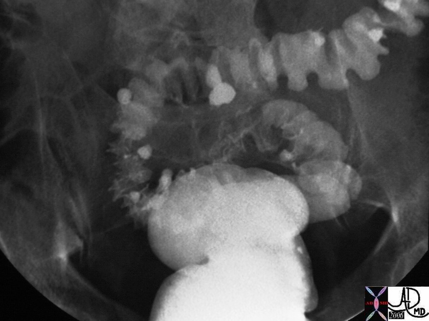
This patient demonstrates barium accumulations in diverticuli of the sigmoid colon associated with prominent circumferential muscle bands and minor degree of luminal narrowing.  Note the absence of diverticulosis in the rectum.
Courtesy Ashley Davidoff MD
31836


    [nodeType] => 1
    [parentNode] => (object value omitted)
    [childNodes] => (object value omitted)
    [firstChild] => (object value omitted)
    [lastChild] => (object value omitted)
    [previousSibling] => (object value omitted)
    [nextSibling] => (object value omitted)
    [attributes] => (object value omitted)
    [ownerDocument] => (object value omitted)
    [namespaceURI] =>
    [prefix] =>
    [localName] => table
    [baseURI] =>
    [textContent] =>


Diverticulosis by barium enema


This patient demonstrates barium accumulations in diverticuli of the sigmoid colon associated with prominent circumferential muscle bands and minor degree of luminal narrowing.  Note the absence of diverticulosis in the rectum.
Courtesy Ashley Davidoff MD
31836


)
DOMElement Object
(
    [schemaTypeInfo] =>
    [tagName] => td
    [firstElementChild] => (object value omitted)
    [lastElementChild] => (object value omitted)
    [childElementCount] => 2
    [previousElementSibling] =>
    [nextElementSibling] =>
    [nodeName] => td
    [nodeValue] => This patient demonstrates barium accumulations in diverticuli of the sigmoid colon associated with prominent circumferential muscle bands and minor degree of luminal narrowing.  Note the absence of diverticulosis in the rectum.
Courtesy Ashley Davidoff MD
31836
    [nodeType] => 1
    [parentNode] => (object value omitted)
    [childNodes] => (object value omitted)
    [firstChild] => (object value omitted)
    [lastChild] => (object value omitted)
    [previousSibling] => (object value omitted)
    [nextSibling] => (object value omitted)
    [attributes] => (object value omitted)
    [ownerDocument] => (object value omitted)
    [namespaceURI] =>
    [prefix] =>
    [localName] => td
    [baseURI] =>
    [textContent] => This patient demonstrates barium accumulations in diverticuli of the sigmoid colon associated with prominent circumferential muscle bands and minor degree of luminal narrowing.  Note the absence of diverticulosis in the rectum.
Courtesy Ashley Davidoff MD
31836
)
DOMElement Object
(
    [schemaTypeInfo] =>
    [tagName] => td
    [firstElementChild] => (object value omitted)
    [lastElementChild] => (object value omitted)
    [childElementCount] => 1
    [previousElementSibling] =>
    [nextElementSibling] =>
    [nodeName] => td
    [nodeValue] => Diverticulosis by barium enema
    [nodeType] => 1
    [parentNode] => (object value omitted)
    [childNodes] => (object value omitted)
    [firstChild] => (object value omitted)
    [lastChild] => (object value omitted)
    [previousSibling] => (object value omitted)
    [nextSibling] => (object value omitted)
    [attributes] => (object value omitted)
    [ownerDocument] => (object value omitted)
    [namespaceURI] =>
    [prefix] =>
    [localName] => td
    [baseURI] =>
    [textContent] => Diverticulosis by barium enema
)
DOMElement Object
(
    [schemaTypeInfo] =>
    [tagName] => table
    [firstElementChild] => (object value omitted)
    [lastElementChild] => (object value omitted)
    [childElementCount] => 1
    [previousElementSibling] => (object value omitted)
    [nextElementSibling] => (object value omitted)
    [nodeName] => table
    [nodeValue] =>


Whole mount – diverticulosis


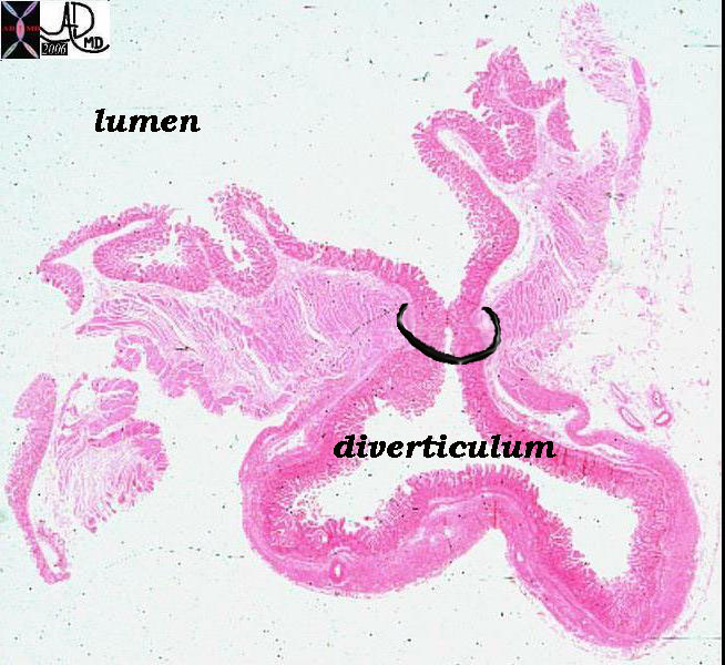
Whole-mount microphotograph of a single diverticulum. Notice that the diverticulum consists of extrusion of the mucosa out through the muscle layer, while it retains the serosal outer covering.  This usually occurs at points where arteries and veins penetrate the muscular layer. The black ring in the image represents the mouth of the diverticulum.
Courtesy Barbara Banner MD
12305b01


    [nodeType] => 1
    [parentNode] => (object value omitted)
    [childNodes] => (object value omitted)
    [firstChild] => (object value omitted)
    [lastChild] => (object value omitted)
    [previousSibling] => (object value omitted)
    [nextSibling] => (object value omitted)
    [attributes] => (object value omitted)
    [ownerDocument] => (object value omitted)
    [namespaceURI] =>
    [prefix] =>
    [localName] => table
    [baseURI] =>
    [textContent] =>


Whole mount – diverticulosis


Whole-mount microphotograph of a single diverticulum. Notice that the diverticulum consists of extrusion of the mucosa out through the muscle layer, while it retains the serosal outer covering.  This usually occurs at points where arteries and veins penetrate the muscular layer. The black ring in the image represents the mouth of the diverticulum.
Courtesy Barbara Banner MD
12305b01


)
DOMElement Object
(
    [schemaTypeInfo] =>
    [tagName] => td
    [firstElementChild] => (object value omitted)
    [lastElementChild] => (object value omitted)
    [childElementCount] => 2
    [previousElementSibling] =>
    [nextElementSibling] =>
    [nodeName] => td
    [nodeValue] => Whole-mount microphotograph of a single diverticulum. Notice that the diverticulum consists of extrusion of the mucosa out through the muscle layer, while it retains the serosal outer covering.  This usually occurs at points where arteries and veins penetrate the muscular layer. The black ring in the image represents the mouth of the diverticulum.
Courtesy Barbara Banner MD
12305b01
    [nodeType] => 1
    [parentNode] => (object value omitted)
    [childNodes] => (object value omitted)
    [firstChild] => (object value omitted)
    [lastChild] => (object value omitted)
    [previousSibling] => (object value omitted)
    [nextSibling] => (object value omitted)
    [attributes] => (object value omitted)
    [ownerDocument] => (object value omitted)
    [namespaceURI] =>
    [prefix] =>
    [localName] => td
    [baseURI] =>
    [textContent] => Whole-mount microphotograph of a single diverticulum. Notice that the diverticulum consists of extrusion of the mucosa out through the muscle layer, while it retains the serosal outer covering.  This usually occurs at points where arteries and veins penetrate the muscular layer. The black ring in the image represents the mouth of the diverticulum.
Courtesy Barbara Banner MD
12305b01
)
DOMElement Object
(
    [schemaTypeInfo] =>
    [tagName] => td
    [firstElementChild] => (object value omitted)
    [lastElementChild] => (object value omitted)
    [childElementCount] => 1
    [previousElementSibling] =>
    [nextElementSibling] =>
    [nodeName] => td
    [nodeValue] => Whole mount – diverticulosis
    [nodeType] => 1
    [parentNode] => (object value omitted)
    [childNodes] => (object value omitted)
    [firstChild] => (object value omitted)
    [lastChild] => (object value omitted)
    [previousSibling] => (object value omitted)
    [nextSibling] => (object value omitted)
    [attributes] => (object value omitted)
    [ownerDocument] => (object value omitted)
    [namespaceURI] =>
    [prefix] =>
    [localName] => td
    [baseURI] =>
    [textContent] => Whole mount – diverticulosis
)
DOMElement Object
(
    [schemaTypeInfo] =>
    [tagName] => table
    [firstElementChild] => (object value omitted)
    [lastElementChild] => (object value omitted)
    [childElementCount] => 1
    [previousElementSibling] => (object value omitted)
    [nextElementSibling] => (object value omitted)
    [nodeName] => table
    [nodeValue] =>


Sigmoid and rectal impaction with megacolon and no obstruction


In this case there is impaction of feces in the sigmoid colon and rectum but there is  no evidence of obstruction since the rest of the colon is not dilated. These findings were of a  chronic nature and unchanged.  A diagnosis of chronic megacolon is likely.
Courtesy Ashley Davidoff MD
45461 45462 45464


    [nodeType] => 1
    [parentNode] => (object value omitted)
    [childNodes] => (object value omitted)
    [firstChild] => (object value omitted)
    [lastChild] => (object value omitted)
    [previousSibling] => (object value omitted)
    [nextSibling] => (object value omitted)
    [attributes] => (object value omitted)
    [ownerDocument] => (object value omitted)
    [namespaceURI] =>
    [prefix] =>
    [localName] => table
    [baseURI] =>
    [textContent] =>


Sigmoid and rectal impaction with megacolon and no obstruction


In this case there is impaction of feces in the sigmoid colon and rectum but there is  no evidence of obstruction since the rest of the colon is not dilated. These findings were of a  chronic nature and unchanged.  A diagnosis of chronic megacolon is likely.
Courtesy Ashley Davidoff MD
45461 45462 45464


)
DOMElement Object
(
    [schemaTypeInfo] =>
    [tagName] => td
    [firstElementChild] => (object value omitted)
    [lastElementChild] => (object value omitted)
    [childElementCount] => 2
    [previousElementSibling] =>
    [nextElementSibling] =>
    [nodeName] => td
    [nodeValue] => In this case there is impaction of feces in the sigmoid colon and rectum but there is  no evidence of obstruction since the rest of the colon is not dilated. These findings were of a  chronic nature and unchanged.  A diagnosis of chronic megacolon is likely.
Courtesy Ashley Davidoff MD
45461 45462 45464
    [nodeType] => 1
    [parentNode] => (object value omitted)
    [childNodes] => (object value omitted)
    [firstChild] => (object value omitted)
    [lastChild] => (object value omitted)
    [previousSibling] => (object value omitted)
    [nextSibling] => (object value omitted)
    [attributes] => (object value omitted)
    [ownerDocument] => (object value omitted)
    [namespaceURI] =>
    [prefix] =>
    [localName] => td
    [baseURI] =>
    [textContent] => In this case there is impaction of feces in the sigmoid colon and rectum but there is  no evidence of obstruction since the rest of the colon is not dilated. These findings were of a  chronic nature and unchanged.  A diagnosis of chronic megacolon is likely.
Courtesy Ashley Davidoff MD
45461 45462 45464
)
DOMElement Object
(
    [schemaTypeInfo] =>
    [tagName] => td
    [firstElementChild] => (object value omitted)
    [lastElementChild] => (object value omitted)
    [childElementCount] => 1
    [previousElementSibling] =>
    [nextElementSibling] =>
    [nodeName] => td
    [nodeValue] => Sigmoid and rectal impaction with megacolon and no obstruction
    [nodeType] => 1
    [parentNode] => (object value omitted)
    [childNodes] => (object value omitted)
    [firstChild] => (object value omitted)
    [lastChild] => (object value omitted)
    [previousSibling] => (object value omitted)
    [nextSibling] => (object value omitted)
    [attributes] => (object value omitted)
    [ownerDocument] => (object value omitted)
    [namespaceURI] =>
    [prefix] =>
    [localName] => td
    [baseURI] =>
    [textContent] => Sigmoid and rectal impaction with megacolon and no obstruction
)
DOMElement Object
(
    [schemaTypeInfo] =>
    [tagName] => table
    [firstElementChild] => (object value omitted)
    [lastElementChild] => (object value omitted)
    [childElementCount] => 1
    [previousElementSibling] => (object value omitted)
    [nextElementSibling] => (object value omitted)
    [nodeName] => table
    [nodeValue] =>


Sigmoid megacolon


A CT performed shows that the sigmoid measured 8.3cms in diameter and was filled with air.
Courtesy Ashley Davidoff MD
45450 45451 45454


    [nodeType] => 1
    [parentNode] => (object value omitted)
    [childNodes] => (object value omitted)
    [firstChild] => (object value omitted)
    [lastChild] => (object value omitted)
    [previousSibling] => (object value omitted)
    [nextSibling] => (object value omitted)
    [attributes] => (object value omitted)
    [ownerDocument] => (object value omitted)
    [namespaceURI] =>
    [prefix] =>
    [localName] => table
    [baseURI] =>
    [textContent] =>


Sigmoid megacolon


A CT performed shows that the sigmoid measured 8.3cms in diameter and was filled with air.
Courtesy Ashley Davidoff MD
45450 45451 45454


)
DOMElement Object
(
    [schemaTypeInfo] =>
    [tagName] => td
    [firstElementChild] => (object value omitted)
    [lastElementChild] => (object value omitted)
    [childElementCount] => 2
    [previousElementSibling] =>
    [nextElementSibling] =>
    [nodeName] => td
    [nodeValue] => A CT performed shows that the sigmoid measured 8.3cms in diameter and was filled with air.
Courtesy Ashley Davidoff MD
45450 45451 45454
    [nodeType] => 1
    [parentNode] => (object value omitted)
    [childNodes] => (object value omitted)
    [firstChild] => (object value omitted)
    [lastChild] => (object value omitted)
    [previousSibling] => (object value omitted)
    [nextSibling] => (object value omitted)
    [attributes] => (object value omitted)
    [ownerDocument] => (object value omitted)
    [namespaceURI] =>
    [prefix] =>
    [localName] => td
    [baseURI] =>
    [textContent] => A CT performed shows that the sigmoid measured 8.3cms in diameter and was filled with air.
Courtesy Ashley Davidoff MD
45450 45451 45454
)
DOMElement Object
(
    [schemaTypeInfo] =>
    [tagName] => td
    [firstElementChild] => (object value omitted)
    [lastElementChild] => (object value omitted)
    [childElementCount] => 1
    [previousElementSibling] =>
    [nextElementSibling] =>
    [nodeName] => td
    [nodeValue] => Sigmoid megacolon
    [nodeType] => 1
    [parentNode] => (object value omitted)
    [childNodes] => (object value omitted)
    [firstChild] => (object value omitted)
    [lastChild] => (object value omitted)
    [previousSibling] => (object value omitted)
    [nextSibling] => (object value omitted)
    [attributes] => (object value omitted)
    [ownerDocument] => (object value omitted)
    [namespaceURI] =>
    [prefix] =>
    [localName] => td
    [baseURI] =>
    [textContent] => Sigmoid megacolon
)
DOMElement Object
(
    [schemaTypeInfo] =>
    [tagName] => table
    [firstElementChild] => (object value omitted)
    [lastElementChild] => (object value omitted)
    [childElementCount] => 1
    [previousElementSibling] => (object value omitted)
    [nextElementSibling] => (object value omitted)
    [nodeName] => table
    [nodeValue] =>


Chronic megacolon


This plain film is from an asymptomatic institutionalized patient.  Talking about an organ that jumps out at you from the film ? diagnosis ? chronic megacolon.  Previous films over a few years had shown similar appearance in this asymptomatic patient (aside from chronic abdominal distension) and hence sigmoid volvulus was not a clinical consideration.
Courtesy Ashley Davidoff MD
16667


    [nodeType] => 1
    [parentNode] => (object value omitted)
    [childNodes] => (object value omitted)
    [firstChild] => (object value omitted)
    [lastChild] => (object value omitted)
    [previousSibling] => (object value omitted)
    [nextSibling] => (object value omitted)
    [attributes] => (object value omitted)
    [ownerDocument] => (object value omitted)
    [namespaceURI] =>
    [prefix] =>
    [localName] => table
    [baseURI] =>
    [textContent] =>


Chronic megacolon


This plain film is from an asymptomatic institutionalized patient.  Talking about an organ that jumps out at you from the film ? diagnosis ? chronic megacolon.  Previous films over a few years had shown similar appearance in this asymptomatic patient (aside from chronic abdominal distension) and hence sigmoid volvulus was not a clinical consideration.
Courtesy Ashley Davidoff MD
16667


)
DOMElement Object
(
    [schemaTypeInfo] =>
    [tagName] => td
    [firstElementChild] => (object value omitted)
    [lastElementChild] => (object value omitted)
    [childElementCount] => 2
    [previousElementSibling] =>
    [nextElementSibling] =>
    [nodeName] => td
    [nodeValue] => This plain film is from an asymptomatic institutionalized patient.  Talking about an organ that jumps out at you from the film ? diagnosis ? chronic megacolon.  Previous films over a few years had shown similar appearance in this asymptomatic patient (aside from chronic abdominal distension) and hence sigmoid volvulus was not a clinical consideration.
Courtesy Ashley Davidoff MD
16667
    [nodeType] => 1
    [parentNode] => (object value omitted)
    [childNodes] => (object value omitted)
    [firstChild] => (object value omitted)
    [lastChild] => (object value omitted)
    [previousSibling] => (object value omitted)
    [nextSibling] => (object value omitted)
    [attributes] => (object value omitted)
    [ownerDocument] => (object value omitted)
    [namespaceURI] =>
    [prefix] =>
    [localName] => td
    [baseURI] =>
    [textContent] => This plain film is from an asymptomatic institutionalized patient.  Talking about an organ that jumps out at you from the film ? diagnosis ? chronic megacolon.  Previous films over a few years had shown similar appearance in this asymptomatic patient (aside from chronic abdominal distension) and hence sigmoid volvulus was not a clinical consideration.
Courtesy Ashley Davidoff MD
16667
)
DOMElement Object
(
    [schemaTypeInfo] =>
    [tagName] => td
    [firstElementChild] => (object value omitted)
    [lastElementChild] => (object value omitted)
    [childElementCount] => 1
    [previousElementSibling] =>
    [nextElementSibling] =>
    [nodeName] => td
    [nodeValue] => Chronic megacolon
    [nodeType] => 1
    [parentNode] => (object value omitted)
    [childNodes] => (object value omitted)
    [firstChild] => (object value omitted)
    [lastChild] => (object value omitted)
    [previousSibling] => (object value omitted)
    [nextSibling] => (object value omitted)
    [attributes] => (object value omitted)
    [ownerDocument] => (object value omitted)
    [namespaceURI] =>
    [prefix] =>
    [localName] => td
    [baseURI] =>
    [textContent] => Chronic megacolon
)
DOMElement Object
(
    [schemaTypeInfo] =>
    [tagName] => table
    [firstElementChild] => (object value omitted)
    [lastElementChild] => (object value omitted)
    [childElementCount] => 1
    [previousElementSibling] => (object value omitted)
    [nextElementSibling] => (object value omitted)
    [nodeName] => table
    [nodeValue] =>


 One of the Sigmoid Curves of the Sigmoid Colon – CT Colonography


75327 sigmoid colon large bowel sigmoid shape normal anatomy air contrast study CT colonography virtual colonoscopy Courtesy Ashley Davidoff MD


    [nodeType] => 1
    [parentNode] => (object value omitted)
    [childNodes] => (object value omitted)
    [firstChild] => (object value omitted)
    [lastChild] => (object value omitted)
    [previousSibling] => (object value omitted)
    [nextSibling] => (object value omitted)
    [attributes] => (object value omitted)
    [ownerDocument] => (object value omitted)
    [namespaceURI] =>
    [prefix] =>
    [localName] => table
    [baseURI] =>
    [textContent] =>


 One of the Sigmoid Curves of the Sigmoid Colon – CT Colonography


75327 sigmoid colon large bowel sigmoid shape normal anatomy air contrast study CT colonography virtual colonoscopy Courtesy Ashley Davidoff MD


)
DOMElement Object
(
    [schemaTypeInfo] =>
    [tagName] => td
    [firstElementChild] =>
    [lastElementChild] =>
    [childElementCount] => 0
    [previousElementSibling] =>
    [nextElementSibling] =>
    [nodeName] => td
    [nodeValue] => 75327 sigmoid colon large bowel sigmoid shape normal anatomy air contrast study CT colonography virtual colonoscopy Courtesy Ashley Davidoff MD
    [nodeType] => 1
    [parentNode] => (object value omitted)
    [childNodes] => (object value omitted)
    [firstChild] => (object value omitted)
    [lastChild] => (object value omitted)
    [previousSibling] => (object value omitted)
    [nextSibling] => (object value omitted)
    [attributes] => (object value omitted)
    [ownerDocument] => (object value omitted)
    [namespaceURI] =>
    [prefix] =>
    [localName] => td
    [baseURI] =>
    [textContent] => 75327 sigmoid colon large bowel sigmoid shape normal anatomy air contrast study CT colonography virtual colonoscopy Courtesy Ashley Davidoff MD
)
DOMElement Object
(
    [schemaTypeInfo] =>
    [tagName] => td
    [firstElementChild] => (object value omitted)
    [lastElementChild] => (object value omitted)
    [childElementCount] => 1
    [previousElementSibling] =>
    [nextElementSibling] =>
    [nodeName] => td
    [nodeValue] =>  One of the Sigmoid Curves of the Sigmoid Colon – CT Colonography
    [nodeType] => 1
    [parentNode] => (object value omitted)
    [childNodes] => (object value omitted)
    [firstChild] => (object value omitted)
    [lastChild] => (object value omitted)
    [previousSibling] => (object value omitted)
    [nextSibling] => (object value omitted)
    [attributes] => (object value omitted)
    [ownerDocument] => (object value omitted)
    [namespaceURI] =>
    [prefix] =>
    [localName] => td
    [baseURI] =>
    [textContent] =>  One of the Sigmoid Curves of the Sigmoid Colon – CT Colonography
)
DOMElement Object
(
    [schemaTypeInfo] =>
    [tagName] => table
    [firstElementChild] => (object value omitted)
    [lastElementChild] => (object value omitted)
    [childElementCount] => 1
    [previousElementSibling] => (object value omitted)
    [nextElementSibling] => (object value omitted)
    [nodeName] => table
    [nodeValue] =>


Rectosigmoid region


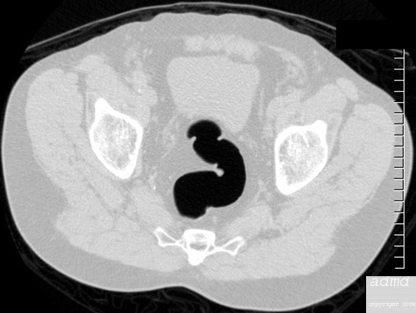
The CTscan shows the morphological differences between the normal sigmoid which is ?S? shaped and rectum which is straight.  The sigmoid colon has taenia coli muscle and so it also has haustra whereas the rectum lacks taenia and therefore lacks a haustral pattern.  The difference is typified in this image.
Courtesy Ashley Davidoff MD
24872


    [nodeType] => 1
    [parentNode] => (object value omitted)
    [childNodes] => (object value omitted)
    [firstChild] => (object value omitted)
    [lastChild] => (object value omitted)
    [previousSibling] => (object value omitted)
    [nextSibling] => (object value omitted)
    [attributes] => (object value omitted)
    [ownerDocument] => (object value omitted)
    [namespaceURI] =>
    [prefix] =>
    [localName] => table
    [baseURI] =>
    [textContent] =>


Rectosigmoid region


The CTscan shows the morphological differences between the normal sigmoid which is ?S? shaped and rectum which is straight.  The sigmoid colon has taenia coli muscle and so it also has haustra whereas the rectum lacks taenia and therefore lacks a haustral pattern.  The difference is typified in this image.
Courtesy Ashley Davidoff MD
24872


)
DOMElement Object
(
    [schemaTypeInfo] =>
    [tagName] => td
    [firstElementChild] => (object value omitted)
    [lastElementChild] => (object value omitted)
    [childElementCount] => 2
    [previousElementSibling] =>
    [nextElementSibling] =>
    [nodeName] => td
    [nodeValue] => The CTscan shows the morphological differences between the normal sigmoid which is ?S? shaped and rectum which is straight.  The sigmoid colon has taenia coli muscle and so it also has haustra whereas the rectum lacks taenia and therefore lacks a haustral pattern.  The difference is typified in this image.
Courtesy Ashley Davidoff MD
24872
    [nodeType] => 1
    [parentNode] => (object value omitted)
    [childNodes] => (object value omitted)
    [firstChild] => (object value omitted)
    [lastChild] => (object value omitted)
    [previousSibling] => (object value omitted)
    [nextSibling] => (object value omitted)
    [attributes] => (object value omitted)
    [ownerDocument] => (object value omitted)
    [namespaceURI] =>
    [prefix] =>
    [localName] => td
    [baseURI] =>
    [textContent] => The CTscan shows the morphological differences between the normal sigmoid which is ?S? shaped and rectum which is straight.  The sigmoid colon has taenia coli muscle and so it also has haustra whereas the rectum lacks taenia and therefore lacks a haustral pattern.  The difference is typified in this image.
Courtesy Ashley Davidoff MD
24872
)
DOMElement Object
(
    [schemaTypeInfo] =>
    [tagName] => td
    [firstElementChild] => (object value omitted)
    [lastElementChild] => (object value omitted)
    [childElementCount] => 1
    [previousElementSibling] =>
    [nextElementSibling] =>
    [nodeName] => td
    [nodeValue] => Rectosigmoid region
    [nodeType] => 1
    [parentNode] => (object value omitted)
    [childNodes] => (object value omitted)
    [firstChild] => (object value omitted)
    [lastChild] => (object value omitted)
    [previousSibling] => (object value omitted)
    [nextSibling] => (object value omitted)
    [attributes] => (object value omitted)
    [ownerDocument] => (object value omitted)
    [namespaceURI] =>
    [prefix] =>
    [localName] => td
    [baseURI] =>
    [textContent] => Rectosigmoid region
)
DOMElement Object
(
    [schemaTypeInfo] =>
    [tagName] => table
    [firstElementChild] => (object value omitted)
    [lastElementChild] => (object value omitted)
    [childElementCount] => 1
    [previousElementSibling] => (object value omitted)
    [nextElementSibling] => (object value omitted)
    [nodeName] => table
    [nodeValue] =>


Sigmoid colon


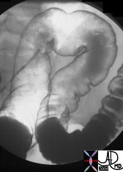
The normal sigmoid colon is demonstrated in this double contrast barium enema.  The shape and length as well as the diameter of the sigmoid colon are variable.
Courtesy Ashley Davidoff MD
20408 20408b01


    [nodeType] => 1
    [parentNode] => (object value omitted)
    [childNodes] => (object value omitted)
    [firstChild] => (object value omitted)
    [lastChild] => (object value omitted)
    [previousSibling] => (object value omitted)
    [nextSibling] => (object value omitted)
    [attributes] => (object value omitted)
    [ownerDocument] => (object value omitted)
    [namespaceURI] =>
    [prefix] =>
    [localName] => table
    [baseURI] =>
    [textContent] =>


Sigmoid colon


The normal sigmoid colon is demonstrated in this double contrast barium enema.  The shape and length as well as the diameter of the sigmoid colon are variable.
Courtesy Ashley Davidoff MD
20408 20408b01


)
DOMElement Object
(
    [schemaTypeInfo] =>
    [tagName] => td
    [firstElementChild] => (object value omitted)
    [lastElementChild] => (object value omitted)
    [childElementCount] => 2
    [previousElementSibling] =>
    [nextElementSibling] =>
    [nodeName] => td
    [nodeValue] => The normal sigmoid colon is demonstrated in this double contrast barium enema.  The shape and length as well as the diameter of the sigmoid colon are variable.
Courtesy Ashley Davidoff MD
20408 20408b01
    [nodeType] => 1
    [parentNode] => (object value omitted)
    [childNodes] => (object value omitted)
    [firstChild] => (object value omitted)
    [lastChild] => (object value omitted)
    [previousSibling] => (object value omitted)
    [nextSibling] => (object value omitted)
    [attributes] => (object value omitted)
    [ownerDocument] => (object value omitted)
    [namespaceURI] =>
    [prefix] =>
    [localName] => td
    [baseURI] =>
    [textContent] => The normal sigmoid colon is demonstrated in this double contrast barium enema.  The shape and length as well as the diameter of the sigmoid colon are variable.
Courtesy Ashley Davidoff MD
20408 20408b01
)
DOMElement Object
(
    [schemaTypeInfo] =>
    [tagName] => td
    [firstElementChild] => (object value omitted)
    [lastElementChild] => (object value omitted)
    [childElementCount] => 1
    [previousElementSibling] =>
    [nextElementSibling] =>
    [nodeName] => td
    [nodeValue] => Sigmoid colon
    [nodeType] => 1
    [parentNode] => (object value omitted)
    [childNodes] => (object value omitted)
    [firstChild] => (object value omitted)
    [lastChild] => (object value omitted)
    [previousSibling] => (object value omitted)
    [nextSibling] => (object value omitted)
    [attributes] => (object value omitted)
    [ownerDocument] => (object value omitted)
    [namespaceURI] =>
    [prefix] =>
    [localName] => td
    [baseURI] =>
    [textContent] => Sigmoid colon
)
DOMElement Object
(
    [schemaTypeInfo] =>
    [tagName] => table
    [firstElementChild] => (object value omitted)
    [lastElementChild] => (object value omitted)
    [childElementCount] => 1
    [previousElementSibling] =>
    [nextElementSibling] =>
    [nodeName] => table
    [nodeValue] =>


The sigmoid colon pounces out of the retroperitoneum like a horse out of the barn, as it gains its freedom from the confines of the retroperitoneum.  It acquires a cloak of long mesentery ? the sigmoid mesocolon that fans out from the base to the apex of the sigmoid colon.  The freedom does come with a price and it is at risk for torsion or volvulus in the same way as the cecum is predisposed, though sigmoid volvulus is far more common than cecal volvulus.  In the perfect world the sigmoid colon holds on to the feces it receives from the straight and narrow descending colon.  Its shape and mobility allows it to store fairly large volumes feces.  Although it usually resides in the pelvis it has the freedom to move up into the abdomen because of its mobility.  When opportunity knocks it passes its product onto the rectum which signals to its owner the need to move on and out. If this message is received at an inconvenient time, the stool is forced back into the sigmoid which holds onto the feces in storage until a more convenient time to evacuate is found.


Sigmoid colon


The normal sigmoid colon is demonstrated in this double contrast barium enema.  The shape and length as well as the diameter of the sigmoid colon are variable.
Courtesy Ashley Davidoff MD
20408 20408b01


The sigmoid is ?s? or sigmoid shaped and hence its name.  It is very different in shape to its predecessor in the chain ? the descending colon which is straight and the rectum which comes after it which is also relatively straight.  It measures about 40 cms. in length but can sometimes assume immense proportions both in diameter and length. (Bhatnagar)  It is large in patients and cultures that have larger volumes of fiber in their diet and thus in Africa for example where dietary fiber is high the sigmoid colon is much larger.  The incidence of sigmoid volvulus in these cultures is also much higher.
The sigmoid colon transitions to the rectum at about the level of the 3rd sacral vertebra.  At the transition point the longitudinal muscles ? taenia coli, spread and incorporate the entire rectum.  The pleating effect therefore disappears and consequently the rectum does not have haustra.


Rectosigmoid region


The CTscan shows the morphological differences between the normal sigmoid which is ?S? shaped and rectum which is straight.  The sigmoid colon has taenia coli muscle and so it also has haustra whereas the rectum lacks taenia and therefore lacks a haustral pattern.  The difference is typified in this image.
Courtesy Ashley Davidoff MD
24872


 One of the Sigmoid Curves of the Sigmoid Colon – CT Colonography


75327 sigmoid colon large bowel sigmoid shape normal anatomy air contrast study CT colonography virtual colonoscopy Courtesy Ashley Davidoff MD


The sigmoid colon is supplied by branches of the inferior mesenteric artery and the venous drainage is into the inferior mesenteric vein.
Applied Anatomy
Megacolon and megarectum

This entity describes an enlarged colon that is not related to mechanical obstruction and is characterized by a cecum that is larger than 12cms, an ascending colon larger than 8cms, or a rectosigmoid that is greater than 6.5cms.
It has three forms.  We have discussed toxic megacolon and the other two are acute megacolon (Ogilvie?s syndrome) and chronic megacolon.  In the cases shown below the megacolon and megarectum are of the chronic form.


Chronic megacolon


This plain film is from an asymptomatic institutionalized patient.  Talking about an organ that jumps out at you from the film ? diagnosis ? chronic megacolon.  Previous films over a few years had shown similar appearance in this asymptomatic patient (aside from chronic abdominal distension) and hence sigmoid volvulus was not a clinical consideration.
Courtesy Ashley Davidoff MD
16667


Sigmoid megacolon


A CT performed shows that the sigmoid measured 8.3cms in diameter and was filled with air.
Courtesy Ashley Davidoff MD
45450 45451 45454


Sigmoid and rectal impaction with megacolon and no obstruction


In this case there is impaction of feces in the sigmoid colon and rectum but there is  no evidence of obstruction since the rest of the colon is not dilated. These findings were of a  chronic nature and unchanged.  A diagnosis of chronic megacolon is likely.
Courtesy Ashley Davidoff MD
45461 45462 45464


Although it is rather surprising that the patient above was not obstructed other complications such as stercoral ulcers due to chronic irritation of the feces on the mucosa, and bowel ischemia from vascular compromise must be considered in the appropriate clinical setting.

Diverticulosis

Diverticulosis is a very common entity in Western civilizations and is thought to be due to the lack of fiber bulk in our diets. In the USA approximately 50-60% of the elderly have evidence of diverticulosis and it is found predominantly on the left side of the colon and most commonly in the sigmoid colon.  In the Asian population interestingly, diverticulosis occurs predominantly on the right side. The rectum is not affected presumably because of the extra layer of longitudinal muscle that completely encompasses the circumference.  The pathogenesis of the entity is thought to relate to excessive pressure build up in the colon due to strong peristaltic action on the low bulk content, causing excessive intraluminal pressure, muscular hypertrophy, mucosal thickening, bowel shortening, and to some degree luminal narrowing.  The increased luminal pressure is transferred to the walls of the bowel and blow out predominantly of the mucosa through weak regions in the bowel wall occurs.  As stated in the histology section above, the regions of weakness occur where the blood vessels enter and leave the bowel wall.  (Cleveland)  (eMedicine)


Whole mount – diverticulosis


Whole-mount microphotograph of a single diverticulum. Notice that the diverticulum consists of extrusion of the mucosa out through the muscle layer, while it retains the serosal outer covering.  This usually occurs at points where arteries and veins penetrate the muscular layer. The black ring in the image represents the mouth of the diverticulum.
Courtesy Barbara Banner MD
12305b01


The diverticuli are easily identified on any of the diagnostic studies performed including endoscopy barium enema and CTscan.


Diverticulosis by barium enema


This patient demonstrates barium accumulations in diverticuli of the sigmoid colon associated with prominent circumferential muscle bands and minor degree of luminal narrowing.  Note the absence of diverticulosis in the rectum.
Courtesy Ashley Davidoff MD
31836


Diverticulosis of the sigmoid colon by CT scan


In this CTscan of the pelvis the diverticular disease is seen as small outpouchings of gas (black) that extend beyond the expected confines of the wall of the bowel.
Courtesy Ashley Davidoff MD
34663


Diverticulitis

Feces and debris may get trapped in the narrow necked diverticulum and result in an inflammatory response.  Mucus secretion and swelling of the mucosa result, which in turn cause further pressure build up in the diverticulum.  The pressure on the walls may cause ischemia with microperforation with extension of the inflammatory process and infection to the surrounding fat.  Complications include abscess formation, peritonitis, and fistulization to the bladder or skin.  CTscan is the study of choice, and the diagnosis is made by finding inflammatory changes in the pericolic fat or of changes within the sigmoid mesocolon.


Mild diverticulitis


In this case a few small air filled diverticuli are noted in the sigmoid colon, but the key to the diagnosis is the mild induration (dirty fat) of the pericolic fat, and the thickening of the base of the sigmoid colon.  The sigmoid mesocolon attaches to the left side of the pelvis (orange line) between the left side of the sigmoid and the external iliac artery (red) and vein (blue).
Courtesy Ashley Davidoff MD
18569


Diverticulitis caused by c difficile colitis


This  72 female with intractable pseudomembranous colitis developed toxic megacolon and required surgical colectomy.  In the descending colon the enhancing mucosa and submucosa is seen herniating through the thickened muscularis and is associated with induration of the fat and the lateral portion of Gerota?s fascia.  This is another type of diverticulitis.
Courtesy Ashley Davidoff MD
45589


    [nodeType] => 1
    [parentNode] => (object value omitted)
    [childNodes] => (object value omitted)
    [firstChild] => (object value omitted)
    [lastChild] => (object value omitted)
    [previousSibling] =>
    [nextSibling] => (object value omitted)
    [attributes] => (object value omitted)
    [ownerDocument] => (object value omitted)
    [namespaceURI] =>
    [prefix] =>
    [localName] => table
    [baseURI] =>
    [textContent] =>


The sigmoid colon pounces out of the retroperitoneum like a horse out of the barn, as it gains its freedom from the confines of the retroperitoneum.  It acquires a cloak of long mesentery ? the sigmoid mesocolon that fans out from the base to the apex of the sigmoid colon.  The freedom does come with a price and it is at risk for torsion or volvulus in the same way as the cecum is predisposed, though sigmoid volvulus is far more common than cecal volvulus.  In the perfect world the sigmoid colon holds on to the feces it receives from the straight and narrow descending colon.  Its shape and mobility allows it to store fairly large volumes feces.  Although it usually resides in the pelvis it has the freedom to move up into the abdomen because of its mobility.  When opportunity knocks it passes its product onto the rectum which signals to its owner the need to move on and out. If this message is received at an inconvenient time, the stool is forced back into the sigmoid which holds onto the feces in storage until a more convenient time to evacuate is found.


Sigmoid colon


The normal sigmoid colon is demonstrated in this double contrast barium enema.  The shape and length as well as the diameter of the sigmoid colon are variable.
Courtesy Ashley Davidoff MD
20408 20408b01


The sigmoid is ?s? or sigmoid shaped and hence its name.  It is very different in shape to its predecessor in the chain ? the descending colon which is straight and the rectum which comes after it which is also relatively straight.  It measures about 40 cms. in length but can sometimes assume immense proportions both in diameter and length. (Bhatnagar)  It is large in patients and cultures that have larger volumes of fiber in their diet and thus in Africa for example where dietary fiber is high the sigmoid colon is much larger.  The incidence of sigmoid volvulus in these cultures is also much higher.
The sigmoid colon transitions to the rectum at about the level of the 3rd sacral vertebra.  At the transition point the longitudinal muscles ? taenia coli, spread and incorporate the entire rectum.  The pleating effect therefore disappears and consequently the rectum does not have haustra.


Rectosigmoid region


The CTscan shows the morphological differences between the normal sigmoid which is ?S? shaped and rectum which is straight.  The sigmoid colon has taenia coli muscle and so it also has haustra whereas the rectum lacks taenia and therefore lacks a haustral pattern.  The difference is typified in this image.
Courtesy Ashley Davidoff MD
24872


 One of the Sigmoid Curves of the Sigmoid Colon – CT Colonography


75327 sigmoid colon large bowel sigmoid shape normal anatomy air contrast study CT colonography virtual colonoscopy Courtesy Ashley Davidoff MD


The sigmoid colon is supplied by branches of the inferior mesenteric artery and the venous drainage is into the inferior mesenteric vein.
Applied Anatomy
Megacolon and megarectum

This entity describes an enlarged colon that is not related to mechanical obstruction and is characterized by a cecum that is larger than 12cms, an ascending colon larger than 8cms, or a rectosigmoid that is greater than 6.5cms.
It has three forms.  We have discussed toxic megacolon and the other two are acute megacolon (Ogilvie?s syndrome) and chronic megacolon.  In the cases shown below the megacolon and megarectum are of the chronic form.


Chronic megacolon


This plain film is from an asymptomatic institutionalized patient.  Talking about an organ that jumps out at you from the film ? diagnosis ? chronic megacolon.  Previous films over a few years had shown similar appearance in this asymptomatic patient (aside from chronic abdominal distension) and hence sigmoid volvulus was not a clinical consideration.
Courtesy Ashley Davidoff MD
16667


Sigmoid megacolon


A CT performed shows that the sigmoid measured 8.3cms in diameter and was filled with air.
Courtesy Ashley Davidoff MD
45450 45451 45454


Sigmoid and rectal impaction with megacolon and no obstruction


In this case there is impaction of feces in the sigmoid colon and rectum but there is  no evidence of obstruction since the rest of the colon is not dilated. These findings were of a  chronic nature and unchanged.  A diagnosis of chronic megacolon is likely.
Courtesy Ashley Davidoff MD
45461 45462 45464


Although it is rather surprising that the patient above was not obstructed other complications such as stercoral ulcers due to chronic irritation of the feces on the mucosa, and bowel ischemia from vascular compromise must be considered in the appropriate clinical setting.

Diverticulosis

Diverticulosis is a very common entity in Western civilizations and is thought to be due to the lack of fiber bulk in our diets. In the USA approximately 50-60% of the elderly have evidence of diverticulosis and it is found predominantly on the left side of the colon and most commonly in the sigmoid colon.  In the Asian population interestingly, diverticulosis occurs predominantly on the right side. The rectum is not affected presumably because of the extra layer of longitudinal muscle that completely encompasses the circumference.  The pathogenesis of the entity is thought to relate to excessive pressure build up in the colon due to strong peristaltic action on the low bulk content, causing excessive intraluminal pressure, muscular hypertrophy, mucosal thickening, bowel shortening, and to some degree luminal narrowing.  The increased luminal pressure is transferred to the walls of the bowel and blow out predominantly of the mucosa through weak regions in the bowel wall occurs.  As stated in the histology section above, the regions of weakness occur where the blood vessels enter and leave the bowel wall.  (Cleveland)  (eMedicine)


Whole mount – diverticulosis


Whole-mount microphotograph of a single diverticulum. Notice that the diverticulum consists of extrusion of the mucosa out through the muscle layer, while it retains the serosal outer covering.  This usually occurs at points where arteries and veins penetrate the muscular layer. The black ring in the image represents the mouth of the diverticulum.
Courtesy Barbara Banner MD
12305b01


The diverticuli are easily identified on any of the diagnostic studies performed including endoscopy barium enema and CTscan.


Diverticulosis by barium enema


This patient demonstrates barium accumulations in diverticuli of the sigmoid colon associated with prominent circumferential muscle bands and minor degree of luminal narrowing.  Note the absence of diverticulosis in the rectum.
Courtesy Ashley Davidoff MD
31836


Diverticulosis of the sigmoid colon by CT scan


In this CTscan of the pelvis the diverticular disease is seen as small outpouchings of gas (black) that extend beyond the expected confines of the wall of the bowel.
Courtesy Ashley Davidoff MD
34663


Diverticulitis

Feces and debris may get trapped in the narrow necked diverticulum and result in an inflammatory response.  Mucus secretion and swelling of the mucosa result, which in turn cause further pressure build up in the diverticulum.  The pressure on the walls may cause ischemia with microperforation with extension of the inflammatory process and infection to the surrounding fat.  Complications include abscess formation, peritonitis, and fistulization to the bladder or skin.  CTscan is the study of choice, and the diagnosis is made by finding inflammatory changes in the pericolic fat or of changes within the sigmoid mesocolon.


Mild diverticulitis


In this case a few small air filled diverticuli are noted in the sigmoid colon, but the key to the diagnosis is the mild induration (dirty fat) of the pericolic fat, and the thickening of the base of the sigmoid colon.  The sigmoid mesocolon attaches to the left side of the pelvis (orange line) between the left side of the sigmoid and the external iliac artery (red) and vein (blue).
Courtesy Ashley Davidoff MD
18569


Diverticulitis caused by c difficile colitis


This  72 female with intractable pseudomembranous colitis developed toxic megacolon and required surgical colectomy.  In the descending colon the enhancing mucosa and submucosa is seen herniating through the thickened muscularis and is associated with induration of the fat and the lateral portion of Gerota?s fascia.  This is another type of diverticulitis.
Courtesy Ashley Davidoff MD
45589


)
DOMElement Object
(
    [schemaTypeInfo] =>
    [tagName] => td
    [firstElementChild] => (object value omitted)
    [lastElementChild] => (object value omitted)
    [childElementCount] => 2
    [previousElementSibling] =>
    [nextElementSibling] =>
    [nodeName] => td
    [nodeValue] => This  72 female with intractable pseudomembranous colitis developed toxic megacolon and required surgical colectomy.  In the descending colon the enhancing mucosa and submucosa is seen herniating through the thickened muscularis and is associated with induration of the fat and the lateral portion of Gerota?s fascia.  This is another type of diverticulitis.
Courtesy Ashley Davidoff MD
45589
    [nodeType] => 1
    [parentNode] => (object value omitted)
    [childNodes] => (object value omitted)
    [firstChild] => (object value omitted)
    [lastChild] => (object value omitted)
    [previousSibling] => (object value omitted)
    [nextSibling] => (object value omitted)
    [attributes] => (object value omitted)
    [ownerDocument] => (object value omitted)
    [namespaceURI] =>
    [prefix] =>
    [localName] => td
    [baseURI] =>
    [textContent] => This  72 female with intractable pseudomembranous colitis developed toxic megacolon and required surgical colectomy.  In the descending colon the enhancing mucosa and submucosa is seen herniating through the thickened muscularis and is associated with induration of the fat and the lateral portion of Gerota?s fascia.  This is another type of diverticulitis.
Courtesy Ashley Davidoff MD
45589
)
DOMElement Object
(
    [schemaTypeInfo] =>
    [tagName] => td
    [firstElementChild] => (object value omitted)
    [lastElementChild] => (object value omitted)
    [childElementCount] => 2
    [previousElementSibling] =>
    [nextElementSibling] =>
    [nodeName] => td
    [nodeValue] => Diverticulitis caused by c difficile colitis
    [nodeType] => 1
    [parentNode] => (object value omitted)
    [childNodes] => (object value omitted)
    [firstChild] => (object value omitted)
    [lastChild] => (object value omitted)
    [previousSibling] => (object value omitted)
    [nextSibling] => (object value omitted)
    [attributes] => (object value omitted)
    [ownerDocument] => (object value omitted)
    [namespaceURI] =>
    [prefix] =>
    [localName] => td
    [baseURI] =>
    [textContent] => Diverticulitis caused by c difficile colitis
)
DOMElement Object
(
    [schemaTypeInfo] =>
    [tagName] => td
    [firstElementChild] => (object value omitted)
    [lastElementChild] => (object value omitted)
    [childElementCount] => 2
    [previousElementSibling] =>
    [nextElementSibling] =>
    [nodeName] => td
    [nodeValue] => In this case a few small air filled diverticuli are noted in the sigmoid colon, but the key to the diagnosis is the mild induration (dirty fat) of the pericolic fat, and the thickening of the base of the sigmoid colon.  The sigmoid mesocolon attaches to the left side of the pelvis (orange line) between the left side of the sigmoid and the external iliac artery (red) and vein (blue).
Courtesy Ashley Davidoff MD
18569
    [nodeType] => 1
    [parentNode] => (object value omitted)
    [childNodes] => (object value omitted)
    [firstChild] => (object value omitted)
    [lastChild] => (object value omitted)
    [previousSibling] => (object value omitted)
    [nextSibling] => (object value omitted)
    [attributes] => (object value omitted)
    [ownerDocument] => (object value omitted)
    [namespaceURI] =>
    [prefix] =>
    [localName] => td
    [baseURI] =>
    [textContent] => In this case a few small air filled diverticuli are noted in the sigmoid colon, but the key to the diagnosis is the mild induration (dirty fat) of the pericolic fat, and the thickening of the base of the sigmoid colon.  The sigmoid mesocolon attaches to the left side of the pelvis (orange line) between the left side of the sigmoid and the external iliac artery (red) and vein (blue).
Courtesy Ashley Davidoff MD
18569
)
DOMElement Object
(
    [schemaTypeInfo] =>
    [tagName] => td
    [firstElementChild] => (object value omitted)
    [lastElementChild] => (object value omitted)
    [childElementCount] => 2
    [previousElementSibling] =>
    [nextElementSibling] =>
    [nodeName] => td
    [nodeValue] => Mild diverticulitis
    [nodeType] => 1
    [parentNode] => (object value omitted)
    [childNodes] => (object value omitted)
    [firstChild] => (object value omitted)
    [lastChild] => (object value omitted)
    [previousSibling] => (object value omitted)
    [nextSibling] => (object value omitted)
    [attributes] => (object value omitted)
    [ownerDocument] => (object value omitted)
    [namespaceURI] =>
    [prefix] =>
    [localName] => td
    [baseURI] =>
    [textContent] => Mild diverticulitis
)
DOMElement Object
(
    [schemaTypeInfo] =>
    [tagName] => td
    [firstElementChild] => (object value omitted)
    [lastElementChild] => (object value omitted)
    [childElementCount] => 2
    [previousElementSibling] =>
    [nextElementSibling] =>
    [nodeName] => td
    [nodeValue] => In this CTscan of the pelvis the diverticular disease is seen as small outpouchings of gas (black) that extend beyond the expected confines of the wall of the bowel.
Courtesy Ashley Davidoff MD
34663
    [nodeType] => 1
    [parentNode] => (object value omitted)
    [childNodes] => (object value omitted)
    [firstChild] => (object value omitted)
    [lastChild] => (object value omitted)
    [previousSibling] => (object value omitted)
    [nextSibling] => (object value omitted)
    [attributes] => (object value omitted)
    [ownerDocument] => (object value omitted)
    [namespaceURI] =>
    [prefix] =>
    [localName] => td
    [baseURI] =>
    [textContent] => In this CTscan of the pelvis the diverticular disease is seen as small outpouchings of gas (black) that extend beyond the expected confines of the wall of the bowel.
Courtesy Ashley Davidoff MD
34663
)
DOMElement Object
(
    [schemaTypeInfo] =>
    [tagName] => td
    [firstElementChild] => (object value omitted)
    [lastElementChild] => (object value omitted)
    [childElementCount] => 2
    [previousElementSibling] =>
    [nextElementSibling] =>
    [nodeName] => td
    [nodeValue] => Diverticulosis of the sigmoid colon by CT scan
    [nodeType] => 1
    [parentNode] => (object value omitted)
    [childNodes] => (object value omitted)
    [firstChild] => (object value omitted)
    [lastChild] => (object value omitted)
    [previousSibling] => (object value omitted)
    [nextSibling] => (object value omitted)
    [attributes] => (object value omitted)
    [ownerDocument] => (object value omitted)
    [namespaceURI] =>
    [prefix] =>
    [localName] => td
    [baseURI] =>
    [textContent] => Diverticulosis of the sigmoid colon by CT scan
)
DOMElement Object
(
    [schemaTypeInfo] =>
    [tagName] => td
    [firstElementChild] => (object value omitted)
    [lastElementChild] => (object value omitted)
    [childElementCount] => 2
    [previousElementSibling] =>
    [nextElementSibling] =>
    [nodeName] => td
    [nodeValue] => This patient demonstrates barium accumulations in diverticuli of the sigmoid colon associated with prominent circumferential muscle bands and minor degree of luminal narrowing.  Note the absence of diverticulosis in the rectum.
Courtesy Ashley Davidoff MD
31836
    [nodeType] => 1
    [parentNode] => (object value omitted)
    [childNodes] => (object value omitted)
    [firstChild] => (object value omitted)
    [lastChild] => (object value omitted)
    [previousSibling] => (object value omitted)
    [nextSibling] => (object value omitted)
    [attributes] => (object value omitted)
    [ownerDocument] => (object value omitted)
    [namespaceURI] =>
    [prefix] =>
    [localName] => td
    [baseURI] =>
    [textContent] => This patient demonstrates barium accumulations in diverticuli of the sigmoid colon associated with prominent circumferential muscle bands and minor degree of luminal narrowing.  Note the absence of diverticulosis in the rectum.
Courtesy Ashley Davidoff MD
31836
)
DOMElement Object
(
    [schemaTypeInfo] =>
    [tagName] => td
    [firstElementChild] => (object value omitted)
    [lastElementChild] => (object value omitted)
    [childElementCount] => 2
    [previousElementSibling] =>
    [nextElementSibling] =>
    [nodeName] => td
    [nodeValue] => Diverticulosis by barium enema
    [nodeType] => 1
    [parentNode] => (object value omitted)
    [childNodes] => (object value omitted)
    [firstChild] => (object value omitted)
    [lastChild] => (object value omitted)
    [previousSibling] => (object value omitted)
    [nextSibling] => (object value omitted)
    [attributes] => (object value omitted)
    [ownerDocument] => (object value omitted)
    [namespaceURI] =>
    [prefix] =>
    [localName] => td
    [baseURI] =>
    [textContent] => Diverticulosis by barium enema
)
DOMElement Object
(
    [schemaTypeInfo] =>
    [tagName] => td
    [firstElementChild] => (object value omitted)
    [lastElementChild] => (object value omitted)
    [childElementCount] => 2
    [previousElementSibling] =>
    [nextElementSibling] =>
    [nodeName] => td
    [nodeValue] => Whole-mount microphotograph of a single diverticulum. Notice that the diverticulum consists of extrusion of the mucosa out through the muscle layer, while it retains the serosal outer covering.  This usually occurs at points where arteries and veins penetrate the muscular layer. The black ring in the image represents the mouth of the diverticulum.
Courtesy Barbara Banner MD
12305b01
    [nodeType] => 1
    [parentNode] => (object value omitted)
    [childNodes] => (object value omitted)
    [firstChild] => (object value omitted)
    [lastChild] => (object value omitted)
    [previousSibling] => (object value omitted)
    [nextSibling] => (object value omitted)
    [attributes] => (object value omitted)
    [ownerDocument] => (object value omitted)
    [namespaceURI] =>
    [prefix] =>
    [localName] => td
    [baseURI] =>
    [textContent] => Whole-mount microphotograph of a single diverticulum. Notice that the diverticulum consists of extrusion of the mucosa out through the muscle layer, while it retains the serosal outer covering.  This usually occurs at points where arteries and veins penetrate the muscular layer. The black ring in the image represents the mouth of the diverticulum.
Courtesy Barbara Banner MD
12305b01
)
DOMElement Object
(
    [schemaTypeInfo] =>
    [tagName] => td
    [firstElementChild] => (object value omitted)
    [lastElementChild] => (object value omitted)
    [childElementCount] => 2
    [previousElementSibling] =>
    [nextElementSibling] =>
    [nodeName] => td
    [nodeValue] => Whole mount – diverticulosis
    [nodeType] => 1
    [parentNode] => (object value omitted)
    [childNodes] => (object value omitted)
    [firstChild] => (object value omitted)
    [lastChild] => (object value omitted)
    [previousSibling] => (object value omitted)
    [nextSibling] => (object value omitted)
    [attributes] => (object value omitted)
    [ownerDocument] => (object value omitted)
    [namespaceURI] =>
    [prefix] =>
    [localName] => td
    [baseURI] =>
    [textContent] => Whole mount – diverticulosis
)
DOMElement Object
(
    [schemaTypeInfo] =>
    [tagName] => td
    [firstElementChild] => (object value omitted)
    [lastElementChild] => (object value omitted)
    [childElementCount] => 2
    [previousElementSibling] =>
    [nextElementSibling] =>
    [nodeName] => td
    [nodeValue] => In this case there is impaction of feces in the sigmoid colon and rectum but there is  no evidence of obstruction since the rest of the colon is not dilated. These findings were of a  chronic nature and unchanged.  A diagnosis of chronic megacolon is likely.
Courtesy Ashley Davidoff MD
45461 45462 45464
    [nodeType] => 1
    [parentNode] => (object value omitted)
    [childNodes] => (object value omitted)
    [firstChild] => (object value omitted)
    [lastChild] => (object value omitted)
    [previousSibling] => (object value omitted)
    [nextSibling] => (object value omitted)
    [attributes] => (object value omitted)
    [ownerDocument] => (object value omitted)
    [namespaceURI] =>
    [prefix] =>
    [localName] => td
    [baseURI] =>
    [textContent] => In this case there is impaction of feces in the sigmoid colon and rectum but there is  no evidence of obstruction since the rest of the colon is not dilated. These findings were of a  chronic nature and unchanged.  A diagnosis of chronic megacolon is likely.
Courtesy Ashley Davidoff MD
45461 45462 45464
)
DOMElement Object
(
    [schemaTypeInfo] =>
    [tagName] => td
    [firstElementChild] => (object value omitted)
    [lastElementChild] => (object value omitted)
    [childElementCount] => 4
    [previousElementSibling] =>
    [nextElementSibling] =>
    [nodeName] => td
    [nodeValue] => Sigmoid and rectal impaction with megacolon and no obstruction
    [nodeType] => 1
    [parentNode] => (object value omitted)
    [childNodes] => (object value omitted)
    [firstChild] => (object value omitted)
    [lastChild] => (object value omitted)
    [previousSibling] => (object value omitted)
    [nextSibling] => (object value omitted)
    [attributes] => (object value omitted)
    [ownerDocument] => (object value omitted)
    [namespaceURI] =>
    [prefix] =>
    [localName] => td
    [baseURI] =>
    [textContent] => Sigmoid and rectal impaction with megacolon and no obstruction
)
DOMElement Object
(
    [schemaTypeInfo] =>
    [tagName] => td
    [firstElementChild] => (object value omitted)
    [lastElementChild] => (object value omitted)
    [childElementCount] => 2
    [previousElementSibling] =>
    [nextElementSibling] =>
    [nodeName] => td
    [nodeValue] => A CT performed shows that the sigmoid measured 8.3cms in diameter and was filled with air.
Courtesy Ashley Davidoff MD
45450 45451 45454
    [nodeType] => 1
    [parentNode] => (object value omitted)
    [childNodes] => (object value omitted)
    [firstChild] => (object value omitted)
    [lastChild] => (object value omitted)
    [previousSibling] => (object value omitted)
    [nextSibling] => (object value omitted)
    [attributes] => (object value omitted)
    [ownerDocument] => (object value omitted)
    [namespaceURI] =>
    [prefix] =>
    [localName] => td
    [baseURI] =>
    [textContent] => A CT performed shows that the sigmoid measured 8.3cms in diameter and was filled with air.
Courtesy Ashley Davidoff MD
45450 45451 45454
)
DOMElement Object
(
    [schemaTypeInfo] =>
    [tagName] => td
    [firstElementChild] => (object value omitted)
    [lastElementChild] => (object value omitted)
    [childElementCount] => 4
    [previousElementSibling] =>
    [nextElementSibling] =>
    [nodeName] => td
    [nodeValue] => Sigmoid megacolon
    [nodeType] => 1
    [parentNode] => (object value omitted)
    [childNodes] => (object value omitted)
    [firstChild] => (object value omitted)
    [lastChild] => (object value omitted)
    [previousSibling] => (object value omitted)
    [nextSibling] => (object value omitted)
    [attributes] => (object value omitted)
    [ownerDocument] => (object value omitted)
    [namespaceURI] =>
    [prefix] =>
    [localName] => td
    [baseURI] =>
    [textContent] => Sigmoid megacolon
)
DOMElement Object
(
    [schemaTypeInfo] =>
    [tagName] => td
    [firstElementChild] => (object value omitted)
    [lastElementChild] => (object value omitted)
    [childElementCount] => 2
    [previousElementSibling] =>
    [nextElementSibling] =>
    [nodeName] => td
    [nodeValue] => This plain film is from an asymptomatic institutionalized patient.  Talking about an organ that jumps out at you from the film ? diagnosis ? chronic megacolon.  Previous films over a few years had shown similar appearance in this asymptomatic patient (aside from chronic abdominal distension) and hence sigmoid volvulus was not a clinical consideration.
Courtesy Ashley Davidoff MD
16667
    [nodeType] => 1
    [parentNode] => (object value omitted)
    [childNodes] => (object value omitted)
    [firstChild] => (object value omitted)
    [lastChild] => (object value omitted)
    [previousSibling] => (object value omitted)
    [nextSibling] => (object value omitted)
    [attributes] => (object value omitted)
    [ownerDocument] => (object value omitted)
    [namespaceURI] =>
    [prefix] =>
    [localName] => td
    [baseURI] =>
    [textContent] => This plain film is from an asymptomatic institutionalized patient.  Talking about an organ that jumps out at you from the film ? diagnosis ? chronic megacolon.  Previous films over a few years had shown similar appearance in this asymptomatic patient (aside from chronic abdominal distension) and hence sigmoid volvulus was not a clinical consideration.
Courtesy Ashley Davidoff MD
16667
)
DOMElement Object
(
    [schemaTypeInfo] =>
    [tagName] => td
    [firstElementChild] => (object value omitted)
    [lastElementChild] => (object value omitted)
    [childElementCount] => 1
    [previousElementSibling] =>
    [nextElementSibling] =>
    [nodeName] => td
    [nodeValue] => Chronic megacolon
    [nodeType] => 1
    [parentNode] => (object value omitted)
    [childNodes] => (object value omitted)
    [firstChild] => (object value omitted)
    [lastChild] => (object value omitted)
    [previousSibling] => (object value omitted)
    [nextSibling] => (object value omitted)
    [attributes] => (object value omitted)
    [ownerDocument] => (object value omitted)
    [namespaceURI] =>
    [prefix] =>
    [localName] => td
    [baseURI] =>
    [textContent] => Chronic megacolon
)
DOMElement Object
(
    [schemaTypeInfo] =>
    [tagName] => td
    [firstElementChild] =>
    [lastElementChild] =>
    [childElementCount] => 0
    [previousElementSibling] =>
    [nextElementSibling] =>
    [nodeName] => td
    [nodeValue] => 75327 sigmoid colon large bowel sigmoid shape normal anatomy air contrast study CT colonography virtual colonoscopy Courtesy Ashley Davidoff MD
    [nodeType] => 1
    [parentNode] => (object value omitted)
    [childNodes] => (object value omitted)
    [firstChild] => (object value omitted)
    [lastChild] => (object value omitted)
    [previousSibling] => (object value omitted)
    [nextSibling] => (object value omitted)
    [attributes] => (object value omitted)
    [ownerDocument] => (object value omitted)
    [namespaceURI] =>
    [prefix] =>
    [localName] => td
    [baseURI] =>
    [textContent] => 75327 sigmoid colon large bowel sigmoid shape normal anatomy air contrast study CT colonography virtual colonoscopy Courtesy Ashley Davidoff MD
)
DOMElement Object
(
    [schemaTypeInfo] =>
    [tagName] => td
    [firstElementChild] => (object value omitted)
    [lastElementChild] => (object value omitted)
    [childElementCount] => 2
    [previousElementSibling] =>
    [nextElementSibling] =>
    [nodeName] => td
    [nodeValue] =>  One of the Sigmoid Curves of the Sigmoid Colon – CT Colonography
    [nodeType] => 1
    [parentNode] => (object value omitted)
    [childNodes] => (object value omitted)
    [firstChild] => (object value omitted)
    [lastChild] => (object value omitted)
    [previousSibling] => (object value omitted)
    [nextSibling] => (object value omitted)
    [attributes] => (object value omitted)
    [ownerDocument] => (object value omitted)
    [namespaceURI] =>
    [prefix] =>
    [localName] => td
    [baseURI] =>
    [textContent] =>  One of the Sigmoid Curves of the Sigmoid Colon – CT Colonography
)
DOMElement Object
(
    [schemaTypeInfo] =>
    [tagName] => td
    [firstElementChild] => (object value omitted)
    [lastElementChild] => (object value omitted)
    [childElementCount] => 2
    [previousElementSibling] =>
    [nextElementSibling] =>
    [nodeName] => td
    [nodeValue] => The CTscan shows the morphological differences between the normal sigmoid which is ?S? shaped and rectum which is straight.  The sigmoid colon has taenia coli muscle and so it also has haustra whereas the rectum lacks taenia and therefore lacks a haustral pattern.  The difference is typified in this image.
Courtesy Ashley Davidoff MD
24872
    [nodeType] => 1
    [parentNode] => (object value omitted)
    [childNodes] => (object value omitted)
    [firstChild] => (object value omitted)
    [lastChild] => (object value omitted)
    [previousSibling] => (object value omitted)
    [nextSibling] => (object value omitted)
    [attributes] => (object value omitted)
    [ownerDocument] => (object value omitted)
    [namespaceURI] =>
    [prefix] =>
    [localName] => td
    [baseURI] =>
    [textContent] => The CTscan shows the morphological differences between the normal sigmoid which is ?S? shaped and rectum which is straight.  The sigmoid colon has taenia coli muscle and so it also has haustra whereas the rectum lacks taenia and therefore lacks a haustral pattern.  The difference is typified in this image.
Courtesy Ashley Davidoff MD
24872
)
DOMElement Object
(
    [schemaTypeInfo] =>
    [tagName] => td
    [firstElementChild] => (object value omitted)
    [lastElementChild] => (object value omitted)
    [childElementCount] => 2
    [previousElementSibling] =>
    [nextElementSibling] =>
    [nodeName] => td
    [nodeValue] => Rectosigmoid region
    [nodeType] => 1
    [parentNode] => (object value omitted)
    [childNodes] => (object value omitted)
    [firstChild] => (object value omitted)
    [lastChild] => (object value omitted)
    [previousSibling] => (object value omitted)
    [nextSibling] => (object value omitted)
    [attributes] => (object value omitted)
    [ownerDocument] => (object value omitted)
    [namespaceURI] =>
    [prefix] =>
    [localName] => td
    [baseURI] =>
    [textContent] => Rectosigmoid region
)
DOMElement Object
(
    [schemaTypeInfo] =>
    [tagName] => td
    [firstElementChild] => (object value omitted)
    [lastElementChild] => (object value omitted)
    [childElementCount] => 2
    [previousElementSibling] =>
    [nextElementSibling] =>
    [nodeName] => td
    [nodeValue] => The normal sigmoid colon is demonstrated in this double contrast barium enema.  The shape and length as well as the diameter of the sigmoid colon are variable.
Courtesy Ashley Davidoff MD
20408 20408b01
    [nodeType] => 1
    [parentNode] => (object value omitted)
    [childNodes] => (object value omitted)
    [firstChild] => (object value omitted)
    [lastChild] => (object value omitted)
    [previousSibling] => (object value omitted)
    [nextSibling] => (object value omitted)
    [attributes] => (object value omitted)
    [ownerDocument] => (object value omitted)
    [namespaceURI] =>
    [prefix] =>
    [localName] => td
    [baseURI] =>
    [textContent] => The normal sigmoid colon is demonstrated in this double contrast barium enema.  The shape and length as well as the diameter of the sigmoid colon are variable.
Courtesy Ashley Davidoff MD
20408 20408b01
)
DOMElement Object
(
    [schemaTypeInfo] =>
    [tagName] => td
    [firstElementChild] => (object value omitted)
    [lastElementChild] => (object value omitted)
    [childElementCount] => 2
    [previousElementSibling] =>
    [nextElementSibling] =>
    [nodeName] => td
    [nodeValue] => Sigmoid colon
    [nodeType] => 1
    [parentNode] => (object value omitted)
    [childNodes] => (object value omitted)
    [firstChild] => (object value omitted)
    [lastChild] => (object value omitted)
    [previousSibling] => (object value omitted)
    [nextSibling] => (object value omitted)
    [attributes] => (object value omitted)
    [ownerDocument] => (object value omitted)
    [namespaceURI] =>
    [prefix] =>
    [localName] => td
    [baseURI] =>
    [textContent] => Sigmoid colon
)
DOMElement Object
(
    [schemaTypeInfo] =>
    [tagName] => td
    [firstElementChild] => (object value omitted)
    [lastElementChild] => (object value omitted)
    [childElementCount] => 45
    [previousElementSibling] =>
    [nextElementSibling] =>
    [nodeName] => td
    [nodeValue] =>

The sigmoid colon pounces out of the retroperitoneum like a horse out of the barn, as it gains its freedom from the confines of the retroperitoneum.  It acquires a cloak of long mesentery ? the sigmoid mesocolon that fans out from the base to the apex of the sigmoid colon.  The freedom does come with a price and it is at risk for torsion or volvulus in the same way as the cecum is predisposed, though sigmoid volvulus is far more common than cecal volvulus.  In the perfect world the sigmoid colon holds on to the feces it receives from the straight and narrow descending colon.  Its shape and mobility allows it to store fairly large volumes feces.  Although it usually resides in the pelvis it has the freedom to move up into the abdomen because of its mobility.  When opportunity knocks it passes its product onto the rectum which signals to its owner the need to move on and out. If this message is received at an inconvenient time, the stool is forced back into the sigmoid which holds onto the feces in storage until a more convenient time to evacuate is found.


Sigmoid colon


The normal sigmoid colon is demonstrated in this double contrast barium enema.  The shape and length as well as the diameter of the sigmoid colon are variable.
Courtesy Ashley Davidoff MD
20408 20408b01


The sigmoid is ?s? or sigmoid shaped and hence its name.  It is very different in shape to its predecessor in the chain ? the descending colon which is straight and the rectum which comes after it which is also relatively straight.  It measures about 40 cms. in length but can sometimes assume immense proportions both in diameter and length. (Bhatnagar)  It is large in patients and cultures that have larger volumes of fiber in their diet and thus in Africa for example where dietary fiber is high the sigmoid colon is much larger.  The incidence of sigmoid volvulus in these cultures is also much higher.
The sigmoid colon transitions to the rectum at about the level of the 3rd sacral vertebra.  At the transition point the longitudinal muscles ? taenia coli, spread and incorporate the entire rectum.  The pleating effect therefore disappears and consequently the rectum does not have haustra.


Rectosigmoid region


The CTscan shows the morphological differences between the normal sigmoid which is ?S? shaped and rectum which is straight.  The sigmoid colon has taenia coli muscle and so it also has haustra whereas the rectum lacks taenia and therefore lacks a haustral pattern.  The difference is typified in this image.
Courtesy Ashley Davidoff MD
24872


 One of the Sigmoid Curves of the Sigmoid Colon – CT Colonography


75327 sigmoid colon large bowel sigmoid shape normal anatomy air contrast study CT colonography virtual colonoscopy Courtesy Ashley Davidoff MD


The sigmoid colon is supplied by branches of the inferior mesenteric artery and the venous drainage is into the inferior mesenteric vein.
Applied Anatomy
Megacolon and megarectum

This entity describes an enlarged colon that is not related to mechanical obstruction and is characterized by a cecum that is larger than 12cms, an ascending colon larger than 8cms, or a rectosigmoid that is greater than 6.5cms.
It has three forms.  We have discussed toxic megacolon and the other two are acute megacolon (Ogilvie?s syndrome) and chronic megacolon.  In the cases shown below the megacolon and megarectum are of the chronic form.


Chronic megacolon


This plain film is from an asymptomatic institutionalized patient.  Talking about an organ that jumps out at you from the film ? diagnosis ? chronic megacolon.  Previous films over a few years had shown similar appearance in this asymptomatic patient (aside from chronic abdominal distension) and hence sigmoid volvulus was not a clinical consideration.
Courtesy Ashley Davidoff MD
16667


Sigmoid megacolon


A CT performed shows that the sigmoid measured 8.3cms in diameter and was filled with air.
Courtesy Ashley Davidoff MD
45450 45451 45454


Sigmoid and rectal impaction with megacolon and no obstruction


In this case there is impaction of feces in the sigmoid colon and rectum but there is  no evidence of obstruction since the rest of the colon is not dilated. These findings were of a  chronic nature and unchanged.  A diagnosis of chronic megacolon is likely.
Courtesy Ashley Davidoff MD
45461 45462 45464


Although it is rather surprising that the patient above was not obstructed other complications such as stercoral ulcers due to chronic irritation of the feces on the mucosa, and bowel ischemia from vascular compromise must be considered in the appropriate clinical setting.

Diverticulosis

Diverticulosis is a very common entity in Western civilizations and is thought to be due to the lack of fiber bulk in our diets. In the USA approximately 50-60% of the elderly have evidence of diverticulosis and it is found predominantly on the left side of the colon and most commonly in the sigmoid colon.  In the Asian population interestingly, diverticulosis occurs predominantly on the right side. The rectum is not affected presumably because of the extra layer of longitudinal muscle that completely encompasses the circumference.  The pathogenesis of the entity is thought to relate to excessive pressure build up in the colon due to strong peristaltic action on the low bulk content, causing excessive intraluminal pressure, muscular hypertrophy, mucosal thickening, bowel shortening, and to some degree luminal narrowing.  The increased luminal pressure is transferred to the walls of the bowel and blow out predominantly of the mucosa through weak regions in the bowel wall occurs.  As stated in the histology section above, the regions of weakness occur where the blood vessels enter and leave the bowel wall.  (Cleveland)  (eMedicine)


Whole mount – diverticulosis


Whole-mount microphotograph of a single diverticulum. Notice that the diverticulum consists of extrusion of the mucosa out through the muscle layer, while it retains the serosal outer covering.  This usually occurs at points where arteries and veins penetrate the muscular layer. The black ring in the image represents the mouth of the diverticulum.
Courtesy Barbara Banner MD
12305b01


The diverticuli are easily identified on any of the diagnostic studies performed including endoscopy barium enema and CTscan.


Diverticulosis by barium enema


This patient demonstrates barium accumulations in diverticuli of the sigmoid colon associated with prominent circumferential muscle bands and minor degree of luminal narrowing.  Note the absence of diverticulosis in the rectum.
Courtesy Ashley Davidoff MD
31836


Diverticulosis of the sigmoid colon by CT scan


In this CTscan of the pelvis the diverticular disease is seen as small outpouchings of gas (black) that extend beyond the expected confines of the wall of the bowel.
Courtesy Ashley Davidoff MD
34663


Diverticulitis

Feces and debris may get trapped in the narrow necked diverticulum and result in an inflammatory response.  Mucus secretion and swelling of the mucosa result, which in turn cause further pressure build up in the diverticulum.  The pressure on the walls may cause ischemia with microperforation with extension of the inflammatory process and infection to the surrounding fat.  Complications include abscess formation, peritonitis, and fistulization to the bladder or skin.  CTscan is the study of choice, and the diagnosis is made by finding inflammatory changes in the pericolic fat or of changes within the sigmoid mesocolon.


Mild diverticulitis


In this case a few small air filled diverticuli are noted in the sigmoid colon, but the key to the diagnosis is the mild induration (dirty fat) of the pericolic fat, and the thickening of the base of the sigmoid colon.  The sigmoid mesocolon attaches to the left side of the pelvis (orange line) between the left side of the sigmoid and the external iliac artery (red) and vein (blue).
Courtesy Ashley Davidoff MD
18569


Diverticulitis caused by c difficile colitis


This  72 female with intractable pseudomembranous colitis developed toxic megacolon and required surgical colectomy.  In the descending colon the enhancing mucosa and submucosa is seen herniating through the thickened muscularis and is associated with induration of the fat and the lateral portion of Gerota?s fascia.  This is another type of diverticulitis.
Courtesy Ashley Davidoff MD
45589


    [nodeType] => 1
    [parentNode] => (object value omitted)
    [childNodes] => (object value omitted)
    [firstChild] => (object value omitted)
    [lastChild] => (object value omitted)
    [previousSibling] => (object value omitted)
    [nextSibling] => (object value omitted)
    [attributes] => (object value omitted)
    [ownerDocument] => (object value omitted)
    [namespaceURI] =>
    [prefix] =>
    [localName] => td
    [baseURI] =>
    [textContent] =>

The sigmoid colon pounces out of the retroperitoneum like a horse out of the barn, as it gains its freedom from the confines of the retroperitoneum.  It acquires a cloak of long mesentery ? the sigmoid mesocolon that fans out from the base to the apex of the sigmoid colon.  The freedom does come with a price and it is at risk for torsion or volvulus in the same way as the cecum is predisposed, though sigmoid volvulus is far more common than cecal volvulus.  In the perfect world the sigmoid colon holds on to the feces it receives from the straight and narrow descending colon.  Its shape and mobility allows it to store fairly large volumes feces.  Although it usually resides in the pelvis it has the freedom to move up into the abdomen because of its mobility.  When opportunity knocks it passes its product onto the rectum which signals to its owner the need to move on and out. If this message is received at an inconvenient time, the stool is forced back into the sigmoid which holds onto the feces in storage until a more convenient time to evacuate is found.


Sigmoid colon


The normal sigmoid colon is demonstrated in this double contrast barium enema.  The shape and length as well as the diameter of the sigmoid colon are variable.
Courtesy Ashley Davidoff MD
20408 20408b01


The sigmoid is ?s? or sigmoid shaped and hence its name.  It is very different in shape to its predecessor in the chain ? the descending colon which is straight and the rectum which comes after it which is also relatively straight.  It measures about 40 cms. in length but can sometimes assume immense proportions both in diameter and length. (Bhatnagar)  It is large in patients and cultures that have larger volumes of fiber in their diet and thus in Africa for example where dietary fiber is high the sigmoid colon is much larger.  The incidence of sigmoid volvulus in these cultures is also much higher.
The sigmoid colon transitions to the rectum at about the level of the 3rd sacral vertebra.  At the transition point the longitudinal muscles ? taenia coli, spread and incorporate the entire rectum.  The pleating effect therefore disappears and consequently the rectum does not have haustra.


Rectosigmoid region


The CTscan shows the morphological differences between the normal sigmoid which is ?S? shaped and rectum which is straight.  The sigmoid colon has taenia coli muscle and so it also has haustra whereas the rectum lacks taenia and therefore lacks a haustral pattern.  The difference is typified in this image.
Courtesy Ashley Davidoff MD
24872


 One of the Sigmoid Curves of the Sigmoid Colon – CT Colonography


75327 sigmoid colon large bowel sigmoid shape normal anatomy air contrast study CT colonography virtual colonoscopy Courtesy Ashley Davidoff MD


The sigmoid colon is supplied by branches of the inferior mesenteric artery and the venous drainage is into the inferior mesenteric vein.
Applied Anatomy
Megacolon and megarectum

This entity describes an enlarged colon that is not related to mechanical obstruction and is characterized by a cecum that is larger than 12cms, an ascending colon larger than 8cms, or a rectosigmoid that is greater than 6.5cms.
It has three forms.  We have discussed toxic megacolon and the other two are acute megacolon (Ogilvie?s syndrome) and chronic megacolon.  In the cases shown below the megacolon and megarectum are of the chronic form.


Chronic megacolon


This plain film is from an asymptomatic institutionalized patient.  Talking about an organ that jumps out at you from the film ? diagnosis ? chronic megacolon.  Previous films over a few years had shown similar appearance in this asymptomatic patient (aside from chronic abdominal distension) and hence sigmoid volvulus was not a clinical consideration.
Courtesy Ashley Davidoff MD
16667


Sigmoid megacolon


A CT performed shows that the sigmoid measured 8.3cms in diameter and was filled with air.
Courtesy Ashley Davidoff MD
45450 45451 45454


Sigmoid and rectal impaction with megacolon and no obstruction


In this case there is impaction of feces in the sigmoid colon and rectum but there is  no evidence of obstruction since the rest of the colon is not dilated. These findings were of a  chronic nature and unchanged.  A diagnosis of chronic megacolon is likely.
Courtesy Ashley Davidoff MD
45461 45462 45464


Although it is rather surprising that the patient above was not obstructed other complications such as stercoral ulcers due to chronic irritation of the feces on the mucosa, and bowel ischemia from vascular compromise must be considered in the appropriate clinical setting.

Diverticulosis

Diverticulosis is a very common entity in Western civilizations and is thought to be due to the lack of fiber bulk in our diets. In the USA approximately 50-60% of the elderly have evidence of diverticulosis and it is found predominantly on the left side of the colon and most commonly in the sigmoid colon.  In the Asian population interestingly, diverticulosis occurs predominantly on the right side. The rectum is not affected presumably because of the extra layer of longitudinal muscle that completely encompasses the circumference.  The pathogenesis of the entity is thought to relate to excessive pressure build up in the colon due to strong peristaltic action on the low bulk content, causing excessive intraluminal pressure, muscular hypertrophy, mucosal thickening, bowel shortening, and to some degree luminal narrowing.  The increased luminal pressure is transferred to the walls of the bowel and blow out predominantly of the mucosa through weak regions in the bowel wall occurs.  As stated in the histology section above, the regions of weakness occur where the blood vessels enter and leave the bowel wall.  (Cleveland)  (eMedicine)


Whole mount – diverticulosis


Whole-mount microphotograph of a single diverticulum. Notice that the diverticulum consists of extrusion of the mucosa out through the muscle layer, while it retains the serosal outer covering.  This usually occurs at points where arteries and veins penetrate the muscular layer. The black ring in the image represents the mouth of the diverticulum.
Courtesy Barbara Banner MD
12305b01


The diverticuli are easily identified on any of the diagnostic studies performed including endoscopy barium enema and CTscan.


Diverticulosis by barium enema


This patient demonstrates barium accumulations in diverticuli of the sigmoid colon associated with prominent circumferential muscle bands and minor degree of luminal narrowing.  Note the absence of diverticulosis in the rectum.
Courtesy Ashley Davidoff MD
31836


Diverticulosis of the sigmoid colon by CT scan


In this CTscan of the pelvis the diverticular disease is seen as small outpouchings of gas (black) that extend beyond the expected confines of the wall of the bowel.
Courtesy Ashley Davidoff MD
34663


Diverticulitis

Feces and debris may get trapped in the narrow necked diverticulum and result in an inflammatory response.  Mucus secretion and swelling of the mucosa result, which in turn cause further pressure build up in the diverticulum.  The pressure on the walls may cause ischemia with microperforation with extension of the inflammatory process and infection to the surrounding fat.  Complications include abscess formation, peritonitis, and fistulization to the bladder or skin.  CTscan is the study of choice, and the diagnosis is made by finding inflammatory changes in the pericolic fat or of changes within the sigmoid mesocolon.


Mild diverticulitis


In this case a few small air filled diverticuli are noted in the sigmoid colon, but the key to the diagnosis is the mild induration (dirty fat) of the pericolic fat, and the thickening of the base of the sigmoid colon.  The sigmoid mesocolon attaches to the left side of the pelvis (orange line) between the left side of the sigmoid and the external iliac artery (red) and vein (blue).
Courtesy Ashley Davidoff MD
18569


Diverticulitis caused by c difficile colitis


This  72 female with intractable pseudomembranous colitis developed toxic megacolon and required surgical colectomy.  In the descending colon the enhancing mucosa and submucosa is seen herniating through the thickened muscularis and is associated with induration of the fat and the lateral portion of Gerota?s fascia.  This is another type of diverticulitis.
Courtesy Ashley Davidoff MD
45589


)

