colon-0017-small-sign-500-72.jpg
-
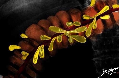
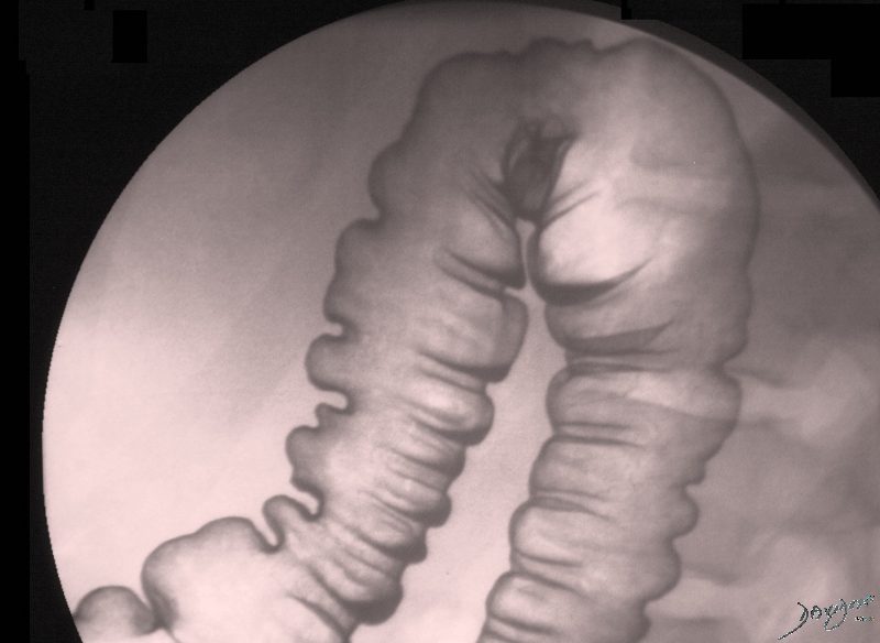
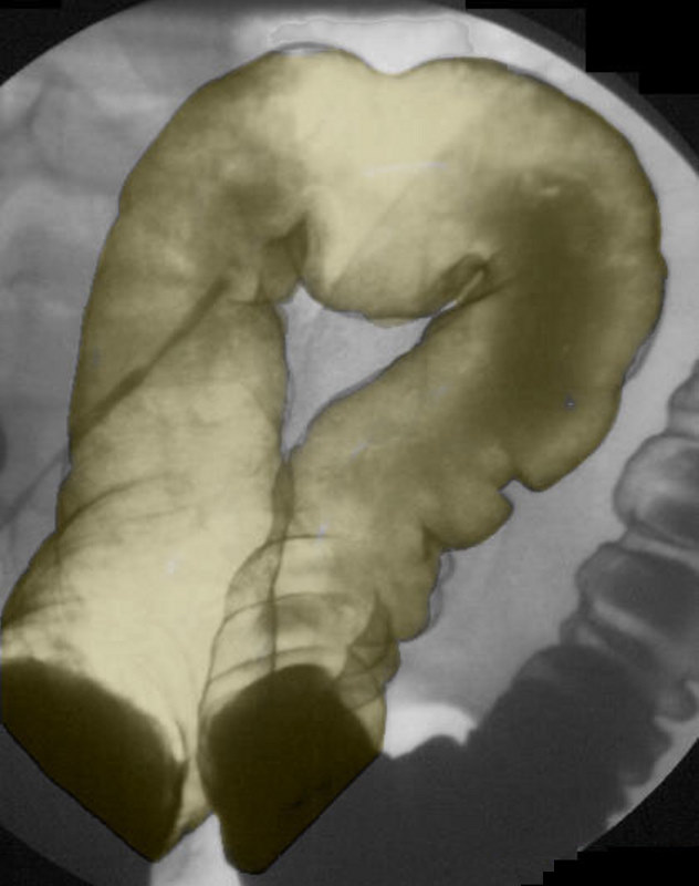
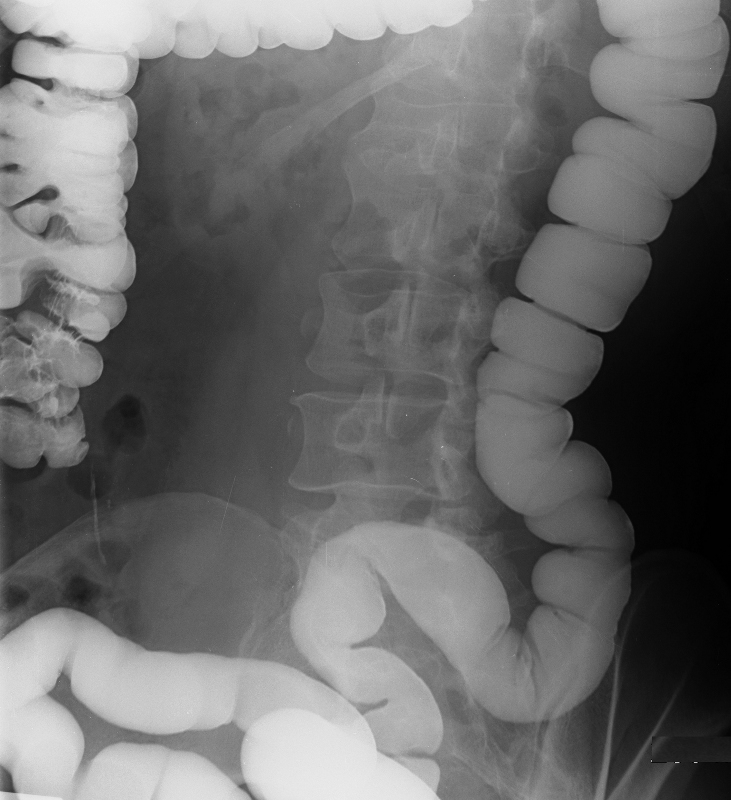
Descending Colon
DOMElement Object
(
    [schemaTypeInfo] =>
    [tagName] => table
    [firstElementChild] => (object value omitted)
    [lastElementChild] => (object value omitted)
    [childElementCount] => 1
    [previousElementSibling] => (object value omitted)
    [nextElementSibling] => (object value omitted)
    [nodeName] => table
    [nodeValue] =>


Ligaments of the transverse colon


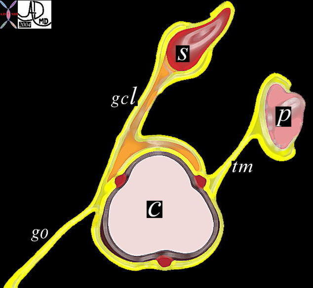
The transverse colon is suspended by two ligaments and acts as a support for a third.  The gastrocolic ligament (gcl) extends from the stomach (S) to the antero-superior aspect of the transverse colon, while the transverse mesocolon ™ extends from the pancreas to the postero-superior aspect.  The greater omentum extends as an apron of fat from the anterior aspect of the transverse colon for a variable distance sometimes reaching all the way to the pelvis.
Courtesy Ashley Davidoff MD
01443b05


    [nodeType] => 1
    [parentNode] => (object value omitted)
    [childNodes] => (object value omitted)
    [firstChild] => (object value omitted)
    [lastChild] => (object value omitted)
    [previousSibling] => (object value omitted)
    [nextSibling] => (object value omitted)
    [attributes] => (object value omitted)
    [ownerDocument] => (object value omitted)
    [namespaceURI] =>
    [prefix] =>
    [localName] => table
    [baseURI] =>
    [textContent] =>


Ligaments of the transverse colon


The transverse colon is suspended by two ligaments and acts as a support for a third.  The gastrocolic ligament (gcl) extends from the stomach (S) to the antero-superior aspect of the transverse colon, while the transverse mesocolon ™ extends from the pancreas to the postero-superior aspect.  The greater omentum extends as an apron of fat from the anterior aspect of the transverse colon for a variable distance sometimes reaching all the way to the pelvis.
Courtesy Ashley Davidoff MD
01443b05


)
DOMElement Object
(
    [schemaTypeInfo] =>
    [tagName] => td
    [firstElementChild] => (object value omitted)
    [lastElementChild] => (object value omitted)
    [childElementCount] => 2
    [previousElementSibling] =>
    [nextElementSibling] =>
    [nodeName] => td
    [nodeValue] => The transverse colon is suspended by two ligaments and acts as a support for a third.  The gastrocolic ligament (gcl) extends from the stomach (S) to the antero-superior aspect of the transverse colon, while the transverse mesocolon ™ extends from the pancreas to the postero-superior aspect.  The greater omentum extends as an apron of fat from the anterior aspect of the transverse colon for a variable distance sometimes reaching all the way to the pelvis.
Courtesy Ashley Davidoff MD
01443b05
    [nodeType] => 1
    [parentNode] => (object value omitted)
    [childNodes] => (object value omitted)
    [firstChild] => (object value omitted)
    [lastChild] => (object value omitted)
    [previousSibling] => (object value omitted)
    [nextSibling] => (object value omitted)
    [attributes] => (object value omitted)
    [ownerDocument] => (object value omitted)
    [namespaceURI] =>
    [prefix] =>
    [localName] => td
    [baseURI] =>
    [textContent] => The transverse colon is suspended by two ligaments and acts as a support for a third.  The gastrocolic ligament (gcl) extends from the stomach (S) to the antero-superior aspect of the transverse colon, while the transverse mesocolon ™ extends from the pancreas to the postero-superior aspect.  The greater omentum extends as an apron of fat from the anterior aspect of the transverse colon for a variable distance sometimes reaching all the way to the pelvis.
Courtesy Ashley Davidoff MD
01443b05
)
DOMElement Object
(
    [schemaTypeInfo] =>
    [tagName] => td
    [firstElementChild] => (object value omitted)
    [lastElementChild] => (object value omitted)
    [childElementCount] => 1
    [previousElementSibling] =>
    [nextElementSibling] =>
    [nodeName] => td
    [nodeValue] => Ligaments of the transverse colon
    [nodeType] => 1
    [parentNode] => (object value omitted)
    [childNodes] => (object value omitted)
    [firstChild] => (object value omitted)
    [lastChild] => (object value omitted)
    [previousSibling] => (object value omitted)
    [nextSibling] => (object value omitted)
    [attributes] => (object value omitted)
    [ownerDocument] => (object value omitted)
    [namespaceURI] =>
    [prefix] =>
    [localName] => td
    [baseURI] =>
    [textContent] => Ligaments of the transverse colon
)
https://beta.thecommonvein.net/wp-content/uploads/2023/04/01443b05.jpg
DOMElement Object
(
    [schemaTypeInfo] =>
    [tagName] => table
    [firstElementChild] => (object value omitted)
    [lastElementChild] => (object value omitted)
    [childElementCount] => 1
    [previousElementSibling] => (object value omitted)
    [nextElementSibling] => (object value omitted)
    [nodeName] => table
    [nodeValue] =>


Parts of the Colon


This drawing shows the teminal ileum entering the colon between the cecum (green) and the ascending colon (light blue).  The appendix in purple lies inferior to the ileocecal valve.  The hepatic flexure (royal blue)connects the acending colon to the transverse colon (pink) while the splenic flexure (yellow) as the highest part of the colon, links the transverse colon to the descending colon (orange).  As the descending colon emerges from the retroperitoneum it becomes the splenic flexure(mustard), but distally it dives back behind the peritoneal cavity to form the rectum. (brown).  The anus (cream) is the terminal portion of the colon and the link to the outside world.
Courtesy Ashley Davidoff MD  12005b07


    [nodeType] => 1
    [parentNode] => (object value omitted)
    [childNodes] => (object value omitted)
    [firstChild] => (object value omitted)
    [lastChild] => (object value omitted)
    [previousSibling] => (object value omitted)
    [nextSibling] => (object value omitted)
    [attributes] => (object value omitted)
    [ownerDocument] => (object value omitted)
    [namespaceURI] =>
    [prefix] =>
    [localName] => table
    [baseURI] =>
    [textContent] =>


Parts of the Colon


This drawing shows the teminal ileum entering the colon between the cecum (green) and the ascending colon (light blue).  The appendix in purple lies inferior to the ileocecal valve.  The hepatic flexure (royal blue)connects the acending colon to the transverse colon (pink) while the splenic flexure (yellow) as the highest part of the colon, links the transverse colon to the descending colon (orange).  As the descending colon emerges from the retroperitoneum it becomes the splenic flexure(mustard), but distally it dives back behind the peritoneal cavity to form the rectum. (brown).  The anus (cream) is the terminal portion of the colon and the link to the outside world.
Courtesy Ashley Davidoff MD  12005b07


)
DOMElement Object
(
    [schemaTypeInfo] =>
    [tagName] => td
    [firstElementChild] => (object value omitted)
    [lastElementChild] => (object value omitted)
    [childElementCount] => 1
    [previousElementSibling] =>
    [nextElementSibling] =>
    [nodeName] => td
    [nodeValue] => This drawing shows the teminal ileum entering the colon between the cecum (green) and the ascending colon (light blue).  The appendix in purple lies inferior to the ileocecal valve.  The hepatic flexure (royal blue)connects the acending colon to the transverse colon (pink) while the splenic flexure (yellow) as the highest part of the colon, links the transverse colon to the descending colon (orange).  As the descending colon emerges from the retroperitoneum it becomes the splenic flexure(mustard), but distally it dives back behind the peritoneal cavity to form the rectum. (brown).  The anus (cream) is the terminal portion of the colon and the link to the outside world.
Courtesy Ashley Davidoff MD  12005b07
    [nodeType] => 1
    [parentNode] => (object value omitted)
    [childNodes] => (object value omitted)
    [firstChild] => (object value omitted)
    [lastChild] => (object value omitted)
    [previousSibling] => (object value omitted)
    [nextSibling] => (object value omitted)
    [attributes] => (object value omitted)
    [ownerDocument] => (object value omitted)
    [namespaceURI] =>
    [prefix] =>
    [localName] => td
    [baseURI] =>
    [textContent] => This drawing shows the teminal ileum entering the colon between the cecum (green) and the ascending colon (light blue).  The appendix in purple lies inferior to the ileocecal valve.  The hepatic flexure (royal blue)connects the acending colon to the transverse colon (pink) while the splenic flexure (yellow) as the highest part of the colon, links the transverse colon to the descending colon (orange).  As the descending colon emerges from the retroperitoneum it becomes the splenic flexure(mustard), but distally it dives back behind the peritoneal cavity to form the rectum. (brown).  The anus (cream) is the terminal portion of the colon and the link to the outside world.
Courtesy Ashley Davidoff MD  12005b07
)
DOMElement Object
(
    [schemaTypeInfo] =>
    [tagName] => td
    [firstElementChild] => (object value omitted)
    [lastElementChild] => (object value omitted)
    [childElementCount] => 1
    [previousElementSibling] =>
    [nextElementSibling] =>
    [nodeName] => td
    [nodeValue] => Parts of the Colon
    [nodeType] => 1
    [parentNode] => (object value omitted)
    [childNodes] => (object value omitted)
    [firstChild] => (object value omitted)
    [lastChild] => (object value omitted)
    [previousSibling] => (object value omitted)
    [nextSibling] => (object value omitted)
    [attributes] => (object value omitted)
    [ownerDocument] => (object value omitted)
    [namespaceURI] =>
    [prefix] =>
    [localName] => td
    [baseURI] =>
    [textContent] => Parts of the Colon
)
https://beta.thecommonvein.net/wp-content/uploads/2023/05/12005.jpg
DOMElement Object
(
    [schemaTypeInfo] =>
    [tagName] => table
    [firstElementChild] => (object value omitted)
    [lastElementChild] => (object value omitted)
    [childElementCount] => 1
    [previousElementSibling] => (object value omitted)
    [nextElementSibling] =>
    [nodeName] => table
    [nodeValue] =>


Parts of the Colon – Introduction
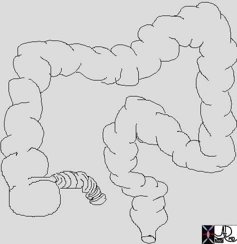
The colon is made up proximally of the cecum which is the widest potion which links the large bowel to the small bowel at the ileocecal junction.  It contains the appendix which is a tubular structure about 3-5 mms in diameter and between 3 and 10cms long.  Thereafter comes the ascending colon which is situated in the retroperitoneal position, followed by the transverse colon.  The transverse colon hangs like a washing line within the peritoneal cavity between the two flexures – hepatic flexure on the right and the splenic flexure on the left.  The descending colon is also in a retroperitoneal position while the sigmoid subtended on the sigmoid mesocolon becomes intraperitoneal.  The rectum is bound down by the peritoneum as it exits at the anus.


Parts of the Colon


This drawing shows the teminal ileum entering the colon between the cecum (green) and the ascending colon (light blue).  The appendix in purple lies inferior to the ileocecal valve.  The hepatic flexure (royal blue)connects the acending colon to the transverse colon (pink) while the splenic flexure (yellow) as the highest part of the colon, links the transverse colon to the descending colon (orange).  As the descending colon emerges from the retroperitoneum it becomes the splenic flexure(mustard), but distally it dives back behind the peritoneal cavity to form the rectum. (brown).  The anus (cream) is the terminal portion of the colon and the link to the outside world.
Courtesy Ashley Davidoff MD  12005b07


Each component of the colon has an inner endothelium, a submucosa, a middle muscular layer called the muscularis and an outer layer called a serosa or adventitia.  This structural makeup was discussed in detail in the histology section.
Minor but characteristic components include the  taenia coli, appendices epiploica.

Support structures of the Colon

The mesocolon supports the intraperitoneal components while the retroperitoneal components are bound down by the peritoneum and are attached by loose areolar tissue to the retroperitoneal tissues.
Ligaments include the transverse mesocolon, greater omentum, sigmoid mesocolon, mesoappendix, phrenicocolic ligament.  The transverse mesocolon supports the transverse colon, and extends from the pancreas to posterosuperior asspect of the transverse colon.  The gastrocolic ligament extends from the stomach to the superior aspect of the transverse colon.  The sigmoid mesocolon supports the sigmoid colon and the mesoappendix supports the appendix.


Ligaments of the transverse colon


The transverse colon is suspended by two ligaments and acts as a support for a third.  The gastrocolic ligament (gcl) extends from the stomach (S) to the antero-superior aspect of the transverse colon, while the transverse mesocolon ™ extends from the pancreas to the postero-superior aspect.  The greater omentum extends as an apron of fat from the anterior aspect of the transverse colon for a variable distance sometimes reaching all the way to the pelvis.
Courtesy Ashley Davidoff MD
01443b05


The phrenicocolic ligament supports the splenic flexure by attaching to the diaphragm. It is a horizontal fold of peritoneum.  The ascending and descending colon have no mesentery but are attached to the retroperitoneum by loose areolar tissue to the muscles such as the diaphragm, psoas, quadratus lumborum and the iliopsoas.


    [nodeType] => 1
    [parentNode] => (object value omitted)
    [childNodes] => (object value omitted)
    [firstChild] => (object value omitted)
    [lastChild] => (object value omitted)
    [previousSibling] => (object value omitted)
    [nextSibling] => (object value omitted)
    [attributes] => (object value omitted)
    [ownerDocument] => (object value omitted)
    [namespaceURI] =>
    [prefix] =>
    [localName] => table
    [baseURI] =>
    [textContent] =>


Parts of the Colon – Introduction
The colon is made up proximally of the cecum which is the widest potion which links the large bowel to the small bowel at the ileocecal junction.  It contains the appendix which is a tubular structure about 3-5 mms in diameter and between 3 and 10cms long.  Thereafter comes the ascending colon which is situated in the retroperitoneal position, followed by the transverse colon.  The transverse colon hangs like a washing line within the peritoneal cavity between the two flexures – hepatic flexure on the right and the splenic flexure on the left.  The descending colon is also in a retroperitoneal position while the sigmoid subtended on the sigmoid mesocolon becomes intraperitoneal.  The rectum is bound down by the peritoneum as it exits at the anus.


Parts of the Colon


This drawing shows the teminal ileum entering the colon between the cecum (green) and the ascending colon (light blue).  The appendix in purple lies inferior to the ileocecal valve.  The hepatic flexure (royal blue)connects the acending colon to the transverse colon (pink) while the splenic flexure (yellow) as the highest part of the colon, links the transverse colon to the descending colon (orange).  As the descending colon emerges from the retroperitoneum it becomes the splenic flexure(mustard), but distally it dives back behind the peritoneal cavity to form the rectum. (brown).  The anus (cream) is the terminal portion of the colon and the link to the outside world.
Courtesy Ashley Davidoff MD  12005b07


Each component of the colon has an inner endothelium, a submucosa, a middle muscular layer called the muscularis and an outer layer called a serosa or adventitia.  This structural makeup was discussed in detail in the histology section.
Minor but characteristic components include the  taenia coli, appendices epiploica.

Support structures of the Colon

The mesocolon supports the intraperitoneal components while the retroperitoneal components are bound down by the peritoneum and are attached by loose areolar tissue to the retroperitoneal tissues.
Ligaments include the transverse mesocolon, greater omentum, sigmoid mesocolon, mesoappendix, phrenicocolic ligament.  The transverse mesocolon supports the transverse colon, and extends from the pancreas to posterosuperior asspect of the transverse colon.  The gastrocolic ligament extends from the stomach to the superior aspect of the transverse colon.  The sigmoid mesocolon supports the sigmoid colon and the mesoappendix supports the appendix.


Ligaments of the transverse colon


The transverse colon is suspended by two ligaments and acts as a support for a third.  The gastrocolic ligament (gcl) extends from the stomach (S) to the antero-superior aspect of the transverse colon, while the transverse mesocolon ™ extends from the pancreas to the postero-superior aspect.  The greater omentum extends as an apron of fat from the anterior aspect of the transverse colon for a variable distance sometimes reaching all the way to the pelvis.
Courtesy Ashley Davidoff MD
01443b05


The phrenicocolic ligament supports the splenic flexure by attaching to the diaphragm. It is a horizontal fold of peritoneum.  The ascending and descending colon have no mesentery but are attached to the retroperitoneum by loose areolar tissue to the muscles such as the diaphragm, psoas, quadratus lumborum and the iliopsoas.


)
DOMElement Object
(
    [schemaTypeInfo] =>
    [tagName] => td
    [firstElementChild] => (object value omitted)
    [lastElementChild] => (object value omitted)
    [childElementCount] => 2
    [previousElementSibling] =>
    [nextElementSibling] =>
    [nodeName] => td
    [nodeValue] => The transverse colon is suspended by two ligaments and acts as a support for a third.  The gastrocolic ligament (gcl) extends from the stomach (S) to the antero-superior aspect of the transverse colon, while the transverse mesocolon ™ extends from the pancreas to the postero-superior aspect.  The greater omentum extends as an apron of fat from the anterior aspect of the transverse colon for a variable distance sometimes reaching all the way to the pelvis.
Courtesy Ashley Davidoff MD
01443b05
    [nodeType] => 1
    [parentNode] => (object value omitted)
    [childNodes] => (object value omitted)
    [firstChild] => (object value omitted)
    [lastChild] => (object value omitted)
    [previousSibling] => (object value omitted)
    [nextSibling] => (object value omitted)
    [attributes] => (object value omitted)
    [ownerDocument] => (object value omitted)
    [namespaceURI] =>
    [prefix] =>
    [localName] => td
    [baseURI] =>
    [textContent] => The transverse colon is suspended by two ligaments and acts as a support for a third.  The gastrocolic ligament (gcl) extends from the stomach (S) to the antero-superior aspect of the transverse colon, while the transverse mesocolon ™ extends from the pancreas to the postero-superior aspect.  The greater omentum extends as an apron of fat from the anterior aspect of the transverse colon for a variable distance sometimes reaching all the way to the pelvis.
Courtesy Ashley Davidoff MD
01443b05
)
DOMElement Object
(
    [schemaTypeInfo] =>
    [tagName] => td
    [firstElementChild] => (object value omitted)
    [lastElementChild] => (object value omitted)
    [childElementCount] => 2
    [previousElementSibling] =>
    [nextElementSibling] =>
    [nodeName] => td
    [nodeValue] => Ligaments of the transverse colon
    [nodeType] => 1
    [parentNode] => (object value omitted)
    [childNodes] => (object value omitted)
    [firstChild] => (object value omitted)
    [lastChild] => (object value omitted)
    [previousSibling] => (object value omitted)
    [nextSibling] => (object value omitted)
    [attributes] => (object value omitted)
    [ownerDocument] => (object value omitted)
    [namespaceURI] =>
    [prefix] =>
    [localName] => td
    [baseURI] =>
    [textContent] => Ligaments of the transverse colon
)
DOMElement Object
(
    [schemaTypeInfo] =>
    [tagName] => td
    [firstElementChild] => (object value omitted)
    [lastElementChild] => (object value omitted)
    [childElementCount] => 1
    [previousElementSibling] =>
    [nextElementSibling] =>
    [nodeName] => td
    [nodeValue] => This drawing shows the teminal ileum entering the colon between the cecum (green) and the ascending colon (light blue).  The appendix in purple lies inferior to the ileocecal valve.  The hepatic flexure (royal blue)connects the acending colon to the transverse colon (pink) while the splenic flexure (yellow) as the highest part of the colon, links the transverse colon to the descending colon (orange).  As the descending colon emerges from the retroperitoneum it becomes the splenic flexure(mustard), but distally it dives back behind the peritoneal cavity to form the rectum. (brown).  The anus (cream) is the terminal portion of the colon and the link to the outside world.
Courtesy Ashley Davidoff MD  12005b07
    [nodeType] => 1
    [parentNode] => (object value omitted)
    [childNodes] => (object value omitted)
    [firstChild] => (object value omitted)
    [lastChild] => (object value omitted)
    [previousSibling] => (object value omitted)
    [nextSibling] => (object value omitted)
    [attributes] => (object value omitted)
    [ownerDocument] => (object value omitted)
    [namespaceURI] =>
    [prefix] =>
    [localName] => td
    [baseURI] =>
    [textContent] => This drawing shows the teminal ileum entering the colon between the cecum (green) and the ascending colon (light blue).  The appendix in purple lies inferior to the ileocecal valve.  The hepatic flexure (royal blue)connects the acending colon to the transverse colon (pink) while the splenic flexure (yellow) as the highest part of the colon, links the transverse colon to the descending colon (orange).  As the descending colon emerges from the retroperitoneum it becomes the splenic flexure(mustard), but distally it dives back behind the peritoneal cavity to form the rectum. (brown).  The anus (cream) is the terminal portion of the colon and the link to the outside world.
Courtesy Ashley Davidoff MD  12005b07
)
https://beta.thecommonvein.net/wp-content/uploads/2023/04/01443b05.jpg
DOMElement Object
(
    [schemaTypeInfo] =>
    [tagName] => td
    [firstElementChild] => (object value omitted)
    [lastElementChild] => (object value omitted)
    [childElementCount] => 2
    [previousElementSibling] =>
    [nextElementSibling] =>
    [nodeName] => td
    [nodeValue] => Parts of the Colon
    [nodeType] => 1
    [parentNode] => (object value omitted)
    [childNodes] => (object value omitted)
    [firstChild] => (object value omitted)
    [lastChild] => (object value omitted)
    [previousSibling] => (object value omitted)
    [nextSibling] => (object value omitted)
    [attributes] => (object value omitted)
    [ownerDocument] => (object value omitted)
    [namespaceURI] =>
    [prefix] =>
    [localName] => td
    [baseURI] =>
    [textContent] => Parts of the Colon
)
https://beta.thecommonvein.net/wp-content/uploads/2023/04/01443b05.jpg
https://beta.thecommonvein.net/wp-content/uploads/2023/05/12005.jpg
DOMElement Object
(
    [schemaTypeInfo] =>
    [tagName] => td
    [firstElementChild] => (object value omitted)
    [lastElementChild] => (object value omitted)
    [childElementCount] => 19
    [previousElementSibling] =>
    [nextElementSibling] =>
    [nodeName] => td
    [nodeValue] => Parts of the Colon – Introduction
The colon is made up proximally of the cecum which is the widest potion which links the large bowel to the small bowel at the ileocecal junction.  It contains the appendix which is a tubular structure about 3-5 mms in diameter and between 3 and 10cms long.  Thereafter comes the ascending colon which is situated in the retroperitoneal position, followed by the transverse colon.  The transverse colon hangs like a washing line within the peritoneal cavity between the two flexures – hepatic flexure on the right and the splenic flexure on the left.  The descending colon is also in a retroperitoneal position while the sigmoid subtended on the sigmoid mesocolon becomes intraperitoneal.  The rectum is bound down by the peritoneum as it exits at the anus.


Parts of the Colon


This drawing shows the teminal ileum entering the colon between the cecum (green) and the ascending colon (light blue).  The appendix in purple lies inferior to the ileocecal valve.  The hepatic flexure (royal blue)connects the acending colon to the transverse colon (pink) while the splenic flexure (yellow) as the highest part of the colon, links the transverse colon to the descending colon (orange).  As the descending colon emerges from the retroperitoneum it becomes the splenic flexure(mustard), but distally it dives back behind the peritoneal cavity to form the rectum. (brown).  The anus (cream) is the terminal portion of the colon and the link to the outside world.
Courtesy Ashley Davidoff MD  12005b07


Each component of the colon has an inner endothelium, a submucosa, a middle muscular layer called the muscularis and an outer layer called a serosa or adventitia.  This structural makeup was discussed in detail in the histology section.
Minor but characteristic components include the  taenia coli, appendices epiploica.

Support structures of the Colon

The mesocolon supports the intraperitoneal components while the retroperitoneal components are bound down by the peritoneum and are attached by loose areolar tissue to the retroperitoneal tissues.
Ligaments include the transverse mesocolon, greater omentum, sigmoid mesocolon, mesoappendix, phrenicocolic ligament.  The transverse mesocolon supports the transverse colon, and extends from the pancreas to posterosuperior asspect of the transverse colon.  The gastrocolic ligament extends from the stomach to the superior aspect of the transverse colon.  The sigmoid mesocolon supports the sigmoid colon and the mesoappendix supports the appendix.


Ligaments of the transverse colon


The transverse colon is suspended by two ligaments and acts as a support for a third.  The gastrocolic ligament (gcl) extends from the stomach (S) to the antero-superior aspect of the transverse colon, while the transverse mesocolon ™ extends from the pancreas to the postero-superior aspect.  The greater omentum extends as an apron of fat from the anterior aspect of the transverse colon for a variable distance sometimes reaching all the way to the pelvis.
Courtesy Ashley Davidoff MD
01443b05


The phrenicocolic ligament supports the splenic flexure by attaching to the diaphragm. It is a horizontal fold of peritoneum.  The ascending and descending colon have no mesentery but are attached to the retroperitoneum by loose areolar tissue to the muscles such as the diaphragm, psoas, quadratus lumborum and the iliopsoas.
    [nodeType] => 1
    [parentNode] => (object value omitted)
    [childNodes] => (object value omitted)
    [firstChild] => (object value omitted)
    [lastChild] => (object value omitted)
    [previousSibling] => (object value omitted)
    [nextSibling] => (object value omitted)
    [attributes] => (object value omitted)
    [ownerDocument] => (object value omitted)
    [namespaceURI] =>
    [prefix] =>
    [localName] => td
    [baseURI] =>
    [textContent] => Parts of the Colon – Introduction
The colon is made up proximally of the cecum which is the widest potion which links the large bowel to the small bowel at the ileocecal junction.  It contains the appendix which is a tubular structure about 3-5 mms in diameter and between 3 and 10cms long.  Thereafter comes the ascending colon which is situated in the retroperitoneal position, followed by the transverse colon.  The transverse colon hangs like a washing line within the peritoneal cavity between the two flexures – hepatic flexure on the right and the splenic flexure on the left.  The descending colon is also in a retroperitoneal position while the sigmoid subtended on the sigmoid mesocolon becomes intraperitoneal.  The rectum is bound down by the peritoneum as it exits at the anus.


Parts of the Colon


This drawing shows the teminal ileum entering the colon between the cecum (green) and the ascending colon (light blue).  The appendix in purple lies inferior to the ileocecal valve.  The hepatic flexure (royal blue)connects the acending colon to the transverse colon (pink) while the splenic flexure (yellow) as the highest part of the colon, links the transverse colon to the descending colon (orange).  As the descending colon emerges from the retroperitoneum it becomes the splenic flexure(mustard), but distally it dives back behind the peritoneal cavity to form the rectum. (brown).  The anus (cream) is the terminal portion of the colon and the link to the outside world.
Courtesy Ashley Davidoff MD  12005b07


Each component of the colon has an inner endothelium, a submucosa, a middle muscular layer called the muscularis and an outer layer called a serosa or adventitia.  This structural makeup was discussed in detail in the histology section.
Minor but characteristic components include the  taenia coli, appendices epiploica.

Support structures of the Colon

The mesocolon supports the intraperitoneal components while the retroperitoneal components are bound down by the peritoneum and are attached by loose areolar tissue to the retroperitoneal tissues.
Ligaments include the transverse mesocolon, greater omentum, sigmoid mesocolon, mesoappendix, phrenicocolic ligament.  The transverse mesocolon supports the transverse colon, and extends from the pancreas to posterosuperior asspect of the transverse colon.  The gastrocolic ligament extends from the stomach to the superior aspect of the transverse colon.  The sigmoid mesocolon supports the sigmoid colon and the mesoappendix supports the appendix.


Ligaments of the transverse colon


The transverse colon is suspended by two ligaments and acts as a support for a third.  The gastrocolic ligament (gcl) extends from the stomach (S) to the antero-superior aspect of the transverse colon, while the transverse mesocolon ™ extends from the pancreas to the postero-superior aspect.  The greater omentum extends as an apron of fat from the anterior aspect of the transverse colon for a variable distance sometimes reaching all the way to the pelvis.
Courtesy Ashley Davidoff MD
01443b05


The phrenicocolic ligament supports the splenic flexure by attaching to the diaphragm. It is a horizontal fold of peritoneum.  The ascending and descending colon have no mesentery but are attached to the retroperitoneum by loose areolar tissue to the muscles such as the diaphragm, psoas, quadratus lumborum and the iliopsoas.
)

