DOMElement Object
(
    [schemaTypeInfo] =>
    [tagName] => table
    [firstElementChild] => (object value omitted)
    [lastElementChild] => (object value omitted)
    [childElementCount] => 1
    [previousElementSibling] => (object value omitted)
    [nextElementSibling] => (object value omitted)
    [nodeName] => table
    [nodeValue] =>


Chilaiditi syndrome – interposition the colon


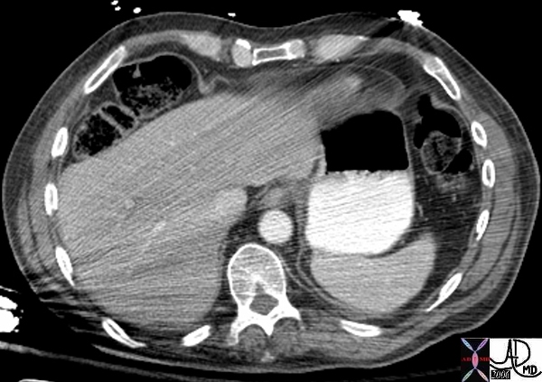
This axial image of the abdomen shows the colon and specifically the hepatic flexure malpositioned anterior to the liver.  The liver is displaced from its usual position by the colon which makes its way anteriorly (in front of the liver), and superiorly (below the diaphragm), so that if there is air within this loop it will masquearades as “free air”  (Image courtesy of Ashley Davidoff M.D.) 45768


    [nodeType] => 1
    [parentNode] => (object value omitted)
    [childNodes] => (object value omitted)
    [firstChild] => (object value omitted)
    [lastChild] => (object value omitted)
    [previousSibling] => (object value omitted)
    [nextSibling] => (object value omitted)
    [attributes] => (object value omitted)
    [ownerDocument] => (object value omitted)
    [namespaceURI] =>
    [prefix] =>
    [localName] => table
    [baseURI] =>
    [textContent] =>


Chilaiditi syndrome – interposition the colon


This axial image of the abdomen shows the colon and specifically the hepatic flexure malpositioned anterior to the liver.  The liver is displaced from its usual position by the colon which makes its way anteriorly (in front of the liver), and superiorly (below the diaphragm), so that if there is air within this loop it will masquearades as “free air”  (Image courtesy of Ashley Davidoff M.D.) 45768


)
DOMElement Object
(
    [schemaTypeInfo] =>
    [tagName] => td
    [firstElementChild] =>
    [lastElementChild] =>
    [childElementCount] => 0
    [previousElementSibling] =>
    [nextElementSibling] =>
    [nodeName] => td
    [nodeValue] => This axial image of the abdomen shows the colon and specifically the hepatic flexure malpositioned anterior to the liver.  The liver is displaced from its usual position by the colon which makes its way anteriorly (in front of the liver), and superiorly (below the diaphragm), so that if there is air within this loop it will masquearades as “free air”  (Image courtesy of Ashley Davidoff M.D.) 45768
    [nodeType] => 1
    [parentNode] => (object value omitted)
    [childNodes] => (object value omitted)
    [firstChild] => (object value omitted)
    [lastChild] => (object value omitted)
    [previousSibling] => (object value omitted)
    [nextSibling] => (object value omitted)
    [attributes] => (object value omitted)
    [ownerDocument] => (object value omitted)
    [namespaceURI] =>
    [prefix] =>
    [localName] => td
    [baseURI] =>
    [textContent] => This axial image of the abdomen shows the colon and specifically the hepatic flexure malpositioned anterior to the liver.  The liver is displaced from its usual position by the colon which makes its way anteriorly (in front of the liver), and superiorly (below the diaphragm), so that if there is air within this loop it will masquearades as “free air”  (Image courtesy of Ashley Davidoff M.D.) 45768
)
DOMElement Object
(
    [schemaTypeInfo] =>
    [tagName] => td
    [firstElementChild] => (object value omitted)
    [lastElementChild] => (object value omitted)
    [childElementCount] => 1
    [previousElementSibling] =>
    [nextElementSibling] =>
    [nodeName] => td
    [nodeValue] => Chilaiditi syndrome – interposition the colon
    [nodeType] => 1
    [parentNode] => (object value omitted)
    [childNodes] => (object value omitted)
    [firstChild] => (object value omitted)
    [lastChild] => (object value omitted)
    [previousSibling] => (object value omitted)
    [nextSibling] => (object value omitted)
    [attributes] => (object value omitted)
    [ownerDocument] => (object value omitted)
    [namespaceURI] =>
    [prefix] =>
    [localName] => td
    [baseURI] =>
    [textContent] => Chilaiditi syndrome – interposition the colon
)
https://beta.thecommonvein.net/wp-content/uploads/2023/06/45768.jpg
DOMElement Object
(
    [schemaTypeInfo] =>
    [tagName] => table
    [firstElementChild] => (object value omitted)
    [lastElementChild] => (object value omitted)
    [childElementCount] => 1
    [previousElementSibling] => (object value omitted)
    [nextElementSibling] => (object value omitted)
    [nodeName] => table
    [nodeValue] =>


Chilaiditi syndrome – interposition the colon


This coronal image of the abdomen shows the colon and specifically the hepatic flexure malpositioned under the diaphragm and mimics the presence of free air under the diaphragm as noted in the above images.  The liver is displaced from its usual position by the colon which makes its way anteriorly (in front of the liver), and superiorly (below the diaphragm), so that if there is air within this loop it will masquerades as “free air”  (Image courtesy of Ashley Davidoff M.D.) 45765


    [nodeType] => 1
    [parentNode] => (object value omitted)
    [childNodes] => (object value omitted)
    [firstChild] => (object value omitted)
    [lastChild] => (object value omitted)
    [previousSibling] => (object value omitted)
    [nextSibling] => (object value omitted)
    [attributes] => (object value omitted)
    [ownerDocument] => (object value omitted)
    [namespaceURI] =>
    [prefix] =>
    [localName] => table
    [baseURI] =>
    [textContent] =>


Chilaiditi syndrome – interposition the colon


This coronal image of the abdomen shows the colon and specifically the hepatic flexure malpositioned under the diaphragm and mimics the presence of free air under the diaphragm as noted in the above images.  The liver is displaced from its usual position by the colon which makes its way anteriorly (in front of the liver), and superiorly (below the diaphragm), so that if there is air within this loop it will masquerades as “free air”  (Image courtesy of Ashley Davidoff M.D.) 45765


)
DOMElement Object
(
    [schemaTypeInfo] =>
    [tagName] => td
    [firstElementChild] =>
    [lastElementChild] =>
    [childElementCount] => 0
    [previousElementSibling] =>
    [nextElementSibling] =>
    [nodeName] => td
    [nodeValue] => This coronal image of the abdomen shows the colon and specifically the hepatic flexure malpositioned under the diaphragm and mimics the presence of free air under the diaphragm as noted in the above images.  The liver is displaced from its usual position by the colon which makes its way anteriorly (in front of the liver), and superiorly (below the diaphragm), so that if there is air within this loop it will masquerades as “free air”  (Image courtesy of Ashley Davidoff M.D.) 45765
    [nodeType] => 1
    [parentNode] => (object value omitted)
    [childNodes] => (object value omitted)
    [firstChild] => (object value omitted)
    [lastChild] => (object value omitted)
    [previousSibling] => (object value omitted)
    [nextSibling] => (object value omitted)
    [attributes] => (object value omitted)
    [ownerDocument] => (object value omitted)
    [namespaceURI] =>
    [prefix] =>
    [localName] => td
    [baseURI] =>
    [textContent] => This coronal image of the abdomen shows the colon and specifically the hepatic flexure malpositioned under the diaphragm and mimics the presence of free air under the diaphragm as noted in the above images.  The liver is displaced from its usual position by the colon which makes its way anteriorly (in front of the liver), and superiorly (below the diaphragm), so that if there is air within this loop it will masquerades as “free air”  (Image courtesy of Ashley Davidoff M.D.) 45765
)
DOMElement Object
(
    [schemaTypeInfo] =>
    [tagName] => td
    [firstElementChild] => (object value omitted)
    [lastElementChild] => (object value omitted)
    [childElementCount] => 1
    [previousElementSibling] =>
    [nextElementSibling] =>
    [nodeName] => td
    [nodeValue] => Chilaiditi syndrome – interposition the colon
    [nodeType] => 1
    [parentNode] => (object value omitted)
    [childNodes] => (object value omitted)
    [firstChild] => (object value omitted)
    [lastChild] => (object value omitted)
    [previousSibling] => (object value omitted)
    [nextSibling] => (object value omitted)
    [attributes] => (object value omitted)
    [ownerDocument] => (object value omitted)
    [namespaceURI] =>
    [prefix] =>
    [localName] => td
    [baseURI] =>
    [textContent] => Chilaiditi syndrome – interposition the colon
)
https://beta.thecommonvein.net/wp-content/uploads/2023/06/45765.jpg
DOMElement Object
(
    [schemaTypeInfo] =>
    [tagName] => table
    [firstElementChild] => (object value omitted)
    [lastElementChild] => (object value omitted)
    [childElementCount] => 1
    [previousElementSibling] => (object value omitted)
    [nextElementSibling] => (object value omitted)
    [nodeName] => table
    [nodeValue] =>


Question free air?


This upright iamge of the abdomen clearly shows that the air under the diapragm is part of air within the lumen of a loop of bowel in the right upper quadrant.  (Image courtesy of Ashley Davidoff M.D.) 45761


    [nodeType] => 1
    [parentNode] => (object value omitted)
    [childNodes] => (object value omitted)
    [firstChild] => (object value omitted)
    [lastChild] => (object value omitted)
    [previousSibling] => (object value omitted)
    [nextSibling] => (object value omitted)
    [attributes] => (object value omitted)
    [ownerDocument] => (object value omitted)
    [namespaceURI] =>
    [prefix] =>
    [localName] => table
    [baseURI] =>
    [textContent] =>


Question free air?


This upright iamge of the abdomen clearly shows that the air under the diapragm is part of air within the lumen of a loop of bowel in the right upper quadrant.  (Image courtesy of Ashley Davidoff M.D.) 45761


)
DOMElement Object
(
    [schemaTypeInfo] =>
    [tagName] => td
    [firstElementChild] =>
    [lastElementChild] =>
    [childElementCount] => 0
    [previousElementSibling] =>
    [nextElementSibling] =>
    [nodeName] => td
    [nodeValue] => This upright iamge of the abdomen clearly shows that the air under the diapragm is part of air within the lumen of a loop of bowel in the right upper quadrant.  (Image courtesy of Ashley Davidoff M.D.) 45761
    [nodeType] => 1
    [parentNode] => (object value omitted)
    [childNodes] => (object value omitted)
    [firstChild] => (object value omitted)
    [lastChild] => (object value omitted)
    [previousSibling] => (object value omitted)
    [nextSibling] => (object value omitted)
    [attributes] => (object value omitted)
    [ownerDocument] => (object value omitted)
    [namespaceURI] =>
    [prefix] =>
    [localName] => td
    [baseURI] =>
    [textContent] => This upright iamge of the abdomen clearly shows that the air under the diapragm is part of air within the lumen of a loop of bowel in the right upper quadrant.  (Image courtesy of Ashley Davidoff M.D.) 45761
)
DOMElement Object
(
    [schemaTypeInfo] =>
    [tagName] => td
    [firstElementChild] => (object value omitted)
    [lastElementChild] => (object value omitted)
    [childElementCount] => 1
    [previousElementSibling] =>
    [nextElementSibling] =>
    [nodeName] => td
    [nodeValue] => Question free air?
    [nodeType] => 1
    [parentNode] => (object value omitted)
    [childNodes] => (object value omitted)
    [firstChild] => (object value omitted)
    [lastChild] => (object value omitted)
    [previousSibling] => (object value omitted)
    [nextSibling] => (object value omitted)
    [attributes] => (object value omitted)
    [ownerDocument] => (object value omitted)
    [namespaceURI] =>
    [prefix] =>
    [localName] => td
    [baseURI] =>
    [textContent] => Question free air?
)
https://beta.thecommonvein.net/wp-content/uploads/2023/06/45761.jpg
DOMElement Object
(
    [schemaTypeInfo] =>
    [tagName] => table
    [firstElementChild] => (object value omitted)
    [lastElementChild] => (object value omitted)
    [childElementCount] => 1
    [previousElementSibling] => (object value omitted)
    [nextElementSibling] => (object value omitted)
    [nodeName] => table
    [nodeValue] =>


Question free air?


This image shows a curvilinear air shadow that suggests free ar under the diaphragm.  The patient was asymptomatic  and so a subsequent upright KUB was performed.  See next image (Image courtesy of Ashley Davidoff M.D.) 45758 45759


    [nodeType] => 1
    [parentNode] => (object value omitted)
    [childNodes] => (object value omitted)
    [firstChild] => (object value omitted)
    [lastChild] => (object value omitted)
    [previousSibling] => (object value omitted)
    [nextSibling] => (object value omitted)
    [attributes] => (object value omitted)
    [ownerDocument] => (object value omitted)
    [namespaceURI] =>
    [prefix] =>
    [localName] => table
    [baseURI] =>
    [textContent] =>


Question free air?


This image shows a curvilinear air shadow that suggests free ar under the diaphragm.  The patient was asymptomatic  and so a subsequent upright KUB was performed.  See next image (Image courtesy of Ashley Davidoff M.D.) 45758 45759


)
DOMElement Object
(
    [schemaTypeInfo] =>
    [tagName] => td
    [firstElementChild] =>
    [lastElementChild] =>
    [childElementCount] => 0
    [previousElementSibling] =>
    [nextElementSibling] =>
    [nodeName] => td
    [nodeValue] => This image shows a curvilinear air shadow that suggests free ar under the diaphragm.  The patient was asymptomatic  and so a subsequent upright KUB was performed.  See next image (Image courtesy of Ashley Davidoff M.D.) 45758 45759
    [nodeType] => 1
    [parentNode] => (object value omitted)
    [childNodes] => (object value omitted)
    [firstChild] => (object value omitted)
    [lastChild] => (object value omitted)
    [previousSibling] => (object value omitted)
    [nextSibling] => (object value omitted)
    [attributes] => (object value omitted)
    [ownerDocument] => (object value omitted)
    [namespaceURI] =>
    [prefix] =>
    [localName] => td
    [baseURI] =>
    [textContent] => This image shows a curvilinear air shadow that suggests free ar under the diaphragm.  The patient was asymptomatic  and so a subsequent upright KUB was performed.  See next image (Image courtesy of Ashley Davidoff M.D.) 45758 45759
)
DOMElement Object
(
    [schemaTypeInfo] =>
    [tagName] => td
    [firstElementChild] => (object value omitted)
    [lastElementChild] => (object value omitted)
    [childElementCount] => 1
    [previousElementSibling] =>
    [nextElementSibling] =>
    [nodeName] => td
    [nodeValue] => Question free air?
    [nodeType] => 1
    [parentNode] => (object value omitted)
    [childNodes] => (object value omitted)
    [firstChild] => (object value omitted)
    [lastChild] => (object value omitted)
    [previousSibling] => (object value omitted)
    [nextSibling] => (object value omitted)
    [attributes] => (object value omitted)
    [ownerDocument] => (object value omitted)
    [namespaceURI] =>
    [prefix] =>
    [localName] => td
    [baseURI] =>
    [textContent] => Question free air?
)
https://beta.thecommonvein.net/wp-content/uploads/2023/06/45759.jpg https://beta.thecommonvein.net/wp-content/uploads/2023/05/45758.jpg
DOMElement Object
(
    [schemaTypeInfo] =>
    [tagName] => table
    [firstElementChild] => (object value omitted)
    [lastElementChild] => (object value omitted)
    [childElementCount] => 1
    [previousElementSibling] => (object value omitted)
    [nextElementSibling] => (object value omitted)
    [nodeName] => table
    [nodeValue] =>


Hepatic flexure mucin secreting adenocarcinoma


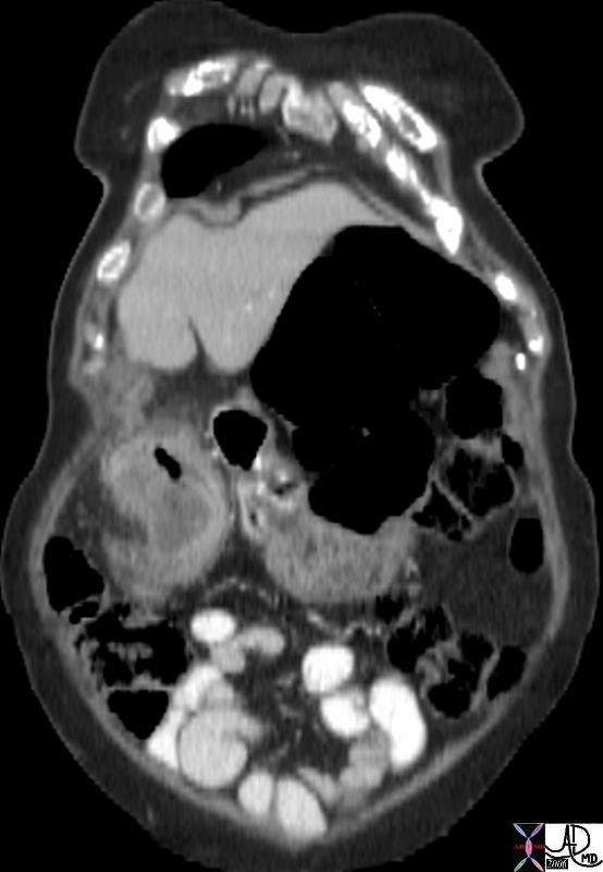
The coronal reformat from a frail and elderly woman shows a thick walled hepatic flexure with transmural extension toward the liver.  Note the fluid filled colon caused by a mucin secreting adenocarcinoma.
Courtesy Ashley Davidoff MD
45083


    [nodeType] => 1
    [parentNode] => (object value omitted)
    [childNodes] => (object value omitted)
    [firstChild] => (object value omitted)
    [lastChild] => (object value omitted)
    [previousSibling] => (object value omitted)
    [nextSibling] => (object value omitted)
    [attributes] => (object value omitted)
    [ownerDocument] => (object value omitted)
    [namespaceURI] =>
    [prefix] =>
    [localName] => table
    [baseURI] =>
    [textContent] =>


Hepatic flexure mucin secreting adenocarcinoma


The coronal reformat from a frail and elderly woman shows a thick walled hepatic flexure with transmural extension toward the liver.  Note the fluid filled colon caused by a mucin secreting adenocarcinoma.
Courtesy Ashley Davidoff MD
45083


)
DOMElement Object
(
    [schemaTypeInfo] =>
    [tagName] => td
    [firstElementChild] => (object value omitted)
    [lastElementChild] => (object value omitted)
    [childElementCount] => 2
    [previousElementSibling] =>
    [nextElementSibling] =>
    [nodeName] => td
    [nodeValue] => The coronal reformat from a frail and elderly woman shows a thick walled hepatic flexure with transmural extension toward the liver.  Note the fluid filled colon caused by a mucin secreting adenocarcinoma.
Courtesy Ashley Davidoff MD
45083
    [nodeType] => 1
    [parentNode] => (object value omitted)
    [childNodes] => (object value omitted)
    [firstChild] => (object value omitted)
    [lastChild] => (object value omitted)
    [previousSibling] => (object value omitted)
    [nextSibling] => (object value omitted)
    [attributes] => (object value omitted)
    [ownerDocument] => (object value omitted)
    [namespaceURI] =>
    [prefix] =>
    [localName] => td
    [baseURI] =>
    [textContent] => The coronal reformat from a frail and elderly woman shows a thick walled hepatic flexure with transmural extension toward the liver.  Note the fluid filled colon caused by a mucin secreting adenocarcinoma.
Courtesy Ashley Davidoff MD
45083
)
DOMElement Object
(
    [schemaTypeInfo] =>
    [tagName] => td
    [firstElementChild] => (object value omitted)
    [lastElementChild] => (object value omitted)
    [childElementCount] => 1
    [previousElementSibling] =>
    [nextElementSibling] =>
    [nodeName] => td
    [nodeValue] => Hepatic flexure mucin secreting adenocarcinoma
    [nodeType] => 1
    [parentNode] => (object value omitted)
    [childNodes] => (object value omitted)
    [firstChild] => (object value omitted)
    [lastChild] => (object value omitted)
    [previousSibling] => (object value omitted)
    [nextSibling] => (object value omitted)
    [attributes] => (object value omitted)
    [ownerDocument] => (object value omitted)
    [namespaceURI] =>
    [prefix] =>
    [localName] => td
    [baseURI] =>
    [textContent] => Hepatic flexure mucin secreting adenocarcinoma
)
https://beta.thecommonvein.net/wp-content/uploads/2023/06/45083.jpg
DOMElement Object
(
    [schemaTypeInfo] =>
    [tagName] => table
    [firstElementChild] => (object value omitted)
    [lastElementChild] => (object value omitted)
    [childElementCount] => 1
    [previousElementSibling] => (object value omitted)
    [nextElementSibling] => (object value omitted)
    [nodeName] => table
    [nodeValue] =>


Redundant hepatic flexure


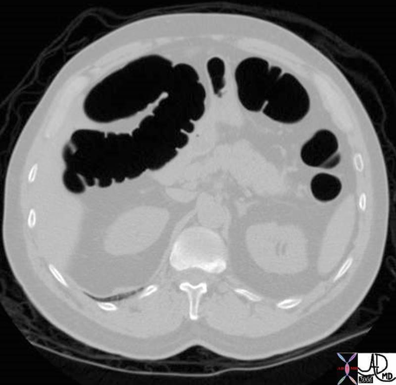
The hepatic flexure is not always a simple 90 degree turn.  It is commonly redundant and it may be difficult to uncoil this redundancy with a barium enema.  This CT study shows a redundant complex turn of the hepatic flexure.
Courtesy Ashley Davidoff MD
38903


    [nodeType] => 1
    [parentNode] => (object value omitted)
    [childNodes] => (object value omitted)
    [firstChild] => (object value omitted)
    [lastChild] => (object value omitted)
    [previousSibling] => (object value omitted)
    [nextSibling] => (object value omitted)
    [attributes] => (object value omitted)
    [ownerDocument] => (object value omitted)
    [namespaceURI] =>
    [prefix] =>
    [localName] => table
    [baseURI] =>
    [textContent] =>


Redundant hepatic flexure


The hepatic flexure is not always a simple 90 degree turn.  It is commonly redundant and it may be difficult to uncoil this redundancy with a barium enema.  This CT study shows a redundant complex turn of the hepatic flexure.
Courtesy Ashley Davidoff MD
38903


)
DOMElement Object
(
    [schemaTypeInfo] =>
    [tagName] => td
    [firstElementChild] => (object value omitted)
    [lastElementChild] => (object value omitted)
    [childElementCount] => 2
    [previousElementSibling] =>
    [nextElementSibling] =>
    [nodeName] => td
    [nodeValue] => The hepatic flexure is not always a simple 90 degree turn.  It is commonly redundant and it may be difficult to uncoil this redundancy with a barium enema.  This CT study shows a redundant complex turn of the hepatic flexure.
Courtesy Ashley Davidoff MD
38903
    [nodeType] => 1
    [parentNode] => (object value omitted)
    [childNodes] => (object value omitted)
    [firstChild] => (object value omitted)
    [lastChild] => (object value omitted)
    [previousSibling] => (object value omitted)
    [nextSibling] => (object value omitted)
    [attributes] => (object value omitted)
    [ownerDocument] => (object value omitted)
    [namespaceURI] =>
    [prefix] =>
    [localName] => td
    [baseURI] =>
    [textContent] => The hepatic flexure is not always a simple 90 degree turn.  It is commonly redundant and it may be difficult to uncoil this redundancy with a barium enema.  This CT study shows a redundant complex turn of the hepatic flexure.
Courtesy Ashley Davidoff MD
38903
)
DOMElement Object
(
    [schemaTypeInfo] =>
    [tagName] => td
    [firstElementChild] => (object value omitted)
    [lastElementChild] => (object value omitted)
    [childElementCount] => 1
    [previousElementSibling] =>
    [nextElementSibling] =>
    [nodeName] => td
    [nodeValue] => Redundant hepatic flexure
    [nodeType] => 1
    [parentNode] => (object value omitted)
    [childNodes] => (object value omitted)
    [firstChild] => (object value omitted)
    [lastChild] => (object value omitted)
    [previousSibling] => (object value omitted)
    [nextSibling] => (object value omitted)
    [attributes] => (object value omitted)
    [ownerDocument] => (object value omitted)
    [namespaceURI] =>
    [prefix] =>
    [localName] => td
    [baseURI] =>
    [textContent] => Redundant hepatic flexure
)
https://beta.thecommonvein.net/wp-content/uploads/2023/05/38903.jpg
DOMElement Object
(
    [schemaTypeInfo] =>
    [tagName] => table
    [firstElementChild] => (object value omitted)
    [lastElementChild] => (object value omitted)
    [childElementCount] => 1
    [previousElementSibling] => (object value omitted)
    [nextElementSibling] => (object value omitted)
    [nodeName] => table
    [nodeValue] =>


Normal hepatic flexure


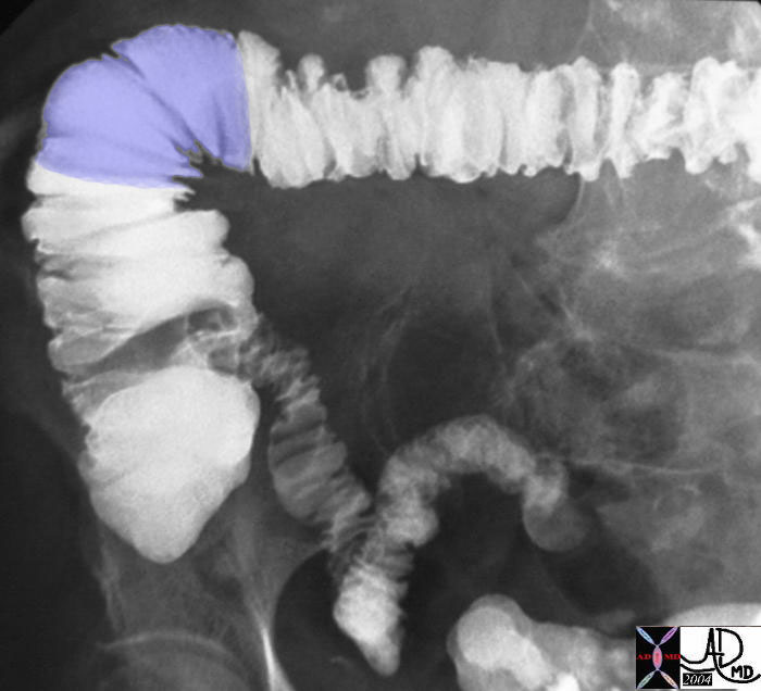
The beginning and end of the hepatic flexure are ill defined, but it connects the ascending colon with the transverse colon.
Courtesy Ashley Davidoff MD
32525b05


    [nodeType] => 1
    [parentNode] => (object value omitted)
    [childNodes] => (object value omitted)
    [firstChild] => (object value omitted)
    [lastChild] => (object value omitted)
    [previousSibling] => (object value omitted)
    [nextSibling] => (object value omitted)
    [attributes] => (object value omitted)
    [ownerDocument] => (object value omitted)
    [namespaceURI] =>
    [prefix] =>
    [localName] => table
    [baseURI] =>
    [textContent] =>


Normal hepatic flexure


The beginning and end of the hepatic flexure are ill defined, but it connects the ascending colon with the transverse colon.
Courtesy Ashley Davidoff MD
32525b05


)
DOMElement Object
(
    [schemaTypeInfo] =>
    [tagName] => td
    [firstElementChild] => (object value omitted)
    [lastElementChild] => (object value omitted)
    [childElementCount] => 2
    [previousElementSibling] =>
    [nextElementSibling] =>
    [nodeName] => td
    [nodeValue] => The beginning and end of the hepatic flexure are ill defined, but it connects the ascending colon with the transverse colon.
Courtesy Ashley Davidoff MD
32525b05
    [nodeType] => 1
    [parentNode] => (object value omitted)
    [childNodes] => (object value omitted)
    [firstChild] => (object value omitted)
    [lastChild] => (object value omitted)
    [previousSibling] => (object value omitted)
    [nextSibling] => (object value omitted)
    [attributes] => (object value omitted)
    [ownerDocument] => (object value omitted)
    [namespaceURI] =>
    [prefix] =>
    [localName] => td
    [baseURI] =>
    [textContent] => The beginning and end of the hepatic flexure are ill defined, but it connects the ascending colon with the transverse colon.
Courtesy Ashley Davidoff MD
32525b05
)
DOMElement Object
(
    [schemaTypeInfo] =>
    [tagName] => td
    [firstElementChild] => (object value omitted)
    [lastElementChild] => (object value omitted)
    [childElementCount] => 1
    [previousElementSibling] =>
    [nextElementSibling] =>
    [nodeName] => td
    [nodeValue] => Normal hepatic flexure
    [nodeType] => 1
    [parentNode] => (object value omitted)
    [childNodes] => (object value omitted)
    [firstChild] => (object value omitted)
    [lastChild] => (object value omitted)
    [previousSibling] => (object value omitted)
    [nextSibling] => (object value omitted)
    [attributes] => (object value omitted)
    [ownerDocument] => (object value omitted)
    [namespaceURI] =>
    [prefix] =>
    [localName] => td
    [baseURI] =>
    [textContent] => Normal hepatic flexure
)
https://beta.thecommonvein.net/wp-content/uploads/2023/05/32525b05.jpg
DOMElement Object
(
    [schemaTypeInfo] =>
    [tagName] => table
    [firstElementChild] => (object value omitted)
    [lastElementChild] => (object value omitted)
    [childElementCount] => 1
    [previousElementSibling] =>
    [nextElementSibling] =>
    [nodeName] => table
    [nodeValue] =>


The hepatic flexure is the junction between the transverse colon and the ascending colon.  There are no defining structural changes, other than for the endoscopist trying to navigate the colon who may be able to see the dark extrinsic impression of the liver on the colon, thus marking the distinction between the end of the ascending and beginning of transverse colon.  The hepatic flexure receives its blood supply in part from the right colic vessels and in part from the middle colic vessels


Normal hepatic flexure


The beginning and end of the hepatic flexure are ill defined, but it connects the ascending colon with the transverse colon.
Courtesy Ashley Davidoff MD
32525b05


The hepatic flexure is not always a simple 90 degree turn. It is commonly redundant and it may be difficult radiologically to uncoil this redundancy with a barium enema making it difficult to fully visualize all parts of the flexure.


Redundant hepatic flexure


The hepatic flexure is not always a simple 90 degree turn.  It is commonly redundant and it may be difficult to uncoil this redundancy with a barium enema.  This CT study shows a redundant complex turn of the hepatic flexure.
Courtesy Ashley Davidoff MD
38903


Applied Anatomy
The close relationship of this part of the colon with the liver and gallbladder is sometimes relevant in disease.  Occasionally an aggressive carcinoma may spread directly from the colon to the liver or gallbladder, making surgical resection difficult.


Hepatic flexure mucin secreting adenocarcinoma


The coronal reformat from a frail and elderly woman shows a thick walled hepatic flexure with transmural extension toward the liver.  Note the fluid filled colon caused by a mucin secreting adenocarcinoma.
Courtesy Ashley Davidoff MD
45083


Question free air?


This image shows a curvilinear air shadow that suggests free ar under the diaphragm.  The patient was asymptomatic  and so a subsequent upright KUB was performed.  See next image (Image courtesy of Ashley Davidoff M.D.) 45758 45759


Question free air?


This upright iamge of the abdomen clearly shows that the air under the diapragm is part of air within the lumen of a loop of bowel in the right upper quadrant.  (Image courtesy of Ashley Davidoff M.D.) 45761


Chilaiditi syndrome – interposition the colon


This coronal image of the abdomen shows the colon and specifically the hepatic flexure malpositioned under the diaphragm and mimics the presence of free air under the diaphragm as noted in the above images.  The liver is displaced from its usual position by the colon which makes its way anteriorly (in front of the liver), and superiorly (below the diaphragm), so that if there is air within this loop it will masquerades as “free air”  (Image courtesy of Ashley Davidoff M.D.) 45765


Chilaiditi syndrome – interposition the colon


This axial image of the abdomen shows the colon and specifically the hepatic flexure malpositioned anterior to the liver.  The liver is displaced from its usual position by the colon which makes its way anteriorly (in front of the liver), and superiorly (below the diaphragm), so that if there is air within this loop it will masquearades as “free air”  (Image courtesy of Ashley Davidoff M.D.) 45768


    [nodeType] => 1
    [parentNode] => (object value omitted)
    [childNodes] => (object value omitted)
    [firstChild] => (object value omitted)
    [lastChild] => (object value omitted)
    [previousSibling] =>
    [nextSibling] => (object value omitted)
    [attributes] => (object value omitted)
    [ownerDocument] => (object value omitted)
    [namespaceURI] =>
    [prefix] =>
    [localName] => table
    [baseURI] =>
    [textContent] =>


The hepatic flexure is the junction between the transverse colon and the ascending colon.  There are no defining structural changes, other than for the endoscopist trying to navigate the colon who may be able to see the dark extrinsic impression of the liver on the colon, thus marking the distinction between the end of the ascending and beginning of transverse colon.  The hepatic flexure receives its blood supply in part from the right colic vessels and in part from the middle colic vessels


Normal hepatic flexure


The beginning and end of the hepatic flexure are ill defined, but it connects the ascending colon with the transverse colon.
Courtesy Ashley Davidoff MD
32525b05


The hepatic flexure is not always a simple 90 degree turn. It is commonly redundant and it may be difficult radiologically to uncoil this redundancy with a barium enema making it difficult to fully visualize all parts of the flexure.


Redundant hepatic flexure


The hepatic flexure is not always a simple 90 degree turn.  It is commonly redundant and it may be difficult to uncoil this redundancy with a barium enema.  This CT study shows a redundant complex turn of the hepatic flexure.
Courtesy Ashley Davidoff MD
38903


Applied Anatomy
The close relationship of this part of the colon with the liver and gallbladder is sometimes relevant in disease.  Occasionally an aggressive carcinoma may spread directly from the colon to the liver or gallbladder, making surgical resection difficult.


Hepatic flexure mucin secreting adenocarcinoma


The coronal reformat from a frail and elderly woman shows a thick walled hepatic flexure with transmural extension toward the liver.  Note the fluid filled colon caused by a mucin secreting adenocarcinoma.
Courtesy Ashley Davidoff MD
45083


Question free air?


This image shows a curvilinear air shadow that suggests free ar under the diaphragm.  The patient was asymptomatic  and so a subsequent upright KUB was performed.  See next image (Image courtesy of Ashley Davidoff M.D.) 45758 45759


Question free air?


This upright iamge of the abdomen clearly shows that the air under the diapragm is part of air within the lumen of a loop of bowel in the right upper quadrant.  (Image courtesy of Ashley Davidoff M.D.) 45761


Chilaiditi syndrome – interposition the colon


This coronal image of the abdomen shows the colon and specifically the hepatic flexure malpositioned under the diaphragm and mimics the presence of free air under the diaphragm as noted in the above images.  The liver is displaced from its usual position by the colon which makes its way anteriorly (in front of the liver), and superiorly (below the diaphragm), so that if there is air within this loop it will masquerades as “free air”  (Image courtesy of Ashley Davidoff M.D.) 45765


Chilaiditi syndrome – interposition the colon


This axial image of the abdomen shows the colon and specifically the hepatic flexure malpositioned anterior to the liver.  The liver is displaced from its usual position by the colon which makes its way anteriorly (in front of the liver), and superiorly (below the diaphragm), so that if there is air within this loop it will masquearades as “free air”  (Image courtesy of Ashley Davidoff M.D.) 45768


)
DOMElement Object
(
    [schemaTypeInfo] =>
    [tagName] => td
    [firstElementChild] =>
    [lastElementChild] =>
    [childElementCount] => 0
    [previousElementSibling] =>
    [nextElementSibling] =>
    [nodeName] => td
    [nodeValue] => This axial image of the abdomen shows the colon and specifically the hepatic flexure malpositioned anterior to the liver.  The liver is displaced from its usual position by the colon which makes its way anteriorly (in front of the liver), and superiorly (below the diaphragm), so that if there is air within this loop it will masquearades as “free air”  (Image courtesy of Ashley Davidoff M.D.) 45768
    [nodeType] => 1
    [parentNode] => (object value omitted)
    [childNodes] => (object value omitted)
    [firstChild] => (object value omitted)
    [lastChild] => (object value omitted)
    [previousSibling] => (object value omitted)
    [nextSibling] => (object value omitted)
    [attributes] => (object value omitted)
    [ownerDocument] => (object value omitted)
    [namespaceURI] =>
    [prefix] =>
    [localName] => td
    [baseURI] =>
    [textContent] => This axial image of the abdomen shows the colon and specifically the hepatic flexure malpositioned anterior to the liver.  The liver is displaced from its usual position by the colon which makes its way anteriorly (in front of the liver), and superiorly (below the diaphragm), so that if there is air within this loop it will masquearades as “free air”  (Image courtesy of Ashley Davidoff M.D.) 45768
)
DOMElement Object
(
    [schemaTypeInfo] =>
    [tagName] => td
    [firstElementChild] => (object value omitted)
    [lastElementChild] => (object value omitted)
    [childElementCount] => 2
    [previousElementSibling] =>
    [nextElementSibling] =>
    [nodeName] => td
    [nodeValue] => Chilaiditi syndrome – interposition the colon
    [nodeType] => 1
    [parentNode] => (object value omitted)
    [childNodes] => (object value omitted)
    [firstChild] => (object value omitted)
    [lastChild] => (object value omitted)
    [previousSibling] => (object value omitted)
    [nextSibling] => (object value omitted)
    [attributes] => (object value omitted)
    [ownerDocument] => (object value omitted)
    [namespaceURI] =>
    [prefix] =>
    [localName] => td
    [baseURI] =>
    [textContent] => Chilaiditi syndrome – interposition the colon
)
DOMElement Object
(
    [schemaTypeInfo] =>
    [tagName] => td
    [firstElementChild] =>
    [lastElementChild] =>
    [childElementCount] => 0
    [previousElementSibling] =>
    [nextElementSibling] =>
    [nodeName] => td
    [nodeValue] => This coronal image of the abdomen shows the colon and specifically the hepatic flexure malpositioned under the diaphragm and mimics the presence of free air under the diaphragm as noted in the above images.  The liver is displaced from its usual position by the colon which makes its way anteriorly (in front of the liver), and superiorly (below the diaphragm), so that if there is air within this loop it will masquerades as “free air”  (Image courtesy of Ashley Davidoff M.D.) 45765
    [nodeType] => 1
    [parentNode] => (object value omitted)
    [childNodes] => (object value omitted)
    [firstChild] => (object value omitted)
    [lastChild] => (object value omitted)
    [previousSibling] => (object value omitted)
    [nextSibling] => (object value omitted)
    [attributes] => (object value omitted)
    [ownerDocument] => (object value omitted)
    [namespaceURI] =>
    [prefix] =>
    [localName] => td
    [baseURI] =>
    [textContent] => This coronal image of the abdomen shows the colon and specifically the hepatic flexure malpositioned under the diaphragm and mimics the presence of free air under the diaphragm as noted in the above images.  The liver is displaced from its usual position by the colon which makes its way anteriorly (in front of the liver), and superiorly (below the diaphragm), so that if there is air within this loop it will masquerades as “free air”  (Image courtesy of Ashley Davidoff M.D.) 45765
)
https://beta.thecommonvein.net/wp-content/uploads/2023/06/45768.jpg
DOMElement Object
(
    [schemaTypeInfo] =>
    [tagName] => td
    [firstElementChild] => (object value omitted)
    [lastElementChild] => (object value omitted)
    [childElementCount] => 2
    [previousElementSibling] =>
    [nextElementSibling] =>
    [nodeName] => td
    [nodeValue] => Chilaiditi syndrome – interposition the colon
    [nodeType] => 1
    [parentNode] => (object value omitted)
    [childNodes] => (object value omitted)
    [firstChild] => (object value omitted)
    [lastChild] => (object value omitted)
    [previousSibling] => (object value omitted)
    [nextSibling] => (object value omitted)
    [attributes] => (object value omitted)
    [ownerDocument] => (object value omitted)
    [namespaceURI] =>
    [prefix] =>
    [localName] => td
    [baseURI] =>
    [textContent] => Chilaiditi syndrome – interposition the colon
)
https://beta.thecommonvein.net/wp-content/uploads/2023/06/45768.jpg
https://beta.thecommonvein.net/wp-content/uploads/2023/06/45765.jpg
DOMElement Object
(
    [schemaTypeInfo] =>
    [tagName] => td
    [firstElementChild] =>
    [lastElementChild] =>
    [childElementCount] => 0
    [previousElementSibling] =>
    [nextElementSibling] =>
    [nodeName] => td
    [nodeValue] => This upright iamge of the abdomen clearly shows that the air under the diapragm is part of air within the lumen of a loop of bowel in the right upper quadrant.  (Image courtesy of Ashley Davidoff M.D.) 45761
    [nodeType] => 1
    [parentNode] => (object value omitted)
    [childNodes] => (object value omitted)
    [firstChild] => (object value omitted)
    [lastChild] => (object value omitted)
    [previousSibling] => (object value omitted)
    [nextSibling] => (object value omitted)
    [attributes] => (object value omitted)
    [ownerDocument] => (object value omitted)
    [namespaceURI] =>
    [prefix] =>
    [localName] => td
    [baseURI] =>
    [textContent] => This upright iamge of the abdomen clearly shows that the air under the diapragm is part of air within the lumen of a loop of bowel in the right upper quadrant.  (Image courtesy of Ashley Davidoff M.D.) 45761
)
https://beta.thecommonvein.net/wp-content/uploads/2023/06/45768.jpg
DOMElement Object
(
    [schemaTypeInfo] =>
    [tagName] => td
    [firstElementChild] => (object value omitted)
    [lastElementChild] => (object value omitted)
    [childElementCount] => 2
    [previousElementSibling] =>
    [nextElementSibling] =>
    [nodeName] => td
    [nodeValue] => Question free air?
    [nodeType] => 1
    [parentNode] => (object value omitted)
    [childNodes] => (object value omitted)
    [firstChild] => (object value omitted)
    [lastChild] => (object value omitted)
    [previousSibling] => (object value omitted)
    [nextSibling] => (object value omitted)
    [attributes] => (object value omitted)
    [ownerDocument] => (object value omitted)
    [namespaceURI] =>
    [prefix] =>
    [localName] => td
    [baseURI] =>
    [textContent] => Question free air?
)
https://beta.thecommonvein.net/wp-content/uploads/2023/06/45768.jpg
https://beta.thecommonvein.net/wp-content/uploads/2023/06/45761.jpg
DOMElement Object
(
    [schemaTypeInfo] =>
    [tagName] => td
    [firstElementChild] =>
    [lastElementChild] =>
    [childElementCount] => 0
    [previousElementSibling] =>
    [nextElementSibling] =>
    [nodeName] => td
    [nodeValue] => This image shows a curvilinear air shadow that suggests free ar under the diaphragm.  The patient was asymptomatic  and so a subsequent upright KUB was performed.  See next image (Image courtesy of Ashley Davidoff M.D.) 45758 45759
    [nodeType] => 1
    [parentNode] => (object value omitted)
    [childNodes] => (object value omitted)
    [firstChild] => (object value omitted)
    [lastChild] => (object value omitted)
    [previousSibling] => (object value omitted)
    [nextSibling] => (object value omitted)
    [attributes] => (object value omitted)
    [ownerDocument] => (object value omitted)
    [namespaceURI] =>
    [prefix] =>
    [localName] => td
    [baseURI] =>
    [textContent] => This image shows a curvilinear air shadow that suggests free ar under the diaphragm.  The patient was asymptomatic  and so a subsequent upright KUB was performed.  See next image (Image courtesy of Ashley Davidoff M.D.) 45758 45759
)
https://beta.thecommonvein.net/wp-content/uploads/2023/06/45768.jpg
DOMElement Object
(
    [schemaTypeInfo] =>
    [tagName] => td
    [firstElementChild] => (object value omitted)
    [lastElementChild] => (object value omitted)
    [childElementCount] => 3
    [previousElementSibling] =>
    [nextElementSibling] =>
    [nodeName] => td
    [nodeValue] => Question free air?
    [nodeType] => 1
    [parentNode] => (object value omitted)
    [childNodes] => (object value omitted)
    [firstChild] => (object value omitted)
    [lastChild] => (object value omitted)
    [previousSibling] => (object value omitted)
    [nextSibling] => (object value omitted)
    [attributes] => (object value omitted)
    [ownerDocument] => (object value omitted)
    [namespaceURI] =>
    [prefix] =>
    [localName] => td
    [baseURI] =>
    [textContent] => Question free air?
)
https://beta.thecommonvein.net/wp-content/uploads/2023/06/45768.jpg
https://beta.thecommonvein.net/wp-content/uploads/2023/05/45758.jpg https://beta.thecommonvein.net/wp-content/uploads/2023/06/45759.jpg
DOMElement Object
(
    [schemaTypeInfo] =>
    [tagName] => td
    [firstElementChild] => (object value omitted)
    [lastElementChild] => (object value omitted)
    [childElementCount] => 2
    [previousElementSibling] =>
    [nextElementSibling] =>
    [nodeName] => td
    [nodeValue] => The coronal reformat from a frail and elderly woman shows a thick walled hepatic flexure with transmural extension toward the liver.  Note the fluid filled colon caused by a mucin secreting adenocarcinoma.
Courtesy Ashley Davidoff MD
45083
    [nodeType] => 1
    [parentNode] => (object value omitted)
    [childNodes] => (object value omitted)
    [firstChild] => (object value omitted)
    [lastChild] => (object value omitted)
    [previousSibling] => (object value omitted)
    [nextSibling] => (object value omitted)
    [attributes] => (object value omitted)
    [ownerDocument] => (object value omitted)
    [namespaceURI] =>
    [prefix] =>
    [localName] => td
    [baseURI] =>
    [textContent] => The coronal reformat from a frail and elderly woman shows a thick walled hepatic flexure with transmural extension toward the liver.  Note the fluid filled colon caused by a mucin secreting adenocarcinoma.
Courtesy Ashley Davidoff MD
45083
)
https://beta.thecommonvein.net/wp-content/uploads/2023/06/45768.jpg
DOMElement Object
(
    [schemaTypeInfo] =>
    [tagName] => td
    [firstElementChild] => (object value omitted)
    [lastElementChild] => (object value omitted)
    [childElementCount] => 2
    [previousElementSibling] =>
    [nextElementSibling] =>
    [nodeName] => td
    [nodeValue] => Hepatic flexure mucin secreting adenocarcinoma
    [nodeType] => 1
    [parentNode] => (object value omitted)
    [childNodes] => (object value omitted)
    [firstChild] => (object value omitted)
    [lastChild] => (object value omitted)
    [previousSibling] => (object value omitted)
    [nextSibling] => (object value omitted)
    [attributes] => (object value omitted)
    [ownerDocument] => (object value omitted)
    [namespaceURI] =>
    [prefix] =>
    [localName] => td
    [baseURI] =>
    [textContent] => Hepatic flexure mucin secreting adenocarcinoma
)
https://beta.thecommonvein.net/wp-content/uploads/2023/06/45768.jpg
https://beta.thecommonvein.net/wp-content/uploads/2023/06/45083.jpg
DOMElement Object
(
    [schemaTypeInfo] =>
    [tagName] => td
    [firstElementChild] => (object value omitted)
    [lastElementChild] => (object value omitted)
    [childElementCount] => 2
    [previousElementSibling] =>
    [nextElementSibling] =>
    [nodeName] => td
    [nodeValue] => The hepatic flexure is not always a simple 90 degree turn.  It is commonly redundant and it may be difficult to uncoil this redundancy with a barium enema.  This CT study shows a redundant complex turn of the hepatic flexure.
Courtesy Ashley Davidoff MD
38903
    [nodeType] => 1
    [parentNode] => (object value omitted)
    [childNodes] => (object value omitted)
    [firstChild] => (object value omitted)
    [lastChild] => (object value omitted)
    [previousSibling] => (object value omitted)
    [nextSibling] => (object value omitted)
    [attributes] => (object value omitted)
    [ownerDocument] => (object value omitted)
    [namespaceURI] =>
    [prefix] =>
    [localName] => td
    [baseURI] =>
    [textContent] => The hepatic flexure is not always a simple 90 degree turn.  It is commonly redundant and it may be difficult to uncoil this redundancy with a barium enema.  This CT study shows a redundant complex turn of the hepatic flexure.
Courtesy Ashley Davidoff MD
38903
)
https://beta.thecommonvein.net/wp-content/uploads/2023/06/45768.jpg
DOMElement Object
(
    [schemaTypeInfo] =>
    [tagName] => td
    [firstElementChild] => (object value omitted)
    [lastElementChild] => (object value omitted)
    [childElementCount] => 2
    [previousElementSibling] =>
    [nextElementSibling] =>
    [nodeName] => td
    [nodeValue] => Redundant hepatic flexure
    [nodeType] => 1
    [parentNode] => (object value omitted)
    [childNodes] => (object value omitted)
    [firstChild] => (object value omitted)
    [lastChild] => (object value omitted)
    [previousSibling] => (object value omitted)
    [nextSibling] => (object value omitted)
    [attributes] => (object value omitted)
    [ownerDocument] => (object value omitted)
    [namespaceURI] =>
    [prefix] =>
    [localName] => td
    [baseURI] =>
    [textContent] => Redundant hepatic flexure
)
https://beta.thecommonvein.net/wp-content/uploads/2023/06/45768.jpg
https://beta.thecommonvein.net/wp-content/uploads/2023/05/38903.jpg
DOMElement Object
(
    [schemaTypeInfo] =>
    [tagName] => td
    [firstElementChild] => (object value omitted)
    [lastElementChild] => (object value omitted)
    [childElementCount] => 2
    [previousElementSibling] =>
    [nextElementSibling] =>
    [nodeName] => td
    [nodeValue] => The beginning and end of the hepatic flexure are ill defined, but it connects the ascending colon with the transverse colon.
Courtesy Ashley Davidoff MD
32525b05
    [nodeType] => 1
    [parentNode] => (object value omitted)
    [childNodes] => (object value omitted)
    [firstChild] => (object value omitted)
    [lastChild] => (object value omitted)
    [previousSibling] => (object value omitted)
    [nextSibling] => (object value omitted)
    [attributes] => (object value omitted)
    [ownerDocument] => (object value omitted)
    [namespaceURI] =>
    [prefix] =>
    [localName] => td
    [baseURI] =>
    [textContent] => The beginning and end of the hepatic flexure are ill defined, but it connects the ascending colon with the transverse colon.
Courtesy Ashley Davidoff MD
32525b05
)
https://beta.thecommonvein.net/wp-content/uploads/2023/06/45768.jpg
DOMElement Object
(
    [schemaTypeInfo] =>
    [tagName] => td
    [firstElementChild] => (object value omitted)
    [lastElementChild] => (object value omitted)
    [childElementCount] => 2
    [previousElementSibling] =>
    [nextElementSibling] =>
    [nodeName] => td
    [nodeValue] => Normal hepatic flexure
    [nodeType] => 1
    [parentNode] => (object value omitted)
    [childNodes] => (object value omitted)
    [firstChild] => (object value omitted)
    [lastChild] => (object value omitted)
    [previousSibling] => (object value omitted)
    [nextSibling] => (object value omitted)
    [attributes] => (object value omitted)
    [ownerDocument] => (object value omitted)
    [namespaceURI] =>
    [prefix] =>
    [localName] => td
    [baseURI] =>
    [textContent] => Normal hepatic flexure
)
https://beta.thecommonvein.net/wp-content/uploads/2023/06/45768.jpg
https://beta.thecommonvein.net/wp-content/uploads/2023/05/32525b05.jpg
DOMElement Object
(
    [schemaTypeInfo] =>
    [tagName] => td
    [firstElementChild] => (object value omitted)
    [lastElementChild] => (object value omitted)
    [childElementCount] => 17
    [previousElementSibling] =>
    [nextElementSibling] =>
    [nodeName] => td
    [nodeValue] =>
The hepatic flexure is the junction between the transverse colon and the ascending colon.  There are no defining structural changes, other than for the endoscopist trying to navigate the colon who may be able to see the dark extrinsic impression of the liver on the colon, thus marking the distinction between the end of the ascending and beginning of transverse colon.  The hepatic flexure receives its blood supply in part from the right colic vessels and in part from the middle colic vessels


Normal hepatic flexure


The beginning and end of the hepatic flexure are ill defined, but it connects the ascending colon with the transverse colon.
Courtesy Ashley Davidoff MD
32525b05


The hepatic flexure is not always a simple 90 degree turn. It is commonly redundant and it may be difficult radiologically to uncoil this redundancy with a barium enema making it difficult to fully visualize all parts of the flexure.


Redundant hepatic flexure


The hepatic flexure is not always a simple 90 degree turn.  It is commonly redundant and it may be difficult to uncoil this redundancy with a barium enema.  This CT study shows a redundant complex turn of the hepatic flexure.
Courtesy Ashley Davidoff MD
38903


Applied Anatomy
The close relationship of this part of the colon with the liver and gallbladder is sometimes relevant in disease.  Occasionally an aggressive carcinoma may spread directly from the colon to the liver or gallbladder, making surgical resection difficult.


Hepatic flexure mucin secreting adenocarcinoma


The coronal reformat from a frail and elderly woman shows a thick walled hepatic flexure with transmural extension toward the liver.  Note the fluid filled colon caused by a mucin secreting adenocarcinoma.
Courtesy Ashley Davidoff MD
45083


Question free air?


This image shows a curvilinear air shadow that suggests free ar under the diaphragm.  The patient was asymptomatic  and so a subsequent upright KUB was performed.  See next image (Image courtesy of Ashley Davidoff M.D.) 45758 45759


Question free air?


This upright iamge of the abdomen clearly shows that the air under the diapragm is part of air within the lumen of a loop of bowel in the right upper quadrant.  (Image courtesy of Ashley Davidoff M.D.) 45761


Chilaiditi syndrome – interposition the colon


This coronal image of the abdomen shows the colon and specifically the hepatic flexure malpositioned under the diaphragm and mimics the presence of free air under the diaphragm as noted in the above images.  The liver is displaced from its usual position by the colon which makes its way anteriorly (in front of the liver), and superiorly (below the diaphragm), so that if there is air within this loop it will masquerades as “free air”  (Image courtesy of Ashley Davidoff M.D.) 45765


Chilaiditi syndrome – interposition the colon


This axial image of the abdomen shows the colon and specifically the hepatic flexure malpositioned anterior to the liver.  The liver is displaced from its usual position by the colon which makes its way anteriorly (in front of the liver), and superiorly (below the diaphragm), so that if there is air within this loop it will masquearades as “free air”  (Image courtesy of Ashley Davidoff M.D.) 45768


    [nodeType] => 1
    [parentNode] => (object value omitted)
    [childNodes] => (object value omitted)
    [firstChild] => (object value omitted)
    [lastChild] => (object value omitted)
    [previousSibling] => (object value omitted)
    [nextSibling] => (object value omitted)
    [attributes] => (object value omitted)
    [ownerDocument] => (object value omitted)
    [namespaceURI] =>
    [prefix] =>
    [localName] => td
    [baseURI] =>
    [textContent] =>
The hepatic flexure is the junction between the transverse colon and the ascending colon.  There are no defining structural changes, other than for the endoscopist trying to navigate the colon who may be able to see the dark extrinsic impression of the liver on the colon, thus marking the distinction between the end of the ascending and beginning of transverse colon.  The hepatic flexure receives its blood supply in part from the right colic vessels and in part from the middle colic vessels


Normal hepatic flexure


The beginning and end of the hepatic flexure are ill defined, but it connects the ascending colon with the transverse colon.
Courtesy Ashley Davidoff MD
32525b05


The hepatic flexure is not always a simple 90 degree turn. It is commonly redundant and it may be difficult radiologically to uncoil this redundancy with a barium enema making it difficult to fully visualize all parts of the flexure.


Redundant hepatic flexure


The hepatic flexure is not always a simple 90 degree turn.  It is commonly redundant and it may be difficult to uncoil this redundancy with a barium enema.  This CT study shows a redundant complex turn of the hepatic flexure.
Courtesy Ashley Davidoff MD
38903


Applied Anatomy
The close relationship of this part of the colon with the liver and gallbladder is sometimes relevant in disease.  Occasionally an aggressive carcinoma may spread directly from the colon to the liver or gallbladder, making surgical resection difficult.


Hepatic flexure mucin secreting adenocarcinoma


The coronal reformat from a frail and elderly woman shows a thick walled hepatic flexure with transmural extension toward the liver.  Note the fluid filled colon caused by a mucin secreting adenocarcinoma.
Courtesy Ashley Davidoff MD
45083


Question free air?


This image shows a curvilinear air shadow that suggests free ar under the diaphragm.  The patient was asymptomatic  and so a subsequent upright KUB was performed.  See next image (Image courtesy of Ashley Davidoff M.D.) 45758 45759


Question free air?


This upright iamge of the abdomen clearly shows that the air under the diapragm is part of air within the lumen of a loop of bowel in the right upper quadrant.  (Image courtesy of Ashley Davidoff M.D.) 45761


Chilaiditi syndrome – interposition the colon


This coronal image of the abdomen shows the colon and specifically the hepatic flexure malpositioned under the diaphragm and mimics the presence of free air under the diaphragm as noted in the above images.  The liver is displaced from its usual position by the colon which makes its way anteriorly (in front of the liver), and superiorly (below the diaphragm), so that if there is air within this loop it will masquerades as “free air”  (Image courtesy of Ashley Davidoff M.D.) 45765


Chilaiditi syndrome – interposition the colon


This axial image of the abdomen shows the colon and specifically the hepatic flexure malpositioned anterior to the liver.  The liver is displaced from its usual position by the colon which makes its way anteriorly (in front of the liver), and superiorly (below the diaphragm), so that if there is air within this loop it will masquearades as “free air”  (Image courtesy of Ashley Davidoff M.D.) 45768


)


