The Common Vein Copyright 2008
Diverticulitis
Diverticulitis is an inflammation of a diverticulum caused by an initial obstruction and then infection of the diverticulum. This results in spread of the inflammation to the surrounding fat and other tissues or organs surrounding the bowel. Diverticulitis can be complicated by abscess formation, bowel perforation, peritonitis or less commonly by fistulous formation to the bladder
Structural considerations
Diverticula occur at sites of the colon wall that are relative weak, typically at sites of the insertions of blood vessels through the muscle wall. Its lumen communicates with the lumen of the colon. Although not completely understood, these outpouchings are much more common with a Western, low fiber diet and appear to be related to increased intraluminal pressure. Diverticulitis is a condition that most commonly involves the sigmoid colon and therefore presents with left lower quadrant pain which can occasionally be confused with other pelvic pathology. Most colonic diverticula are 0.5 to 1 cm in diameter, typically located in the sigmoid colon.
Clinically the patient classically presents with left lower quadrant pain because the sigmoid colon is the most common site of involvement. The descending colon is sometimes affected in which case the patient will present with left upper quadrant or left mid or lateral pain. Diverticulitis is a condition that most commonly involves the sigmoid colon and therefore presents with left lower quadrant pain which can occasionally be confused with other pelvic pathology. Pain is often present for several days, and there may be a history of prior similar episodes. Fever and elevated white count are common accompaniments. It is sometimes associated with changes in bowel movements.
The diagnosis is confirmed by CT scan which is the study of choice. Oral and intravenous contrast are preferred, but not essential if there is a contraindication. Administration of rectal contrast may be considered in such cases and may slightly improve sensitivity by dilating the colon. The reported sensitivity of CT for diagnosing acute diverticulitis is 79-99%. CT is excellent for detection of complications of diverticulitis including abscess and fistula formation; furthermore, it may detect other non-colonic causes of abdominal pain. CT is able to direct management since uncomplicated diverticulitis would be managed conservatively and when complications such as abscess formation or perforation are identified they would be managed surgically.
Diverticulitis is commonly treated with antibiotics, but sometimes surgery is required. Depending on the severity, the condition is medically managed with oral or parenteral antibiotics which provide broad coverage. This coverage needs to include anaerobes. Sometimes this can be done as an outpatient with oral medications. But for more severe cases, including those associated with vomiting, intractable pain, or associated abscess, are managed in hospital with IV antibiotics. Large abscesses can be drained percutaneously while surgery is sometimes required for abscesses associated with perforation and free air. For frequent recurrences, sometimes the portion of affected bowel is removed electively, after the acute inflammatory process settles down.
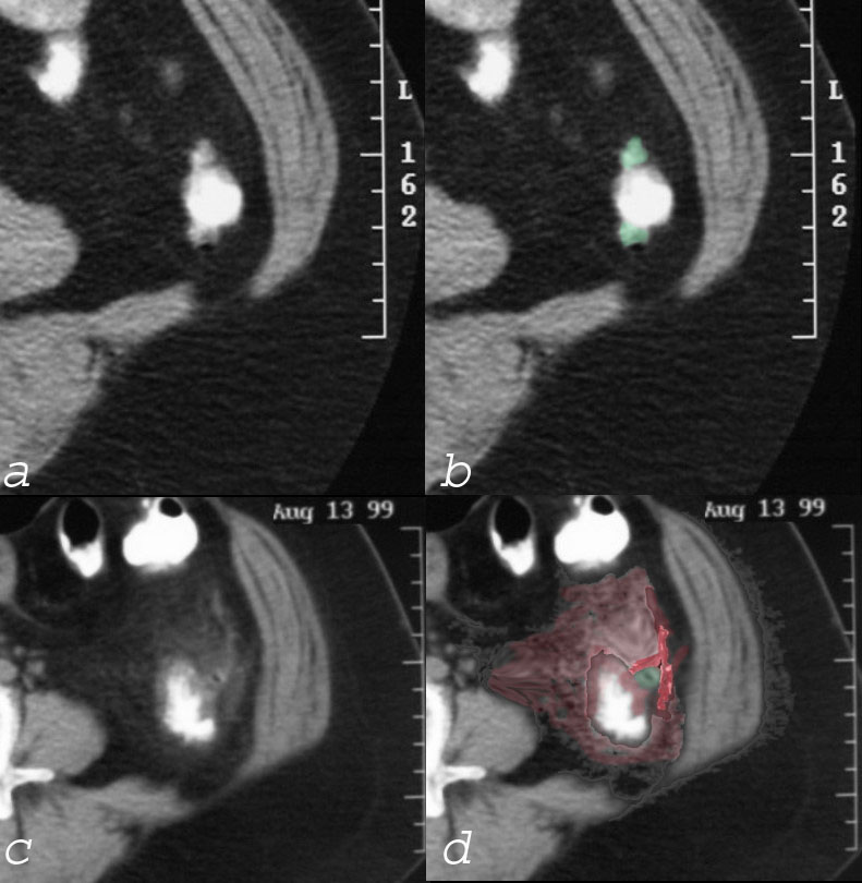
 Non inflammed diverticulum (a, b -green), Diverticulitis (c,d), and Pericolic inflammation (red) Non inflammed diverticulum (a, b -green), Diverticulitis (c,d), and Pericolic inflammation (red) |
| The CT scan shows evidence of both diverticulosis and diverticulitis. In images a and b, two small non inflammed outpouchings are seen. The diverticula are outlined in green in b. In the same patient another diverticulum has become inflammed, and diverticulitis is present. This is characterized by induration of the fat around the diverticulum and the colon (maroon) and extension of the inflammatory process to the peritoneal lining (bright red in d). Inflammation of the colon and colonic wall induces visceral pain which is an ache and poorly localized, and inflammation of the peritoneal lining causes a sharp well localised somatic pain that is sensitive to deep palpation.
28582c02 colon descending colon diverticulum diverticulitis pericolic induration acute inflammation acute diverticulitis diverticulosis CTscan Courtesy Ashley Davidoff MD |
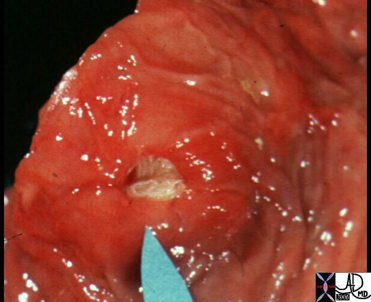
 Acute Diverticulitis – Mouth of Diverticulum is Filled with Pus Acute Diverticulitis – Mouth of Diverticulum is Filled with Pus |
| This pathological specimen shows the mouth of a diverticulum filled with yellow pus in this patient who had complicated diverticulitis. The tissues surrounding the diverticulum are swollen and red.
12077 colon large bowel fx pus filled purulent diverticulum fx mucosal hyperemia reddening dx acute diverticulitis grosspathology Courtesy Barbara Banner MD |
DOMElement Object
(
    [schemaTypeInfo] =>
    [tagName] => table
    [firstElementChild] => (object value omitted)
    [lastElementChild] => (object value omitted)
    [childElementCount] => 1
    [previousElementSibling] => (object value omitted)
    [nextElementSibling] =>
    [nodeName] => table
    [nodeValue] =>


Acute Diverticulitis – Mouth of Diverticulum is Filled with Pus


This pathological specimen shows the mouth of a diverticulum filled with yellow pus in this patient who had complicated diverticulitis.  The tissues surrounding the diverticulum are swollen and red.
12077 colon large bowel fx pus filled purulent diverticulum fx mucosal hyperemia reddening dx acute diverticulitis grosspathology Courtesy Barbara Banner MD


    [nodeType] => 1
    [parentNode] => (object value omitted)
    [childNodes] => (object value omitted)
    [firstChild] => (object value omitted)
    [lastChild] => (object value omitted)
    [previousSibling] => (object value omitted)
    [nextSibling] => (object value omitted)
    [attributes] => (object value omitted)
    [ownerDocument] => (object value omitted)
    [namespaceURI] =>
    [prefix] =>
    [localName] => table
    [baseURI] =>
    [textContent] =>


Acute Diverticulitis – Mouth of Diverticulum is Filled with Pus


This pathological specimen shows the mouth of a diverticulum filled with yellow pus in this patient who had complicated diverticulitis.  The tissues surrounding the diverticulum are swollen and red.
12077 colon large bowel fx pus filled purulent diverticulum fx mucosal hyperemia reddening dx acute diverticulitis grosspathology Courtesy Barbara Banner MD


)
DOMElement Object
(
    [schemaTypeInfo] =>
    [tagName] => td
    [firstElementChild] => (object value omitted)
    [lastElementChild] => (object value omitted)
    [childElementCount] => 1
    [previousElementSibling] =>
    [nextElementSibling] =>
    [nodeName] => td
    [nodeValue] => This pathological specimen shows the mouth of a diverticulum filled with yellow pus in this patient who had complicated diverticulitis.  The tissues surrounding the diverticulum are swollen and red.
12077 colon large bowel fx pus filled purulent diverticulum fx mucosal hyperemia reddening dx acute diverticulitis grosspathology Courtesy Barbara Banner MD
    [nodeType] => 1
    [parentNode] => (object value omitted)
    [childNodes] => (object value omitted)
    [firstChild] => (object value omitted)
    [lastChild] => (object value omitted)
    [previousSibling] => (object value omitted)
    [nextSibling] => (object value omitted)
    [attributes] => (object value omitted)
    [ownerDocument] => (object value omitted)
    [namespaceURI] =>
    [prefix] =>
    [localName] => td
    [baseURI] =>
    [textContent] => This pathological specimen shows the mouth of a diverticulum filled with yellow pus in this patient who had complicated diverticulitis.  The tissues surrounding the diverticulum are swollen and red.
12077 colon large bowel fx pus filled purulent diverticulum fx mucosal hyperemia reddening dx acute diverticulitis grosspathology Courtesy Barbara Banner MD
)
DOMElement Object
(
    [schemaTypeInfo] =>
    [tagName] => td
    [firstElementChild] => (object value omitted)
    [lastElementChild] => (object value omitted)
    [childElementCount] => 1
    [previousElementSibling] =>
    [nextElementSibling] =>
    [nodeName] => td
    [nodeValue] => Acute Diverticulitis – Mouth of Diverticulum is Filled with Pus
    [nodeType] => 1
    [parentNode] => (object value omitted)
    [childNodes] => (object value omitted)
    [firstChild] => (object value omitted)
    [lastChild] => (object value omitted)
    [previousSibling] => (object value omitted)
    [nextSibling] => (object value omitted)
    [attributes] => (object value omitted)
    [ownerDocument] => (object value omitted)
    [namespaceURI] =>
    [prefix] =>
    [localName] => td
    [baseURI] =>
    [textContent] => Acute Diverticulitis – Mouth of Diverticulum is Filled with Pus
)
DOMElement Object
(
    [schemaTypeInfo] =>
    [tagName] => table
    [firstElementChild] => (object value omitted)
    [lastElementChild] => (object value omitted)
    [childElementCount] => 1
    [previousElementSibling] => (object value omitted)
    [nextElementSibling] => (object value omitted)
    [nodeName] => table
    [nodeValue] =>


Non inflammed diverticulum (a, b -green), Diverticulitis (c,d), and Pericolic inflammation (red)


The CT scan shows evidence of both diverticulosis and diverticulitis.  In images a and b, two small non inflammed outpouchings are seen.  The diverticula are outlined in green in b.  In the same patient another diverticulum has become inflammed, and diverticulitis is present.  This is characterized by induration of the fat around the diverticulum and  the colon (maroon) and extension of the inflammatory process to the peritoneal lining (bright red in d).  Inflammation of the colon and colonic wall induces visceral pain which is an ache and poorly localized, and inflammation of the peritoneal lining causes a sharp well localised somatic pain that is sensitive to deep palpation.

28582c02 colon descending colon diverticulum diverticulitis pericolic induration acute inflammation acute diverticulitis diverticulosis CTscan Courtesy Ashley Davidoff MD


    [nodeType] => 1
    [parentNode] => (object value omitted)
    [childNodes] => (object value omitted)
    [firstChild] => (object value omitted)
    [lastChild] => (object value omitted)
    [previousSibling] => (object value omitted)
    [nextSibling] => (object value omitted)
    [attributes] => (object value omitted)
    [ownerDocument] => (object value omitted)
    [namespaceURI] =>
    [prefix] =>
    [localName] => table
    [baseURI] =>
    [textContent] =>


Non inflammed diverticulum (a, b -green), Diverticulitis (c,d), and Pericolic inflammation (red)


The CT scan shows evidence of both diverticulosis and diverticulitis.  In images a and b, two small non inflammed outpouchings are seen.  The diverticula are outlined in green in b.  In the same patient another diverticulum has become inflammed, and diverticulitis is present.  This is characterized by induration of the fat around the diverticulum and  the colon (maroon) and extension of the inflammatory process to the peritoneal lining (bright red in d).  Inflammation of the colon and colonic wall induces visceral pain which is an ache and poorly localized, and inflammation of the peritoneal lining causes a sharp well localised somatic pain that is sensitive to deep palpation.

28582c02 colon descending colon diverticulum diverticulitis pericolic induration acute inflammation acute diverticulitis diverticulosis CTscan Courtesy Ashley Davidoff MD


)
DOMElement Object
(
    [schemaTypeInfo] =>
    [tagName] => td
    [firstElementChild] => (object value omitted)
    [lastElementChild] => (object value omitted)
    [childElementCount] => 2
    [previousElementSibling] =>
    [nextElementSibling] =>
    [nodeName] => td
    [nodeValue] => The CT scan shows evidence of both diverticulosis and diverticulitis.  In images a and b, two small non inflammed outpouchings are seen.  The diverticula are outlined in green in b.  In the same patient another diverticulum has become inflammed, and diverticulitis is present.  This is characterized by induration of the fat around the diverticulum and  the colon (maroon) and extension of the inflammatory process to the peritoneal lining (bright red in d).  Inflammation of the colon and colonic wall induces visceral pain which is an ache and poorly localized, and inflammation of the peritoneal lining causes a sharp well localised somatic pain that is sensitive to deep palpation.

28582c02 colon descending colon diverticulum diverticulitis pericolic induration acute inflammation acute diverticulitis diverticulosis CTscan Courtesy Ashley Davidoff MD
    [nodeType] => 1
    [parentNode] => (object value omitted)
    [childNodes] => (object value omitted)
    [firstChild] => (object value omitted)
    [lastChild] => (object value omitted)
    [previousSibling] => (object value omitted)
    [nextSibling] => (object value omitted)
    [attributes] => (object value omitted)
    [ownerDocument] => (object value omitted)
    [namespaceURI] =>
    [prefix] =>
    [localName] => td
    [baseURI] =>
    [textContent] => The CT scan shows evidence of both diverticulosis and diverticulitis.  In images a and b, two small non inflammed outpouchings are seen.  The diverticula are outlined in green in b.  In the same patient another diverticulum has become inflammed, and diverticulitis is present.  This is characterized by induration of the fat around the diverticulum and  the colon (maroon) and extension of the inflammatory process to the peritoneal lining (bright red in d).  Inflammation of the colon and colonic wall induces visceral pain which is an ache and poorly localized, and inflammation of the peritoneal lining causes a sharp well localised somatic pain that is sensitive to deep palpation.

28582c02 colon descending colon diverticulum diverticulitis pericolic induration acute inflammation acute diverticulitis diverticulosis CTscan Courtesy Ashley Davidoff MD
)
DOMElement Object
(
    [schemaTypeInfo] =>
    [tagName] => td
    [firstElementChild] => (object value omitted)
    [lastElementChild] => (object value omitted)
    [childElementCount] => 1
    [previousElementSibling] =>
    [nextElementSibling] =>
    [nodeName] => td
    [nodeValue] => Non inflammed diverticulum (a, b -green), Diverticulitis (c,d), and Pericolic inflammation (red)
    [nodeType] => 1
    [parentNode] => (object value omitted)
    [childNodes] => (object value omitted)
    [firstChild] => (object value omitted)
    [lastChild] => (object value omitted)
    [previousSibling] => (object value omitted)
    [nextSibling] => (object value omitted)
    [attributes] => (object value omitted)
    [ownerDocument] => (object value omitted)
    [namespaceURI] =>
    [prefix] =>
    [localName] => td
    [baseURI] =>
    [textContent] => Non inflammed diverticulum (a, b -green), Diverticulitis (c,d), and Pericolic inflammation (red)
)


