Subthalamus
Ashley Davidoff Md
The Common Vein Copyright 2010
Definition
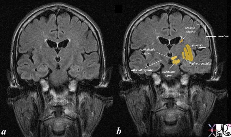
The subtthalamus is a cvomponent of the diencephalon and part of the group of structures loosely associated with the thalamus. Its major structure is the subthalamic nucleus which is a lens shaped structure functionally linked to the basal ganglia. It is situated anterior to the thalamusposterior to the substantia nigra and medial to the internal capsule.

Subthalamic Nuclii in the Subthalamus |
| 38610c02b brain forebrain basal ganglia basal ganglion subthalamic nucleus subthalamic nuclii MRI coronal FLAIR Davidoff MD |
DOMElement Object
(
    [schemaTypeInfo] =>
    [tagName] => table
    [firstElementChild] => (object value omitted)
    [lastElementChild] => (object value omitted)
    [childElementCount] => 1
    [previousElementSibling] => (object value omitted)
    [nextElementSibling] =>
    [nodeName] => table
    [nodeValue] =>


Functional Relationships
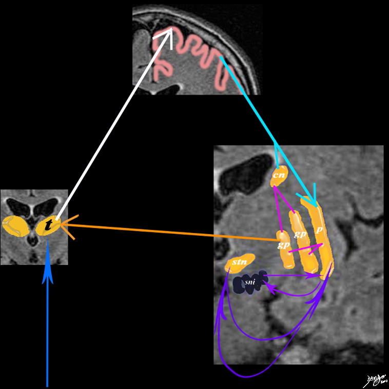
Subthalamus and Subthalamic Nucleus (stn) Integrated With the Basal Ganglia Thalamus and Cortex


A simplified drawing of the connections between the caudate nucleus (orange c), the sensory cortex (salmon pink) and the basal ganglia is shown. After the stimulus has reached the sensory cortex for quantification and qualification it connects to the basal ganglia through the caudate nucleus and putamen. Each of these connects with the two parts of the globus pallidus (gp) which feed back to the thalamus. The caudate nucleus also feeds back and forth to the substantia nigra (sni) and the subthalamic nucleus (stn) brain basal ganglia connections functional thalamus sensory cortex putamen= p caudate nucleus = cn globus pallidus = gp substantia nigra = sni subthalamic nucleus = snu
Courtesy Ashley Davidoff MD Copyright 2010 38610d09e02.8s


    [nodeType] => 1
    [parentNode] => (object value omitted)
    [childNodes] => (object value omitted)
    [firstChild] => (object value omitted)
    [lastChild] => (object value omitted)
    [previousSibling] => (object value omitted)
    [nextSibling] => (object value omitted)
    [attributes] => (object value omitted)
    [ownerDocument] => (object value omitted)
    [namespaceURI] =>
    [prefix] =>
    [localName] => table
    [baseURI] =>
    [textContent] =>


Functional Relationships
Subthalamus and Subthalamic Nucleus (stn) Integrated With the Basal Ganglia Thalamus and Cortex


A simplified drawing of the connections between the caudate nucleus (orange c), the sensory cortex (salmon pink) and the basal ganglia is shown. After the stimulus has reached the sensory cortex for quantification and qualification it connects to the basal ganglia through the caudate nucleus and putamen. Each of these connects with the two parts of the globus pallidus (gp) which feed back to the thalamus. The caudate nucleus also feeds back and forth to the substantia nigra (sni) and the subthalamic nucleus (stn) brain basal ganglia connections functional thalamus sensory cortex putamen= p caudate nucleus = cn globus pallidus = gp substantia nigra = sni subthalamic nucleus = snu
Courtesy Ashley Davidoff MD Copyright 2010 38610d09e02.8s


)
DOMElement Object
(
    [schemaTypeInfo] =>
    [tagName] => td
    [firstElementChild] => (object value omitted)
    [lastElementChild] => (object value omitted)
    [childElementCount] => 2
    [previousElementSibling] =>
    [nextElementSibling] =>
    [nodeName] => td
    [nodeValue] =>
A simplified drawing of the connections between the caudate nucleus (orange c), the sensory cortex (salmon pink) and the basal ganglia is shown. After the stimulus has reached the sensory cortex for quantification and qualification it connects to the basal ganglia through the caudate nucleus and putamen. Each of these connects with the two parts of the globus pallidus (gp) which feed back to the thalamus. The caudate nucleus also feeds back and forth to the substantia nigra (sni) and the subthalamic nucleus (stn) brain basal ganglia connections functional thalamus sensory cortex putamen= p caudate nucleus = cn globus pallidus = gp substantia nigra = sni subthalamic nucleus = snu
Courtesy Ashley Davidoff MD Copyright 2010 38610d09e02.8s

    [nodeType] => 1
    [parentNode] => (object value omitted)
    [childNodes] => (object value omitted)
    [firstChild] => (object value omitted)
    [lastChild] => (object value omitted)
    [previousSibling] => (object value omitted)
    [nextSibling] => (object value omitted)
    [attributes] => (object value omitted)
    [ownerDocument] => (object value omitted)
    [namespaceURI] =>
    [prefix] =>
    [localName] => td
    [baseURI] =>
    [textContent] =>
A simplified drawing of the connections between the caudate nucleus (orange c), the sensory cortex (salmon pink) and the basal ganglia is shown. After the stimulus has reached the sensory cortex for quantification and qualification it connects to the basal ganglia through the caudate nucleus and putamen. Each of these connects with the two parts of the globus pallidus (gp) which feed back to the thalamus. The caudate nucleus also feeds back and forth to the substantia nigra (sni) and the subthalamic nucleus (stn) brain basal ganglia connections functional thalamus sensory cortex putamen= p caudate nucleus = cn globus pallidus = gp substantia nigra = sni subthalamic nucleus = snu
Courtesy Ashley Davidoff MD Copyright 2010 38610d09e02.8s

)
DOMElement Object
(
    [schemaTypeInfo] =>
    [tagName] => td
    [firstElementChild] => (object value omitted)
    [lastElementChild] => (object value omitted)
    [childElementCount] => 3
    [previousElementSibling] =>
    [nextElementSibling] =>
    [nodeName] => td
    [nodeValue] =>
Functional Relationships
Subthalamus and Subthalamic Nucleus (stn) Integrated With the Basal Ganglia Thalamus and Cortex
    [nodeType] => 1
    [parentNode] => (object value omitted)
    [childNodes] => (object value omitted)
    [firstChild] => (object value omitted)
    [lastChild] => (object value omitted)
    [previousSibling] => (object value omitted)
    [nextSibling] => (object value omitted)
    [attributes] => (object value omitted)
    [ownerDocument] => (object value omitted)
    [namespaceURI] =>
    [prefix] =>
    [localName] => td
    [baseURI] =>
    [textContent] =>
Functional Relationships
Subthalamus and Subthalamic Nucleus (stn) Integrated With the Basal Ganglia Thalamus and Cortex
)
DOMElement Object
(
    [schemaTypeInfo] =>
    [tagName] => table
    [firstElementChild] => (object value omitted)
    [lastElementChild] => (object value omitted)
    [childElementCount] => 1
    [previousElementSibling] => (object value omitted)
    [nextElementSibling] => (object value omitted)
    [nodeName] => table
    [nodeValue] =>


Structural Relationships of the Subthalamic Nucleus and Subthalamus


The coronal T1 weighted image reveals the structures that are functionally related to basal ganglial function. These include the caudate nucleus, globus pallidus, putamen, substantia nigra and subthalamus. The caudate nucleus and the putamen are the doorway to the basal ganglia and they receive input from both the sensory cortex and motor cortex. They distribute the signals to the globus pallidus substantia nigra and subthalamic nuclii. The latter (two subthalamic nuclii and substantia nigra) process the signal and send the result back to the globus pallidus which in turn sends the signal back to the thalamus.
Courtesy Ashley Davidoff Md Copyright 2010 All rights reserved 38610c06c06d.81s


    [nodeType] => 1
    [parentNode] => (object value omitted)
    [childNodes] => (object value omitted)
    [firstChild] => (object value omitted)
    [lastChild] => (object value omitted)
    [previousSibling] => (object value omitted)
    [nextSibling] => (object value omitted)
    [attributes] => (object value omitted)
    [ownerDocument] => (object value omitted)
    [namespaceURI] =>
    [prefix] =>
    [localName] => table
    [baseURI] =>
    [textContent] =>


Structural Relationships of the Subthalamic Nucleus and Subthalamus


The coronal T1 weighted image reveals the structures that are functionally related to basal ganglial function. These include the caudate nucleus, globus pallidus, putamen, substantia nigra and subthalamus. The caudate nucleus and the putamen are the doorway to the basal ganglia and they receive input from both the sensory cortex and motor cortex. They distribute the signals to the globus pallidus substantia nigra and subthalamic nuclii. The latter (two subthalamic nuclii and substantia nigra) process the signal and send the result back to the globus pallidus which in turn sends the signal back to the thalamus.
Courtesy Ashley Davidoff Md Copyright 2010 All rights reserved 38610c06c06d.81s


)
DOMElement Object
(
    [schemaTypeInfo] =>
    [tagName] => td
    [firstElementChild] => (object value omitted)
    [lastElementChild] => (object value omitted)
    [childElementCount] => 2
    [previousElementSibling] =>
    [nextElementSibling] =>
    [nodeName] => td
    [nodeValue] =>
The coronal T1 weighted image reveals the structures that are functionally related to basal ganglial function. These include the caudate nucleus, globus pallidus, putamen, substantia nigra and subthalamus. The caudate nucleus and the putamen are the doorway to the basal ganglia and they receive input from both the sensory cortex and motor cortex. They distribute the signals to the globus pallidus substantia nigra and subthalamic nuclii. The latter (two subthalamic nuclii and substantia nigra) process the signal and send the result back to the globus pallidus which in turn sends the signal back to the thalamus.
Courtesy Ashley Davidoff Md Copyright 2010 All rights reserved 38610c06c06d.81s

    [nodeType] => 1
    [parentNode] => (object value omitted)
    [childNodes] => (object value omitted)
    [firstChild] => (object value omitted)
    [lastChild] => (object value omitted)
    [previousSibling] => (object value omitted)
    [nextSibling] => (object value omitted)
    [attributes] => (object value omitted)
    [ownerDocument] => (object value omitted)
    [namespaceURI] =>
    [prefix] =>
    [localName] => td
    [baseURI] =>
    [textContent] =>
The coronal T1 weighted image reveals the structures that are functionally related to basal ganglial function. These include the caudate nucleus, globus pallidus, putamen, substantia nigra and subthalamus. The caudate nucleus and the putamen are the doorway to the basal ganglia and they receive input from both the sensory cortex and motor cortex. They distribute the signals to the globus pallidus substantia nigra and subthalamic nuclii. The latter (two subthalamic nuclii and substantia nigra) process the signal and send the result back to the globus pallidus which in turn sends the signal back to the thalamus.
Courtesy Ashley Davidoff Md Copyright 2010 All rights reserved 38610c06c06d.81s

)
DOMElement Object
(
    [schemaTypeInfo] =>
    [tagName] => td
    [firstElementChild] => (object value omitted)
    [lastElementChild] => (object value omitted)
    [childElementCount] => 2
    [previousElementSibling] =>
    [nextElementSibling] =>
    [nodeName] => td
    [nodeValue] =>
Structural Relationships of the Subthalamic Nucleus and Subthalamus

    [nodeType] => 1
    [parentNode] => (object value omitted)
    [childNodes] => (object value omitted)
    [firstChild] => (object value omitted)
    [lastChild] => (object value omitted)
    [previousSibling] => (object value omitted)
    [nextSibling] => (object value omitted)
    [attributes] => (object value omitted)
    [ownerDocument] => (object value omitted)
    [namespaceURI] =>
    [prefix] =>
    [localName] => td
    [baseURI] =>
    [textContent] =>
Structural Relationships of the Subthalamic Nucleus and Subthalamus

)
DOMElement Object
(
    [schemaTypeInfo] =>
    [tagName] => table
    [firstElementChild] => (object value omitted)
    [lastElementChild] => (object value omitted)
    [childElementCount] => 1
    [previousElementSibling] => (object value omitted)
    [nextElementSibling] => (object value omitted)
    [nodeName] => table
    [nodeValue] =>


Subthalamic Nuclii in the Subthalamus


38610c02b brain forebrain basal ganglia basal ganglion subthalamic nucleus subthalamic nuclii MRI coronal FLAIR Davidoff MD


    [nodeType] => 1
    [parentNode] => (object value omitted)
    [childNodes] => (object value omitted)
    [firstChild] => (object value omitted)
    [lastChild] => (object value omitted)
    [previousSibling] => (object value omitted)
    [nextSibling] => (object value omitted)
    [attributes] => (object value omitted)
    [ownerDocument] => (object value omitted)
    [namespaceURI] =>
    [prefix] =>
    [localName] => table
    [baseURI] =>
    [textContent] =>


Subthalamic Nuclii in the Subthalamus


38610c02b brain forebrain basal ganglia basal ganglion subthalamic nucleus subthalamic nuclii MRI coronal FLAIR Davidoff MD


)
DOMElement Object
(
    [schemaTypeInfo] =>
    [tagName] => td
    [firstElementChild] =>
    [lastElementChild] =>
    [childElementCount] => 0
    [previousElementSibling] =>
    [nextElementSibling] =>
    [nodeName] => td
    [nodeValue] => 38610c02b brain forebrain basal ganglia basal ganglion subthalamic nucleus subthalamic nuclii MRI coronal FLAIR Davidoff MD
    [nodeType] => 1
    [parentNode] => (object value omitted)
    [childNodes] => (object value omitted)
    [firstChild] => (object value omitted)
    [lastChild] => (object value omitted)
    [previousSibling] => (object value omitted)
    [nextSibling] => (object value omitted)
    [attributes] => (object value omitted)
    [ownerDocument] => (object value omitted)
    [namespaceURI] =>
    [prefix] =>
    [localName] => td
    [baseURI] =>
    [textContent] => 38610c02b brain forebrain basal ganglia basal ganglion subthalamic nucleus subthalamic nuclii MRI coronal FLAIR Davidoff MD
)
DOMElement Object
(
    [schemaTypeInfo] =>
    [tagName] => td
    [firstElementChild] => (object value omitted)
    [lastElementChild] => (object value omitted)
    [childElementCount] => 2
    [previousElementSibling] =>
    [nextElementSibling] =>
    [nodeName] => td
    [nodeValue] =>
Subthalamic Nuclii in the Subthalamus
    [nodeType] => 1
    [parentNode] => (object value omitted)
    [childNodes] => (object value omitted)
    [firstChild] => (object value omitted)
    [lastChild] => (object value omitted)
    [previousSibling] => (object value omitted)
    [nextSibling] => (object value omitted)
    [attributes] => (object value omitted)
    [ownerDocument] => (object value omitted)
    [namespaceURI] =>
    [prefix] =>
    [localName] => td
    [baseURI] =>
    [textContent] =>
Subthalamic Nuclii in the Subthalamus
)


