Fourth Ventricle
Sumit Karia MD
The Common vein Copyright 2010
Definition
The fourth ventricle is a middle cavity located behind the brain stem.
It constitutes a dilation of the ependymal cavity at the medullar-pontine level and extends into the cerbellum. It extends from the cerebral aqueduct to the obex(separates the medulla oblongata from the spinal cord) . The cerebellum forms the roof of the fourth ventricle and the rhomboid fossa acts as the floor. The cerebral penduncles forma the posterolateral walls of the fourth ventricl. . The vermis and cerebellum form the roof of the fourth ventricle.
Like all the ventricles, it is lined by an ependymal epithelium that secretes CSF through the choroid plexus.
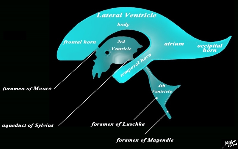
It communicates above with the third ventricle through the aqueduct of Sylvius, and also communicates with the subarachnoid spaces the foramens of Magendie and Luschka. Below, it continues with the central canal of the spinal cord via the cisterna magna.
Shape – It has a flattened form from front to rear and is rhomboid in shape.
Position Its anterior face is formed by the dorsal surfaces of the pons and the medulla; a posterior that corresponds to the hilum of the cerebellum.
Character Essentially a cavity whose walls are covered with ependyma, filled with CSF.
Sagittal Concepts
|
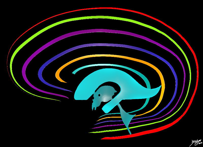
The Major Components of the Ventricles |
| The diagram in the sagital projection reveals the horizontal portion called the lateral ventricle. It is a paired structure.The vertical portion (light green consists of the midline 3rd and 4th ventricle.
Courtesy Ashley Davidoff MD copyright 2010 all rights reserved 94459b06b.8s |
|
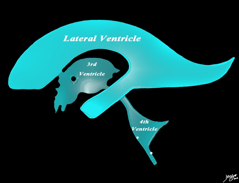
Sagittal View of the Ventricles |
|
The diagram in the sagittal projection reveals the horizontal portion called the lateral ventricle. It is a paired structure that houses the frontal horn, body, and the vertical portion which is composed of the 3rd ventricle, cerebral aqueduct and the 4th ventricle. The lateral ventricle consists of the frontal horn, body, occipital horn, atrium and the temporal horn The foramen of Monro connects the lateral ventricles with the third ventricle. The paired foramina of Luschka are sitiuated anteriorly in the 4th ventricle and they allow CSF to circulate in the subarachnoid spaces. The foramen of Magendie is a single structure and is situated posteriorly and it also enables CSF to enter the subarachnoid space. Courtesy Ashley Davidoff MD copyright 2010 all rights reserved 94459b10b02.82s |
Axial Projection
|
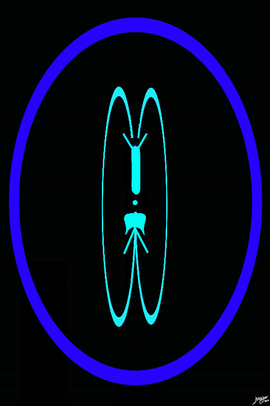
The 4th Ventricle |
|
The 4th ventricle is the last landmark of the ventricular system seen as we proceed inferiorly. It is seen as a puffed crown or the pontifs hat (mitre), which lies posterior to the pons (ponntiff /pons) inferior to the aqueduct of Sylvius and anterior to the cerebellum. It lies within the hinbrain. Courtesy Ashley Davidoff copyright 2010 all rights reserved 93914.3kd06bd03.8s |
|
The Distribution of the ventricles in the Brain Forebrain (green) Midbrain (orange) and hindbrain (salmon) |
|
This diagram shows the positioning of each of the components of the ventriclar system . The lateral ventricles and third ventricle lie in the forebrain (green), the aqueduct of Sylvius (orange) lies in the midbrain, and the 4th ventricle (salmon) lies in the hinbrain. Courtesy Ashley Davidoff copyright 2010 all rights reserved 93914.3kd06bd04.8s |
Coronal Plane
|
4th Ventricle |
|
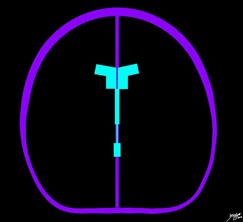
The 4th ventricle has been added to the coronal diagram of the brain. Courtesy Ashley DAvidoff MD copyright 2010 all rights reserved 93914b04d05.8s |
|
4th Ventricle |
|
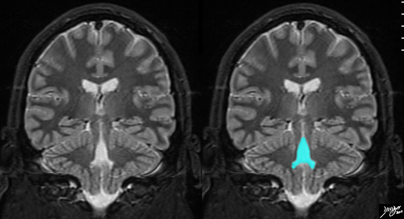
The 4th ventricle is demonstrated in this coronal MRI using a STIR sequence. It is easily recognized and takes on a variety of shapes. In this instance it has the shape of a medieval hat or perhaps an elongated bell. It is a midline structure and is situated anterior to the cerebellum and posterior to the pons. Courtesy Ashley Davidoff MD copyright 2010 all rights reserved concepts 89741cb01.8s |
Applied Biology – Diseases
DOMElement Object
(
    [schemaTypeInfo] =>
    [tagName] => table
    [firstElementChild] => (object value omitted)
    [lastElementChild] => (object value omitted)
    [childElementCount] => 1
    [previousElementSibling] => (object value omitted)
    [nextElementSibling] =>
    [nodeName] => table
    [nodeValue] =>


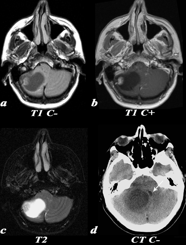
Medulloblastoma Causing Obstruction at the level of the 4th Ventricle


This 26 year old male presented with a three week history of progressive headache, nausea and vomiting. T1 pre (a) and post (b,): The exact origin of this mass can be difficult to ascertain given its large size. It clearly grows into the fourth ventricle, widening the lower portion of the Sylvian aqueduct. Note the smooth interface with the posterior aspect of the pons with associated mass effect verses the more irregular margin posteriorly where it arises from the cerebellum. T2 (c): The solid component of the medulloblastoma matches gray matter signal. Cystic or necrotic areas are demonstrated by higher T2 signal areas. CT(d): The unenhanced CT scan demonstrates a predominately hyperdense mass with punctuate calcifications in the region of the fourth ventricle . The low density area posteriorly is a cystic or necrotic component of the mass while the fourth ventricle is completely effaced.
Image Courtesy Elisa Flower MD and Asim Mian MD 97668c01.81


    [nodeType] => 1
    [parentNode] => (object value omitted)
    [childNodes] => (object value omitted)
    [firstChild] => (object value omitted)
    [lastChild] => (object value omitted)
    [previousSibling] => (object value omitted)
    [nextSibling] => (object value omitted)
    [attributes] => (object value omitted)
    [ownerDocument] => (object value omitted)
    [namespaceURI] =>
    [prefix] =>
    [localName] => table
    [baseURI] =>
    [textContent] =>


Medulloblastoma Causing Obstruction at the level of the 4th Ventricle


This 26 year old male presented with a three week history of progressive headache, nausea and vomiting. T1 pre (a) and post (b,): The exact origin of this mass can be difficult to ascertain given its large size. It clearly grows into the fourth ventricle, widening the lower portion of the Sylvian aqueduct. Note the smooth interface with the posterior aspect of the pons with associated mass effect verses the more irregular margin posteriorly where it arises from the cerebellum. T2 (c): The solid component of the medulloblastoma matches gray matter signal. Cystic or necrotic areas are demonstrated by higher T2 signal areas. CT(d): The unenhanced CT scan demonstrates a predominately hyperdense mass with punctuate calcifications in the region of the fourth ventricle . The low density area posteriorly is a cystic or necrotic component of the mass while the fourth ventricle is completely effaced.
Image Courtesy Elisa Flower MD and Asim Mian MD 97668c01.81


)
DOMElement Object
(
    [schemaTypeInfo] =>
    [tagName] => td
    [firstElementChild] => (object value omitted)
    [lastElementChild] => (object value omitted)
    [childElementCount] => 2
    [previousElementSibling] =>
    [nextElementSibling] =>
    [nodeName] => td
    [nodeValue] =>
This 26 year old male presented with a three week history of progressive headache, nausea and vomiting. T1 pre (a) and post (b,): The exact origin of this mass can be difficult to ascertain given its large size. It clearly grows into the fourth ventricle, widening the lower portion of the Sylvian aqueduct. Note the smooth interface with the posterior aspect of the pons with associated mass effect verses the more irregular margin posteriorly where it arises from the cerebellum. T2 (c): The solid component of the medulloblastoma matches gray matter signal. Cystic or necrotic areas are demonstrated by higher T2 signal areas. CT(d): The unenhanced CT scan demonstrates a predominately hyperdense mass with punctuate calcifications in the region of the fourth ventricle . The low density area posteriorly is a cystic or necrotic component of the mass while the fourth ventricle is completely effaced.
Image Courtesy Elisa Flower MD and Asim Mian MD 97668c01.81

    [nodeType] => 1
    [parentNode] => (object value omitted)
    [childNodes] => (object value omitted)
    [firstChild] => (object value omitted)
    [lastChild] => (object value omitted)
    [previousSibling] => (object value omitted)
    [nextSibling] => (object value omitted)
    [attributes] => (object value omitted)
    [ownerDocument] => (object value omitted)
    [namespaceURI] =>
    [prefix] =>
    [localName] => td
    [baseURI] =>
    [textContent] =>
This 26 year old male presented with a three week history of progressive headache, nausea and vomiting. T1 pre (a) and post (b,): The exact origin of this mass can be difficult to ascertain given its large size. It clearly grows into the fourth ventricle, widening the lower portion of the Sylvian aqueduct. Note the smooth interface with the posterior aspect of the pons with associated mass effect verses the more irregular margin posteriorly where it arises from the cerebellum. T2 (c): The solid component of the medulloblastoma matches gray matter signal. Cystic or necrotic areas are demonstrated by higher T2 signal areas. CT(d): The unenhanced CT scan demonstrates a predominately hyperdense mass with punctuate calcifications in the region of the fourth ventricle . The low density area posteriorly is a cystic or necrotic component of the mass while the fourth ventricle is completely effaced.
Image Courtesy Elisa Flower MD and Asim Mian MD 97668c01.81

)
DOMElement Object
(
    [schemaTypeInfo] =>
    [tagName] => td
    [firstElementChild] => (object value omitted)
    [lastElementChild] => (object value omitted)
    [childElementCount] => 2
    [previousElementSibling] =>
    [nextElementSibling] =>
    [nodeName] => td
    [nodeValue] =>
Medulloblastoma Causing Obstruction at the level of the 4th Ventricle

    [nodeType] => 1
    [parentNode] => (object value omitted)
    [childNodes] => (object value omitted)
    [firstChild] => (object value omitted)
    [lastChild] => (object value omitted)
    [previousSibling] => (object value omitted)
    [nextSibling] => (object value omitted)
    [attributes] => (object value omitted)
    [ownerDocument] => (object value omitted)
    [namespaceURI] =>
    [prefix] =>
    [localName] => td
    [baseURI] =>
    [textContent] =>
Medulloblastoma Causing Obstruction at the level of the 4th Ventricle

)
DOMElement Object
(
    [schemaTypeInfo] =>
    [tagName] => table
    [firstElementChild] => (object value omitted)
    [lastElementChild] => (object value omitted)
    [childElementCount] => 1
    [previousElementSibling] => (object value omitted)
    [nextElementSibling] => (object value omitted)
    [nodeName] => table
    [nodeValue] =>


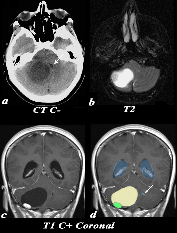
Hemangioblastoma Compressing the 4th ventricle – Hydrocephalus


This 55 year old female complained of an occipital headache for one month with increased difficulty ambulating. CT: Noncontrast CT image (a) demonstrates a large low density lesion in the right cerebellum. This lesion causes significant mass effect on the fourth ventricle which is displaced towards the left (white arrow). This mass effect caused obstruction of the CSF pathway resulting in obstructive hydrocephalus and subsequent symptoms. MRI: The T2 weighted image (b) demonstrates a predominantly T2 hyperintense cystic lesion with a nodular T2 isointense component seen laterally. Post contrast T1 weighted coronal images (identified by high signal in the venous sinuses demonstrate the classic cyst (light yellow in d) with mural nodule (green in d) appearance of a hemangioblastoma. Note the enlarged lateral ventricles (blue) and the displaced and compressed 4th ventricle (white arrow d).
Image Courtesy Elisa Flower MD and Asim Mian MD 97652c01.81


    [nodeType] => 1
    [parentNode] => (object value omitted)
    [childNodes] => (object value omitted)
    [firstChild] => (object value omitted)
    [lastChild] => (object value omitted)
    [previousSibling] => (object value omitted)
    [nextSibling] => (object value omitted)
    [attributes] => (object value omitted)
    [ownerDocument] => (object value omitted)
    [namespaceURI] =>
    [prefix] =>
    [localName] => table
    [baseURI] =>
    [textContent] =>


Hemangioblastoma Compressing the 4th ventricle – Hydrocephalus


This 55 year old female complained of an occipital headache for one month with increased difficulty ambulating. CT: Noncontrast CT image (a) demonstrates a large low density lesion in the right cerebellum. This lesion causes significant mass effect on the fourth ventricle which is displaced towards the left (white arrow). This mass effect caused obstruction of the CSF pathway resulting in obstructive hydrocephalus and subsequent symptoms. MRI: The T2 weighted image (b) demonstrates a predominantly T2 hyperintense cystic lesion with a nodular T2 isointense component seen laterally. Post contrast T1 weighted coronal images (identified by high signal in the venous sinuses demonstrate the classic cyst (light yellow in d) with mural nodule (green in d) appearance of a hemangioblastoma. Note the enlarged lateral ventricles (blue) and the displaced and compressed 4th ventricle (white arrow d).
Image Courtesy Elisa Flower MD and Asim Mian MD 97652c01.81


)
DOMElement Object
(
    [schemaTypeInfo] =>
    [tagName] => td
    [firstElementChild] => (object value omitted)
    [lastElementChild] => (object value omitted)
    [childElementCount] => 2
    [previousElementSibling] =>
    [nextElementSibling] =>
    [nodeName] => td
    [nodeValue] =>
This 55 year old female complained of an occipital headache for one month with increased difficulty ambulating. CT: Noncontrast CT image (a) demonstrates a large low density lesion in the right cerebellum. This lesion causes significant mass effect on the fourth ventricle which is displaced towards the left (white arrow). This mass effect caused obstruction of the CSF pathway resulting in obstructive hydrocephalus and subsequent symptoms. MRI: The T2 weighted image (b) demonstrates a predominantly T2 hyperintense cystic lesion with a nodular T2 isointense component seen laterally. Post contrast T1 weighted coronal images (identified by high signal in the venous sinuses demonstrate the classic cyst (light yellow in d) with mural nodule (green in d) appearance of a hemangioblastoma. Note the enlarged lateral ventricles (blue) and the displaced and compressed 4th ventricle (white arrow d).
Image Courtesy Elisa Flower MD and Asim Mian MD 97652c01.81

    [nodeType] => 1
    [parentNode] => (object value omitted)
    [childNodes] => (object value omitted)
    [firstChild] => (object value omitted)
    [lastChild] => (object value omitted)
    [previousSibling] => (object value omitted)
    [nextSibling] => (object value omitted)
    [attributes] => (object value omitted)
    [ownerDocument] => (object value omitted)
    [namespaceURI] =>
    [prefix] =>
    [localName] => td
    [baseURI] =>
    [textContent] =>
This 55 year old female complained of an occipital headache for one month with increased difficulty ambulating. CT: Noncontrast CT image (a) demonstrates a large low density lesion in the right cerebellum. This lesion causes significant mass effect on the fourth ventricle which is displaced towards the left (white arrow). This mass effect caused obstruction of the CSF pathway resulting in obstructive hydrocephalus and subsequent symptoms. MRI: The T2 weighted image (b) demonstrates a predominantly T2 hyperintense cystic lesion with a nodular T2 isointense component seen laterally. Post contrast T1 weighted coronal images (identified by high signal in the venous sinuses demonstrate the classic cyst (light yellow in d) with mural nodule (green in d) appearance of a hemangioblastoma. Note the enlarged lateral ventricles (blue) and the displaced and compressed 4th ventricle (white arrow d).
Image Courtesy Elisa Flower MD and Asim Mian MD 97652c01.81

)
DOMElement Object
(
    [schemaTypeInfo] =>
    [tagName] => td
    [firstElementChild] => (object value omitted)
    [lastElementChild] => (object value omitted)
    [childElementCount] => 2
    [previousElementSibling] =>
    [nextElementSibling] =>
    [nodeName] => td
    [nodeValue] =>
Hemangioblastoma Compressing the 4th ventricle – Hydrocephalus
    [nodeType] => 1
    [parentNode] => (object value omitted)
    [childNodes] => (object value omitted)
    [firstChild] => (object value omitted)
    [lastChild] => (object value omitted)
    [previousSibling] => (object value omitted)
    [nextSibling] => (object value omitted)
    [attributes] => (object value omitted)
    [ownerDocument] => (object value omitted)
    [namespaceURI] =>
    [prefix] =>
    [localName] => td
    [baseURI] =>
    [textContent] =>
Hemangioblastoma Compressing the 4th ventricle – Hydrocephalus
)
DOMElement Object
(
    [schemaTypeInfo] =>
    [tagName] => table
    [firstElementChild] => (object value omitted)
    [lastElementChild] => (object value omitted)
    [childElementCount] => 1
    [previousElementSibling] => (object value omitted)
    [nextElementSibling] => (object value omitted)
    [nodeName] => table
    [nodeValue] =>


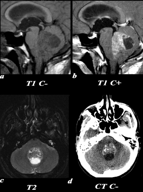
Hemangioblastoma Causing Leftward Shift of the 4th ventricle and Hydrocephalus


This 55 year old female complained of an occipital headache for one month with increased difficulty ambulating. MRI: T1 weighted images without contrast (a) demonstrate a T1 isointense nodule at the periphery of the lesion. Post contrast T1 weighted images (identified by high signal in the venous sinuses and high signal in the nasal mucosa due to normal enhancement) demonstrate enhancement of the nodular component of this tumor. The T2 weighted image demonstrates a predominantly T2 hyperintense cystic lesion with a nodular T2 isointense component seen laterally. CT: Noncontrast CT image demonstrates a large low density lesion in the right cerebellum. This lesion causes significant mass effect on the fourth ventricle which is displaced towards the left (white arrow). This mass effect caused obstruction of the CSF pathway resulting in obstructive hydrocephalus and subsequent symptoms.
Image Courtesy Elisa Flower MD and Asim Mian MD 97652c.81


    [nodeType] => 1
    [parentNode] => (object value omitted)
    [childNodes] => (object value omitted)
    [firstChild] => (object value omitted)
    [lastChild] => (object value omitted)
    [previousSibling] => (object value omitted)
    [nextSibling] => (object value omitted)
    [attributes] => (object value omitted)
    [ownerDocument] => (object value omitted)
    [namespaceURI] =>
    [prefix] =>
    [localName] => table
    [baseURI] =>
    [textContent] =>


Hemangioblastoma Causing Leftward Shift of the 4th ventricle and Hydrocephalus


This 55 year old female complained of an occipital headache for one month with increased difficulty ambulating. MRI: T1 weighted images without contrast (a) demonstrate a T1 isointense nodule at the periphery of the lesion. Post contrast T1 weighted images (identified by high signal in the venous sinuses and high signal in the nasal mucosa due to normal enhancement) demonstrate enhancement of the nodular component of this tumor. The T2 weighted image demonstrates a predominantly T2 hyperintense cystic lesion with a nodular T2 isointense component seen laterally. CT: Noncontrast CT image demonstrates a large low density lesion in the right cerebellum. This lesion causes significant mass effect on the fourth ventricle which is displaced towards the left (white arrow). This mass effect caused obstruction of the CSF pathway resulting in obstructive hydrocephalus and subsequent symptoms.
Image Courtesy Elisa Flower MD and Asim Mian MD 97652c.81


)
DOMElement Object
(
    [schemaTypeInfo] =>
    [tagName] => td
    [firstElementChild] => (object value omitted)
    [lastElementChild] => (object value omitted)
    [childElementCount] => 2
    [previousElementSibling] =>
    [nextElementSibling] =>
    [nodeName] => td
    [nodeValue] =>
This 55 year old female complained of an occipital headache for one month with increased difficulty ambulating. MRI: T1 weighted images without contrast (a) demonstrate a T1 isointense nodule at the periphery of the lesion. Post contrast T1 weighted images (identified by high signal in the venous sinuses and high signal in the nasal mucosa due to normal enhancement) demonstrate enhancement of the nodular component of this tumor. The T2 weighted image demonstrates a predominantly T2 hyperintense cystic lesion with a nodular T2 isointense component seen laterally. CT: Noncontrast CT image demonstrates a large low density lesion in the right cerebellum. This lesion causes significant mass effect on the fourth ventricle which is displaced towards the left (white arrow). This mass effect caused obstruction of the CSF pathway resulting in obstructive hydrocephalus and subsequent symptoms.
Image Courtesy Elisa Flower MD and Asim Mian MD 97652c.81

    [nodeType] => 1
    [parentNode] => (object value omitted)
    [childNodes] => (object value omitted)
    [firstChild] => (object value omitted)
    [lastChild] => (object value omitted)
    [previousSibling] => (object value omitted)
    [nextSibling] => (object value omitted)
    [attributes] => (object value omitted)
    [ownerDocument] => (object value omitted)
    [namespaceURI] =>
    [prefix] =>
    [localName] => td
    [baseURI] =>
    [textContent] =>
This 55 year old female complained of an occipital headache for one month with increased difficulty ambulating. MRI: T1 weighted images without contrast (a) demonstrate a T1 isointense nodule at the periphery of the lesion. Post contrast T1 weighted images (identified by high signal in the venous sinuses and high signal in the nasal mucosa due to normal enhancement) demonstrate enhancement of the nodular component of this tumor. The T2 weighted image demonstrates a predominantly T2 hyperintense cystic lesion with a nodular T2 isointense component seen laterally. CT: Noncontrast CT image demonstrates a large low density lesion in the right cerebellum. This lesion causes significant mass effect on the fourth ventricle which is displaced towards the left (white arrow). This mass effect caused obstruction of the CSF pathway resulting in obstructive hydrocephalus and subsequent symptoms.
Image Courtesy Elisa Flower MD and Asim Mian MD 97652c.81

)
DOMElement Object
(
    [schemaTypeInfo] =>
    [tagName] => td
    [firstElementChild] => (object value omitted)
    [lastElementChild] => (object value omitted)
    [childElementCount] => 2
    [previousElementSibling] =>
    [nextElementSibling] =>
    [nodeName] => td
    [nodeValue] =>
Hemangioblastoma Causing Leftward Shift of the 4th ventricle and Hydrocephalus
    [nodeType] => 1
    [parentNode] => (object value omitted)
    [childNodes] => (object value omitted)
    [firstChild] => (object value omitted)
    [lastChild] => (object value omitted)
    [previousSibling] => (object value omitted)
    [nextSibling] => (object value omitted)
    [attributes] => (object value omitted)
    [ownerDocument] => (object value omitted)
    [namespaceURI] =>
    [prefix] =>
    [localName] => td
    [baseURI] =>
    [textContent] =>
Hemangioblastoma Causing Leftward Shift of the 4th ventricle and Hydrocephalus
)
DOMElement Object
(
    [schemaTypeInfo] =>
    [tagName] => table
    [firstElementChild] => (object value omitted)
    [lastElementChild] => (object value omitted)
    [childElementCount] => 1
    [previousElementSibling] => (object value omitted)
    [nextElementSibling] => (object value omitted)
    [nodeName] => table
    [nodeValue] =>


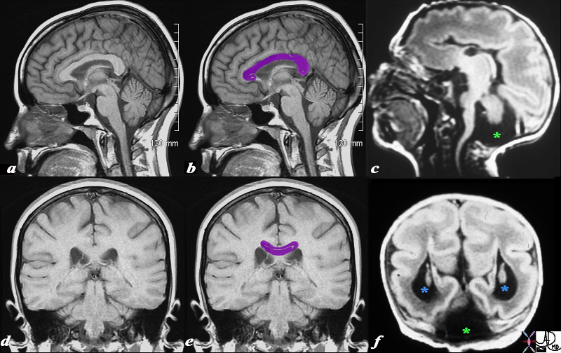
Dandy Walker Syndrom (c,f) Expanded 4th Ventricle


 This T1 weighted MRI is from a normal patient juxtaposed on a T1 weighted MRI from a neonate with agenesis of the corpus callosum, congenital lissencephaly and Dandy Walker syndrome. The normal corpus callosum (purple overlay ) is seen above the ventricles between the ventricles and the supracallosul gyrus and cingulate gyrus. . In the neonate (c,f) the there is no white matter in the expected position of the corpus callosum. These findings are consistent with a diagnosis of agenesis of the corpus callosum In addition, the posterior fossa does not show a normal cerebellum. Instead it is mostly filled with CSF (dark T1 green asterisk) likely due to a dilated 4th ventricle and atrophy of the vermis. Only a small amount of posterior fossa soft tissue is seen. In addition the posterior horns are dilated (blue asterisk). This finding is consistent with a diagnosis of Dandy-Walker syndrome associated with hydrocephalus. Lastly the patient has fewer gyri and sulci than normal. The gyri that are present are also more shallow than normal. These findings are consistent with lissencephaly.
Courtesy James Donnelly MD Copyright 2010 All rights reserved 95466c01L03.8s


    [nodeType] => 1
    [parentNode] => (object value omitted)
    [childNodes] => (object value omitted)
    [firstChild] => (object value omitted)
    [lastChild] => (object value omitted)
    [previousSibling] => (object value omitted)
    [nextSibling] => (object value omitted)
    [attributes] => (object value omitted)
    [ownerDocument] => (object value omitted)
    [namespaceURI] =>
    [prefix] =>
    [localName] => table
    [baseURI] =>
    [textContent] =>


Dandy Walker Syndrom (c,f) Expanded 4th Ventricle


 This T1 weighted MRI is from a normal patient juxtaposed on a T1 weighted MRI from a neonate with agenesis of the corpus callosum, congenital lissencephaly and Dandy Walker syndrome. The normal corpus callosum (purple overlay ) is seen above the ventricles between the ventricles and the supracallosul gyrus and cingulate gyrus. . In the neonate (c,f) the there is no white matter in the expected position of the corpus callosum. These findings are consistent with a diagnosis of agenesis of the corpus callosum In addition, the posterior fossa does not show a normal cerebellum. Instead it is mostly filled with CSF (dark T1 green asterisk) likely due to a dilated 4th ventricle and atrophy of the vermis. Only a small amount of posterior fossa soft tissue is seen. In addition the posterior horns are dilated (blue asterisk). This finding is consistent with a diagnosis of Dandy-Walker syndrome associated with hydrocephalus. Lastly the patient has fewer gyri and sulci than normal. The gyri that are present are also more shallow than normal. These findings are consistent with lissencephaly.
Courtesy James Donnelly MD Copyright 2010 All rights reserved 95466c01L03.8s


)
DOMElement Object
(
    [schemaTypeInfo] =>
    [tagName] => td
    [firstElementChild] => (object value omitted)
    [lastElementChild] => (object value omitted)
    [childElementCount] => 2
    [previousElementSibling] =>
    [nextElementSibling] =>
    [nodeName] => td
    [nodeValue] =>
 This T1 weighted MRI is from a normal patient juxtaposed on a T1 weighted MRI from a neonate with agenesis of the corpus callosum, congenital lissencephaly and Dandy Walker syndrome. The normal corpus callosum (purple overlay ) is seen above the ventricles between the ventricles and the supracallosul gyrus and cingulate gyrus. . In the neonate (c,f) the there is no white matter in the expected position of the corpus callosum. These findings are consistent with a diagnosis of agenesis of the corpus callosum In addition, the posterior fossa does not show a normal cerebellum. Instead it is mostly filled with CSF (dark T1 green asterisk) likely due to a dilated 4th ventricle and atrophy of the vermis. Only a small amount of posterior fossa soft tissue is seen. In addition the posterior horns are dilated (blue asterisk). This finding is consistent with a diagnosis of Dandy-Walker syndrome associated with hydrocephalus. Lastly the patient has fewer gyri and sulci than normal. The gyri that are present are also more shallow than normal. These findings are consistent with lissencephaly.
Courtesy James Donnelly MD Copyright 2010 All rights reserved 95466c01L03.8s

    [nodeType] => 1
    [parentNode] => (object value omitted)
    [childNodes] => (object value omitted)
    [firstChild] => (object value omitted)
    [lastChild] => (object value omitted)
    [previousSibling] => (object value omitted)
    [nextSibling] => (object value omitted)
    [attributes] => (object value omitted)
    [ownerDocument] => (object value omitted)
    [namespaceURI] =>
    [prefix] =>
    [localName] => td
    [baseURI] =>
    [textContent] =>
 This T1 weighted MRI is from a normal patient juxtaposed on a T1 weighted MRI from a neonate with agenesis of the corpus callosum, congenital lissencephaly and Dandy Walker syndrome. The normal corpus callosum (purple overlay ) is seen above the ventricles between the ventricles and the supracallosul gyrus and cingulate gyrus. . In the neonate (c,f) the there is no white matter in the expected position of the corpus callosum. These findings are consistent with a diagnosis of agenesis of the corpus callosum In addition, the posterior fossa does not show a normal cerebellum. Instead it is mostly filled with CSF (dark T1 green asterisk) likely due to a dilated 4th ventricle and atrophy of the vermis. Only a small amount of posterior fossa soft tissue is seen. In addition the posterior horns are dilated (blue asterisk). This finding is consistent with a diagnosis of Dandy-Walker syndrome associated with hydrocephalus. Lastly the patient has fewer gyri and sulci than normal. The gyri that are present are also more shallow than normal. These findings are consistent with lissencephaly.
Courtesy James Donnelly MD Copyright 2010 All rights reserved 95466c01L03.8s

)
DOMElement Object
(
    [schemaTypeInfo] =>
    [tagName] => td
    [firstElementChild] => (object value omitted)
    [lastElementChild] => (object value omitted)
    [childElementCount] => 2
    [previousElementSibling] =>
    [nextElementSibling] =>
    [nodeName] => td
    [nodeValue] =>
Dandy Walker Syndrom (c,f) Expanded 4th Ventricle
    [nodeType] => 1
    [parentNode] => (object value omitted)
    [childNodes] => (object value omitted)
    [firstChild] => (object value omitted)
    [lastChild] => (object value omitted)
    [previousSibling] => (object value omitted)
    [nextSibling] => (object value omitted)
    [attributes] => (object value omitted)
    [ownerDocument] => (object value omitted)
    [namespaceURI] =>
    [prefix] =>
    [localName] => td
    [baseURI] =>
    [textContent] =>
Dandy Walker Syndrom (c,f) Expanded 4th Ventricle
)
DOMElement Object
(
    [schemaTypeInfo] =>
    [tagName] => table
    [firstElementChild] => (object value omitted)
    [lastElementChild] => (object value omitted)
    [childElementCount] => 1
    [previousElementSibling] => (object value omitted)
    [nextElementSibling] => (object value omitted)
    [nodeName] => table
    [nodeValue] =>


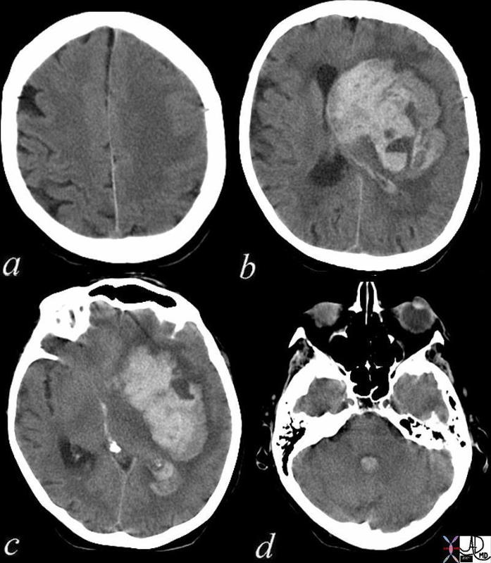
Acute Cerebral Hemorrhage with Mass Effect and Rupture into the Ventricles


This CT shows an acute hemorrhagic event originating in the frontoparietal region of the left cerebral hemisphere causing significant mass effect by compressing and displacing the ipsilateral lateral ventricle with significant midline shift.
The hemorrhage has ruptured into the ipsilateral lateral ventricle and blood can be seen within the choroid plexus (b) in the posterior horn (c) as well as the 4th ventricle (d).
The ipsilateral edema has caused loss of the gray white matter interface in the left parietal lobe
Courtesy Ashley Davidoff MD Copyright 2010 72143c01


    [nodeType] => 1
    [parentNode] => (object value omitted)
    [childNodes] => (object value omitted)
    [firstChild] => (object value omitted)
    [lastChild] => (object value omitted)
    [previousSibling] => (object value omitted)
    [nextSibling] => (object value omitted)
    [attributes] => (object value omitted)
    [ownerDocument] => (object value omitted)
    [namespaceURI] =>
    [prefix] =>
    [localName] => table
    [baseURI] =>
    [textContent] =>


Acute Cerebral Hemorrhage with Mass Effect and Rupture into the Ventricles


This CT shows an acute hemorrhagic event originating in the frontoparietal region of the left cerebral hemisphere causing significant mass effect by compressing and displacing the ipsilateral lateral ventricle with significant midline shift.
The hemorrhage has ruptured into the ipsilateral lateral ventricle and blood can be seen within the choroid plexus (b) in the posterior horn (c) as well as the 4th ventricle (d).
The ipsilateral edema has caused loss of the gray white matter interface in the left parietal lobe
Courtesy Ashley Davidoff MD Copyright 2010 72143c01


)
DOMElement Object
(
    [schemaTypeInfo] =>
    [tagName] => td
    [firstElementChild] => (object value omitted)
    [lastElementChild] => (object value omitted)
    [childElementCount] => 4
    [previousElementSibling] =>
    [nextElementSibling] =>
    [nodeName] => td
    [nodeValue] => This CT shows an acute hemorrhagic event originating in the frontoparietal region of the left cerebral hemisphere causing significant mass effect by compressing and displacing the ipsilateral lateral ventricle with significant midline shift.
The hemorrhage has ruptured into the ipsilateral lateral ventricle and blood can be seen within the choroid plexus (b) in the posterior horn (c) as well as the 4th ventricle (d).
The ipsilateral edema has caused loss of the gray white matter interface in the left parietal lobe
Courtesy Ashley Davidoff MD Copyright 2010 72143c01
    [nodeType] => 1
    [parentNode] => (object value omitted)
    [childNodes] => (object value omitted)
    [firstChild] => (object value omitted)
    [lastChild] => (object value omitted)
    [previousSibling] => (object value omitted)
    [nextSibling] => (object value omitted)
    [attributes] => (object value omitted)
    [ownerDocument] => (object value omitted)
    [namespaceURI] =>
    [prefix] =>
    [localName] => td
    [baseURI] =>
    [textContent] => This CT shows an acute hemorrhagic event originating in the frontoparietal region of the left cerebral hemisphere causing significant mass effect by compressing and displacing the ipsilateral lateral ventricle with significant midline shift.
The hemorrhage has ruptured into the ipsilateral lateral ventricle and blood can be seen within the choroid plexus (b) in the posterior horn (c) as well as the 4th ventricle (d).
The ipsilateral edema has caused loss of the gray white matter interface in the left parietal lobe
Courtesy Ashley Davidoff MD Copyright 2010 72143c01
)
DOMElement Object
(
    [schemaTypeInfo] =>
    [tagName] => td
    [firstElementChild] => (object value omitted)
    [lastElementChild] => (object value omitted)
    [childElementCount] => 2
    [previousElementSibling] =>
    [nextElementSibling] =>
    [nodeName] => td
    [nodeValue] =>
Acute Cerebral Hemorrhage with Mass Effect and Rupture into the Ventricles
    [nodeType] => 1
    [parentNode] => (object value omitted)
    [childNodes] => (object value omitted)
    [firstChild] => (object value omitted)
    [lastChild] => (object value omitted)
    [previousSibling] => (object value omitted)
    [nextSibling] => (object value omitted)
    [attributes] => (object value omitted)
    [ownerDocument] => (object value omitted)
    [namespaceURI] =>
    [prefix] =>
    [localName] => td
    [baseURI] =>
    [textContent] =>
Acute Cerebral Hemorrhage with Mass Effect and Rupture into the Ventricles
)
DOMElement Object
(
    [schemaTypeInfo] =>
    [tagName] => table
    [firstElementChild] => (object value omitted)
    [lastElementChild] => (object value omitted)
    [childElementCount] => 1
    [previousElementSibling] => (object value omitted)
    [nextElementSibling] => (object value omitted)
    [nodeName] => table
    [nodeValue] =>


4th Ventricle


The 4th ventricle is demonstrated in this coronal MRI using a STIR sequence. It is easily recognized and takes on a variety of shapes. In this instance it has the shape of a medieval hat or perhaps an elongated bell. It is a midline structure and is situated anterior to the cerebellum and posterior to the pons.
Courtesy Ashley Davidoff MD copyright 2010 all rights reserved concepts 89741cb01.8s


    [nodeType] => 1
    [parentNode] => (object value omitted)
    [childNodes] => (object value omitted)
    [firstChild] => (object value omitted)
    [lastChild] => (object value omitted)
    [previousSibling] => (object value omitted)
    [nextSibling] => (object value omitted)
    [attributes] => (object value omitted)
    [ownerDocument] => (object value omitted)
    [namespaceURI] =>
    [prefix] =>
    [localName] => table
    [baseURI] =>
    [textContent] =>


4th Ventricle


The 4th ventricle is demonstrated in this coronal MRI using a STIR sequence. It is easily recognized and takes on a variety of shapes. In this instance it has the shape of a medieval hat or perhaps an elongated bell. It is a midline structure and is situated anterior to the cerebellum and posterior to the pons.
Courtesy Ashley Davidoff MD copyright 2010 all rights reserved concepts 89741cb01.8s


)
DOMElement Object
(
    [schemaTypeInfo] =>
    [tagName] => td
    [firstElementChild] => (object value omitted)
    [lastElementChild] => (object value omitted)
    [childElementCount] => 2
    [previousElementSibling] =>
    [nextElementSibling] =>
    [nodeName] => td
    [nodeValue] =>
The 4th ventricle is demonstrated in this coronal MRI using a STIR sequence. It is easily recognized and takes on a variety of shapes. In this instance it has the shape of a medieval hat or perhaps an elongated bell. It is a midline structure and is situated anterior to the cerebellum and posterior to the pons.
Courtesy Ashley Davidoff MD copyright 2010 all rights reserved concepts 89741cb01.8s

    [nodeType] => 1
    [parentNode] => (object value omitted)
    [childNodes] => (object value omitted)
    [firstChild] => (object value omitted)
    [lastChild] => (object value omitted)
    [previousSibling] => (object value omitted)
    [nextSibling] => (object value omitted)
    [attributes] => (object value omitted)
    [ownerDocument] => (object value omitted)
    [namespaceURI] =>
    [prefix] =>
    [localName] => td
    [baseURI] =>
    [textContent] =>
The 4th ventricle is demonstrated in this coronal MRI using a STIR sequence. It is easily recognized and takes on a variety of shapes. In this instance it has the shape of a medieval hat or perhaps an elongated bell. It is a midline structure and is situated anterior to the cerebellum and posterior to the pons.
Courtesy Ashley Davidoff MD copyright 2010 all rights reserved concepts 89741cb01.8s

)
DOMElement Object
(
    [schemaTypeInfo] =>
    [tagName] => td
    [firstElementChild] => (object value omitted)
    [lastElementChild] => (object value omitted)
    [childElementCount] => 2
    [previousElementSibling] =>
    [nextElementSibling] =>
    [nodeName] => td
    [nodeValue] =>

4th Ventricle
    [nodeType] => 1
    [parentNode] => (object value omitted)
    [childNodes] => (object value omitted)
    [firstChild] => (object value omitted)
    [lastChild] => (object value omitted)
    [previousSibling] => (object value omitted)
    [nextSibling] => (object value omitted)
    [attributes] => (object value omitted)
    [ownerDocument] => (object value omitted)
    [namespaceURI] =>
    [prefix] =>
    [localName] => td
    [baseURI] =>
    [textContent] =>

4th Ventricle
)
DOMElement Object
(
    [schemaTypeInfo] =>
    [tagName] => table
    [firstElementChild] => (object value omitted)
    [lastElementChild] => (object value omitted)
    [childElementCount] => 1
    [previousElementSibling] => (object value omitted)
    [nextElementSibling] => (object value omitted)
    [nodeName] => table
    [nodeValue] =>


4th Ventricle


The 4th ventricle has been added to the coronal diagram of the brain.
Courtesy Ashley DAvidoff MD copyright 2010 all rights reserved 93914b04d05.8s


    [nodeType] => 1
    [parentNode] => (object value omitted)
    [childNodes] => (object value omitted)
    [firstChild] => (object value omitted)
    [lastChild] => (object value omitted)
    [previousSibling] => (object value omitted)
    [nextSibling] => (object value omitted)
    [attributes] => (object value omitted)
    [ownerDocument] => (object value omitted)
    [namespaceURI] =>
    [prefix] =>
    [localName] => table
    [baseURI] =>
    [textContent] =>


4th Ventricle


The 4th ventricle has been added to the coronal diagram of the brain.
Courtesy Ashley DAvidoff MD copyright 2010 all rights reserved 93914b04d05.8s


)
DOMElement Object
(
    [schemaTypeInfo] =>
    [tagName] => td
    [firstElementChild] => (object value omitted)
    [lastElementChild] => (object value omitted)
    [childElementCount] => 2
    [previousElementSibling] =>
    [nextElementSibling] =>
    [nodeName] => td
    [nodeValue] =>
The 4th ventricle has been added to the coronal diagram of the brain.
Courtesy Ashley DAvidoff MD copyright 2010 all rights reserved 93914b04d05.8s

    [nodeType] => 1
    [parentNode] => (object value omitted)
    [childNodes] => (object value omitted)
    [firstChild] => (object value omitted)
    [lastChild] => (object value omitted)
    [previousSibling] => (object value omitted)
    [nextSibling] => (object value omitted)
    [attributes] => (object value omitted)
    [ownerDocument] => (object value omitted)
    [namespaceURI] =>
    [prefix] =>
    [localName] => td
    [baseURI] =>
    [textContent] =>
The 4th ventricle has been added to the coronal diagram of the brain.
Courtesy Ashley DAvidoff MD copyright 2010 all rights reserved 93914b04d05.8s

)
DOMElement Object
(
    [schemaTypeInfo] =>
    [tagName] => td
    [firstElementChild] => (object value omitted)
    [lastElementChild] => (object value omitted)
    [childElementCount] => 2
    [previousElementSibling] =>
    [nextElementSibling] =>
    [nodeName] => td
    [nodeValue] =>

4th Ventricle
    [nodeType] => 1
    [parentNode] => (object value omitted)
    [childNodes] => (object value omitted)
    [firstChild] => (object value omitted)
    [lastChild] => (object value omitted)
    [previousSibling] => (object value omitted)
    [nextSibling] => (object value omitted)
    [attributes] => (object value omitted)
    [ownerDocument] => (object value omitted)
    [namespaceURI] =>
    [prefix] =>
    [localName] => td
    [baseURI] =>
    [textContent] =>

4th Ventricle
)
DOMElement Object
(
    [schemaTypeInfo] =>
    [tagName] => table
    [firstElementChild] => (object value omitted)
    [lastElementChild] => (object value omitted)
    [childElementCount] => 1
    [previousElementSibling] => (object value omitted)
    [nextElementSibling] => (object value omitted)
    [nodeName] => table
    [nodeValue] =>


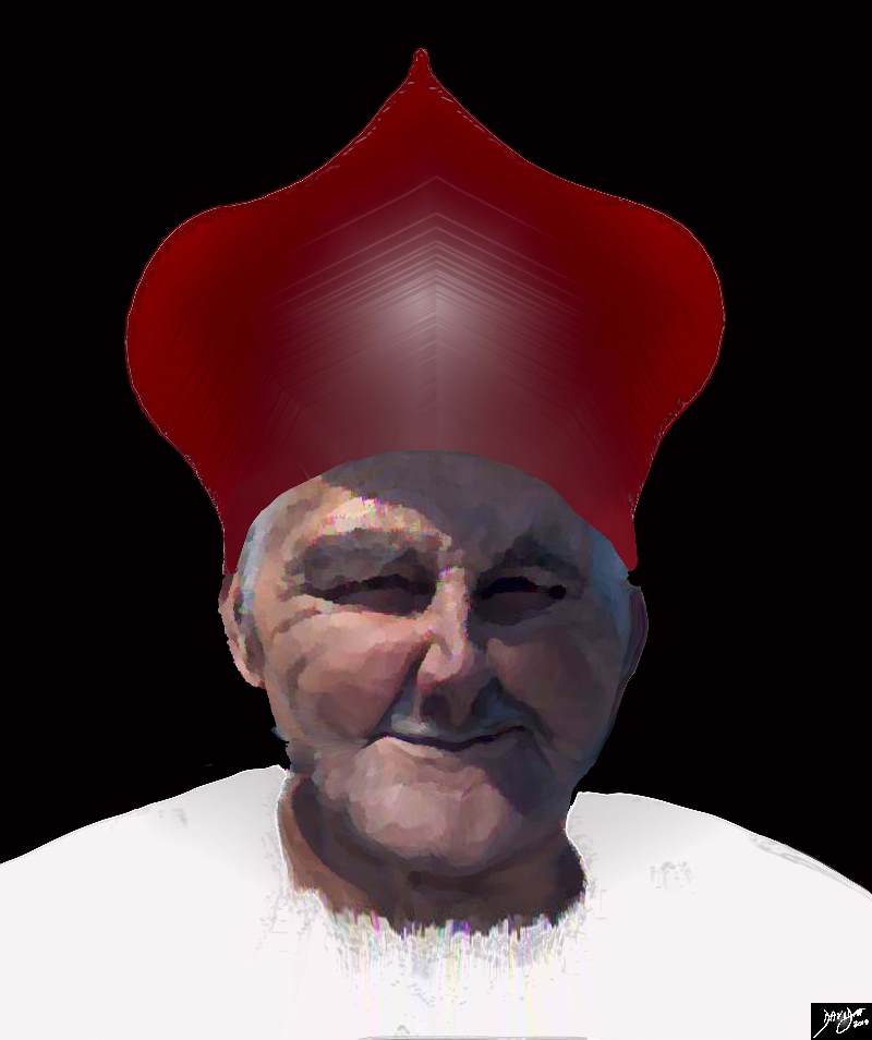
A Mitre – The Pontiffs Hat that Sits on the Pons


The 4th ventricvle in the axial plane looks like the pontiff’s hat which is called the mitre A pmnemonic you may want to use to remind you of the structure that is posteriorly related to the 4th ventricle may be “The pontiff’s hat that sits on the pons”
Copyright Ashley Davidoff MD Copyright 2010 all rights reserved 82407b14d05.8s


    [nodeType] => 1
    [parentNode] => (object value omitted)
    [childNodes] => (object value omitted)
    [firstChild] => (object value omitted)
    [lastChild] => (object value omitted)
    [previousSibling] => (object value omitted)
    [nextSibling] => (object value omitted)
    [attributes] => (object value omitted)
    [ownerDocument] => (object value omitted)
    [namespaceURI] =>
    [prefix] =>
    [localName] => table
    [baseURI] =>
    [textContent] =>


A Mitre – The Pontiffs Hat that Sits on the Pons


The 4th ventricvle in the axial plane looks like the pontiff’s hat which is called the mitre A pmnemonic you may want to use to remind you of the structure that is posteriorly related to the 4th ventricle may be “The pontiff’s hat that sits on the pons”
Copyright Ashley Davidoff MD Copyright 2010 all rights reserved 82407b14d05.8s


)
DOMElement Object
(
    [schemaTypeInfo] =>
    [tagName] => td
    [firstElementChild] => (object value omitted)
    [lastElementChild] => (object value omitted)
    [childElementCount] => 2
    [previousElementSibling] =>
    [nextElementSibling] =>
    [nodeName] => td
    [nodeValue] =>
The 4th ventricvle in the axial plane looks like the pontiff’s hat which is called the mitre A pmnemonic you may want to use to remind you of the structure that is posteriorly related to the 4th ventricle may be “The pontiff’s hat that sits on the pons”
Copyright Ashley Davidoff MD Copyright 2010 all rights reserved 82407b14d05.8s

    [nodeType] => 1
    [parentNode] => (object value omitted)
    [childNodes] => (object value omitted)
    [firstChild] => (object value omitted)
    [lastChild] => (object value omitted)
    [previousSibling] => (object value omitted)
    [nextSibling] => (object value omitted)
    [attributes] => (object value omitted)
    [ownerDocument] => (object value omitted)
    [namespaceURI] =>
    [prefix] =>
    [localName] => td
    [baseURI] =>
    [textContent] =>
The 4th ventricvle in the axial plane looks like the pontiff’s hat which is called the mitre A pmnemonic you may want to use to remind you of the structure that is posteriorly related to the 4th ventricle may be “The pontiff’s hat that sits on the pons”
Copyright Ashley Davidoff MD Copyright 2010 all rights reserved 82407b14d05.8s

)
DOMElement Object
(
    [schemaTypeInfo] =>
    [tagName] => td
    [firstElementChild] => (object value omitted)
    [lastElementChild] => (object value omitted)
    [childElementCount] => 2
    [previousElementSibling] =>
    [nextElementSibling] =>
    [nodeName] => td
    [nodeValue] =>
A Mitre – The Pontiffs Hat that Sits on the Pons
    [nodeType] => 1
    [parentNode] => (object value omitted)
    [childNodes] => (object value omitted)
    [firstChild] => (object value omitted)
    [lastChild] => (object value omitted)
    [previousSibling] => (object value omitted)
    [nextSibling] => (object value omitted)
    [attributes] => (object value omitted)
    [ownerDocument] => (object value omitted)
    [namespaceURI] =>
    [prefix] =>
    [localName] => td
    [baseURI] =>
    [textContent] =>
A Mitre – The Pontiffs Hat that Sits on the Pons
)
DOMElement Object
(
    [schemaTypeInfo] =>
    [tagName] => table
    [firstElementChild] => (object value omitted)
    [lastElementChild] => (object value omitted)
    [childElementCount] => 1
    [previousElementSibling] => (object value omitted)
    [nextElementSibling] => (object value omitted)
    [nodeName] => table
    [nodeValue] =>


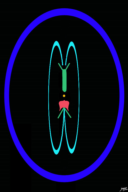
The Distribution of the ventricles in the Brain
Forebrain (green) Midbrain (orange) and hindbrain (salmon)


This diagram shows the positioning of each of the components of the ventriclar system . The lateral ventricles and third ventricle lie in the forebrain (green), the aqueduct of Sylvius (orange) lies in the midbrain, and the 4th ventricle (salmon) lies in the hinbrain.
Courtesy Ashley Davidoff copyright 2010 all rights reserved 93914.3kd06bd04.8s


    [nodeType] => 1
    [parentNode] => (object value omitted)
    [childNodes] => (object value omitted)
    [firstChild] => (object value omitted)
    [lastChild] => (object value omitted)
    [previousSibling] => (object value omitted)
    [nextSibling] => (object value omitted)
    [attributes] => (object value omitted)
    [ownerDocument] => (object value omitted)
    [namespaceURI] =>
    [prefix] =>
    [localName] => table
    [baseURI] =>
    [textContent] =>


The Distribution of the ventricles in the Brain
Forebrain (green) Midbrain (orange) and hindbrain (salmon)


This diagram shows the positioning of each of the components of the ventriclar system . The lateral ventricles and third ventricle lie in the forebrain (green), the aqueduct of Sylvius (orange) lies in the midbrain, and the 4th ventricle (salmon) lies in the hinbrain.
Courtesy Ashley Davidoff copyright 2010 all rights reserved 93914.3kd06bd04.8s


)
DOMElement Object
(
    [schemaTypeInfo] =>
    [tagName] => td
    [firstElementChild] => (object value omitted)
    [lastElementChild] => (object value omitted)
    [childElementCount] => 2
    [previousElementSibling] =>
    [nextElementSibling] =>
    [nodeName] => td
    [nodeValue] =>
This diagram shows the positioning of each of the components of the ventriclar system . The lateral ventricles and third ventricle lie in the forebrain (green), the aqueduct of Sylvius (orange) lies in the midbrain, and the 4th ventricle (salmon) lies in the hinbrain.
Courtesy Ashley Davidoff copyright 2010 all rights reserved 93914.3kd06bd04.8s

    [nodeType] => 1
    [parentNode] => (object value omitted)
    [childNodes] => (object value omitted)
    [firstChild] => (object value omitted)
    [lastChild] => (object value omitted)
    [previousSibling] => (object value omitted)
    [nextSibling] => (object value omitted)
    [attributes] => (object value omitted)
    [ownerDocument] => (object value omitted)
    [namespaceURI] =>
    [prefix] =>
    [localName] => td
    [baseURI] =>
    [textContent] =>
This diagram shows the positioning of each of the components of the ventriclar system . The lateral ventricles and third ventricle lie in the forebrain (green), the aqueduct of Sylvius (orange) lies in the midbrain, and the 4th ventricle (salmon) lies in the hinbrain.
Courtesy Ashley Davidoff copyright 2010 all rights reserved 93914.3kd06bd04.8s

)
DOMElement Object
(
    [schemaTypeInfo] =>
    [tagName] => td
    [firstElementChild] => (object value omitted)
    [lastElementChild] => (object value omitted)
    [childElementCount] => 3
    [previousElementSibling] =>
    [nextElementSibling] =>
    [nodeName] => td
    [nodeValue] =>

The Distribution of the ventricles in the Brain
Forebrain (green) Midbrain (orange) and hindbrain (salmon)
    [nodeType] => 1
    [parentNode] => (object value omitted)
    [childNodes] => (object value omitted)
    [firstChild] => (object value omitted)
    [lastChild] => (object value omitted)
    [previousSibling] => (object value omitted)
    [nextSibling] => (object value omitted)
    [attributes] => (object value omitted)
    [ownerDocument] => (object value omitted)
    [namespaceURI] =>
    [prefix] =>
    [localName] => td
    [baseURI] =>
    [textContent] =>

The Distribution of the ventricles in the Brain
Forebrain (green) Midbrain (orange) and hindbrain (salmon)
)
DOMElement Object
(
    [schemaTypeInfo] =>
    [tagName] => table
    [firstElementChild] => (object value omitted)
    [lastElementChild] => (object value omitted)
    [childElementCount] => 1
    [previousElementSibling] => (object value omitted)
    [nextElementSibling] => (object value omitted)
    [nodeName] => table
    [nodeValue] =>


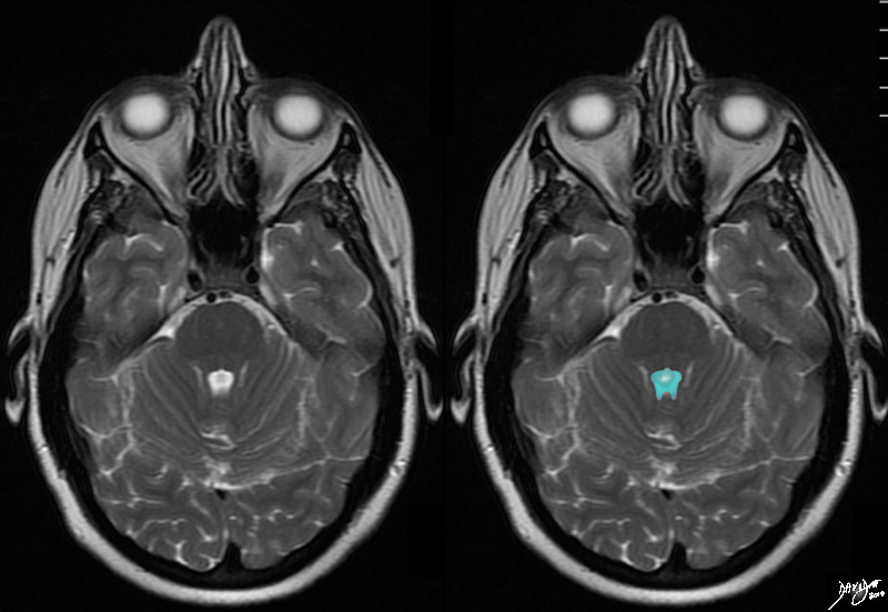
The 4th Ventricle


The T2 weighted axial image through the 4th ventricle is chatracterised by the “puffed crown” or pontiffs hat of the pons of this ventricle. Identifying the 4th ventricle allows localisation of the cut to the hindbrain. The 4th ventricle is anterior to the cerebellum and posterior to the pons.
Courtesy Ashley Davidoff MD copyright 2010 all rights reserved 94084cd05.81s


    [nodeType] => 1
    [parentNode] => (object value omitted)
    [childNodes] => (object value omitted)
    [firstChild] => (object value omitted)
    [lastChild] => (object value omitted)
    [previousSibling] => (object value omitted)
    [nextSibling] => (object value omitted)
    [attributes] => (object value omitted)
    [ownerDocument] => (object value omitted)
    [namespaceURI] =>
    [prefix] =>
    [localName] => table
    [baseURI] =>
    [textContent] =>


The 4th Ventricle


The T2 weighted axial image through the 4th ventricle is chatracterised by the “puffed crown” or pontiffs hat of the pons of this ventricle. Identifying the 4th ventricle allows localisation of the cut to the hindbrain. The 4th ventricle is anterior to the cerebellum and posterior to the pons.
Courtesy Ashley Davidoff MD copyright 2010 all rights reserved 94084cd05.81s


)
DOMElement Object
(
    [schemaTypeInfo] =>
    [tagName] => td
    [firstElementChild] => (object value omitted)
    [lastElementChild] => (object value omitted)
    [childElementCount] => 2
    [previousElementSibling] =>
    [nextElementSibling] =>
    [nodeName] => td
    [nodeValue] =>
The T2 weighted axial image through the 4th ventricle is chatracterised by the “puffed crown” or pontiffs hat of the pons of this ventricle. Identifying the 4th ventricle allows localisation of the cut to the hindbrain. The 4th ventricle is anterior to the cerebellum and posterior to the pons.
Courtesy Ashley Davidoff MD copyright 2010 all rights reserved 94084cd05.81s

    [nodeType] => 1
    [parentNode] => (object value omitted)
    [childNodes] => (object value omitted)
    [firstChild] => (object value omitted)
    [lastChild] => (object value omitted)
    [previousSibling] => (object value omitted)
    [nextSibling] => (object value omitted)
    [attributes] => (object value omitted)
    [ownerDocument] => (object value omitted)
    [namespaceURI] =>
    [prefix] =>
    [localName] => td
    [baseURI] =>
    [textContent] =>
The T2 weighted axial image through the 4th ventricle is chatracterised by the “puffed crown” or pontiffs hat of the pons of this ventricle. Identifying the 4th ventricle allows localisation of the cut to the hindbrain. The 4th ventricle is anterior to the cerebellum and posterior to the pons.
Courtesy Ashley Davidoff MD copyright 2010 all rights reserved 94084cd05.81s

)
DOMElement Object
(
    [schemaTypeInfo] =>
    [tagName] => td
    [firstElementChild] => (object value omitted)
    [lastElementChild] => (object value omitted)
    [childElementCount] => 2
    [previousElementSibling] =>
    [nextElementSibling] =>
    [nodeName] => td
    [nodeValue] =>
The 4th Ventricle
    [nodeType] => 1
    [parentNode] => (object value omitted)
    [childNodes] => (object value omitted)
    [firstChild] => (object value omitted)
    [lastChild] => (object value omitted)
    [previousSibling] => (object value omitted)
    [nextSibling] => (object value omitted)
    [attributes] => (object value omitted)
    [ownerDocument] => (object value omitted)
    [namespaceURI] =>
    [prefix] =>
    [localName] => td
    [baseURI] =>
    [textContent] =>
The 4th Ventricle
)
DOMElement Object
(
    [schemaTypeInfo] =>
    [tagName] => table
    [firstElementChild] => (object value omitted)
    [lastElementChild] => (object value omitted)
    [childElementCount] => 1
    [previousElementSibling] => (object value omitted)
    [nextElementSibling] => (object value omitted)
    [nodeName] => table
    [nodeValue] =>


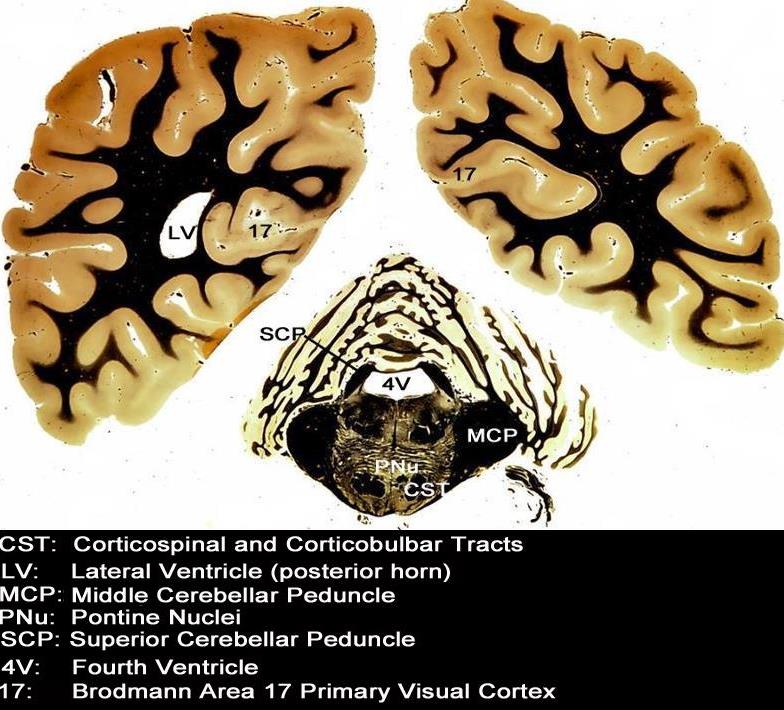
4th ventricle (4V) Coronal View


The fourth ventricle is seen in in relation to tghe cerebellum and the pons
Courtesy Department of Anatomy and Neurobiology at Boston University School of Medicine Dr. Jennifer Luebke , and Dr. Douglas Rosene 97356.C17.9L01


    [nodeType] => 1
    [parentNode] => (object value omitted)
    [childNodes] => (object value omitted)
    [firstChild] => (object value omitted)
    [lastChild] => (object value omitted)
    [previousSibling] => (object value omitted)
    [nextSibling] => (object value omitted)
    [attributes] => (object value omitted)
    [ownerDocument] => (object value omitted)
    [namespaceURI] =>
    [prefix] =>
    [localName] => table
    [baseURI] =>
    [textContent] =>


4th ventricle (4V) Coronal View


The fourth ventricle is seen in in relation to tghe cerebellum and the pons
Courtesy Department of Anatomy and Neurobiology at Boston University School of Medicine Dr. Jennifer Luebke , and Dr. Douglas Rosene 97356.C17.9L01


)
DOMElement Object
(
    [schemaTypeInfo] =>
    [tagName] => td
    [firstElementChild] => (object value omitted)
    [lastElementChild] => (object value omitted)
    [childElementCount] => 2
    [previousElementSibling] =>
    [nextElementSibling] =>
    [nodeName] => td
    [nodeValue] =>
The fourth ventricle is seen in in relation to tghe cerebellum and the pons
Courtesy Department of Anatomy and Neurobiology at Boston University School of Medicine Dr. Jennifer Luebke , and Dr. Douglas Rosene 97356.C17.9L01

    [nodeType] => 1
    [parentNode] => (object value omitted)
    [childNodes] => (object value omitted)
    [firstChild] => (object value omitted)
    [lastChild] => (object value omitted)
    [previousSibling] => (object value omitted)
    [nextSibling] => (object value omitted)
    [attributes] => (object value omitted)
    [ownerDocument] => (object value omitted)
    [namespaceURI] =>
    [prefix] =>
    [localName] => td
    [baseURI] =>
    [textContent] =>
The fourth ventricle is seen in in relation to tghe cerebellum and the pons
Courtesy Department of Anatomy and Neurobiology at Boston University School of Medicine Dr. Jennifer Luebke , and Dr. Douglas Rosene 97356.C17.9L01

)
DOMElement Object
(
    [schemaTypeInfo] =>
    [tagName] => td
    [firstElementChild] => (object value omitted)
    [lastElementChild] => (object value omitted)
    [childElementCount] => 2
    [previousElementSibling] =>
    [nextElementSibling] =>
    [nodeName] => td
    [nodeValue] =>
4th ventricle (4V) Coronal View
    [nodeType] => 1
    [parentNode] => (object value omitted)
    [childNodes] => (object value omitted)
    [firstChild] => (object value omitted)
    [lastChild] => (object value omitted)
    [previousSibling] => (object value omitted)
    [nextSibling] => (object value omitted)
    [attributes] => (object value omitted)
    [ownerDocument] => (object value omitted)
    [namespaceURI] =>
    [prefix] =>
    [localName] => td
    [baseURI] =>
    [textContent] =>
4th ventricle (4V) Coronal View
)
DOMElement Object
(
    [schemaTypeInfo] =>
    [tagName] => table
    [firstElementChild] => (object value omitted)
    [lastElementChild] => (object value omitted)
    [childElementCount] => 1
    [previousElementSibling] => (object value omitted)
    [nextElementSibling] => (object value omitted)
    [nodeName] => table
    [nodeValue] =>


The 4th Ventricle


The 4th ventricle is the last landmark of the ventricular system seen as we proceed inferiorly. It is seen as a puffed crown or the pontifs hat (mitre), which lies posterior to the pons (ponntiff /pons) inferior to the aqueduct of Sylvius and anterior to the cerebellum. It lies within the hinbrain.
Courtesy Ashley Davidoff copyright 2010 all rights reserved 93914.3kd06bd03.8s


    [nodeType] => 1
    [parentNode] => (object value omitted)
    [childNodes] => (object value omitted)
    [firstChild] => (object value omitted)
    [lastChild] => (object value omitted)
    [previousSibling] => (object value omitted)
    [nextSibling] => (object value omitted)
    [attributes] => (object value omitted)
    [ownerDocument] => (object value omitted)
    [namespaceURI] =>
    [prefix] =>
    [localName] => table
    [baseURI] =>
    [textContent] =>


The 4th Ventricle


The 4th ventricle is the last landmark of the ventricular system seen as we proceed inferiorly. It is seen as a puffed crown or the pontifs hat (mitre), which lies posterior to the pons (ponntiff /pons) inferior to the aqueduct of Sylvius and anterior to the cerebellum. It lies within the hinbrain.
Courtesy Ashley Davidoff copyright 2010 all rights reserved 93914.3kd06bd03.8s


)
DOMElement Object
(
    [schemaTypeInfo] =>
    [tagName] => td
    [firstElementChild] => (object value omitted)
    [lastElementChild] => (object value omitted)
    [childElementCount] => 2
    [previousElementSibling] =>
    [nextElementSibling] =>
    [nodeName] => td
    [nodeValue] =>
The 4th ventricle is the last landmark of the ventricular system seen as we proceed inferiorly. It is seen as a puffed crown or the pontifs hat (mitre), which lies posterior to the pons (ponntiff /pons) inferior to the aqueduct of Sylvius and anterior to the cerebellum. It lies within the hinbrain.
Courtesy Ashley Davidoff copyright 2010 all rights reserved 93914.3kd06bd03.8s

    [nodeType] => 1
    [parentNode] => (object value omitted)
    [childNodes] => (object value omitted)
    [firstChild] => (object value omitted)
    [lastChild] => (object value omitted)
    [previousSibling] => (object value omitted)
    [nextSibling] => (object value omitted)
    [attributes] => (object value omitted)
    [ownerDocument] => (object value omitted)
    [namespaceURI] =>
    [prefix] =>
    [localName] => td
    [baseURI] =>
    [textContent] =>
The 4th ventricle is the last landmark of the ventricular system seen as we proceed inferiorly. It is seen as a puffed crown or the pontifs hat (mitre), which lies posterior to the pons (ponntiff /pons) inferior to the aqueduct of Sylvius and anterior to the cerebellum. It lies within the hinbrain.
Courtesy Ashley Davidoff copyright 2010 all rights reserved 93914.3kd06bd03.8s

)
DOMElement Object
(
    [schemaTypeInfo] =>
    [tagName] => td
    [firstElementChild] => (object value omitted)
    [lastElementChild] => (object value omitted)
    [childElementCount] => 2
    [previousElementSibling] =>
    [nextElementSibling] =>
    [nodeName] => td
    [nodeValue] =>

The 4th Ventricle
    [nodeType] => 1
    [parentNode] => (object value omitted)
    [childNodes] => (object value omitted)
    [firstChild] => (object value omitted)
    [lastChild] => (object value omitted)
    [previousSibling] => (object value omitted)
    [nextSibling] => (object value omitted)
    [attributes] => (object value omitted)
    [ownerDocument] => (object value omitted)
    [namespaceURI] =>
    [prefix] =>
    [localName] => td
    [baseURI] =>
    [textContent] =>

The 4th Ventricle
)
DOMElement Object
(
    [schemaTypeInfo] =>
    [tagName] => table
    [firstElementChild] => (object value omitted)
    [lastElementChild] => (object value omitted)
    [childElementCount] => 1
    [previousElementSibling] => (object value omitted)
    [nextElementSibling] => (object value omitted)
    [nodeName] => table
    [nodeValue] =>


The Recesses


The diagram in the sagittal projection reveals the horizontal portion called the lateral ventricle. It is a paired structure that houses the frontal horn, body, and the vertical portion which is composed of the 3rd ventricle, cerebral aqueduct and the 4th ventricle. The lateral ventricle consists of the frontal horn, body, occipital horn, atrium and the temporal horn The foramen of Monro connects the lateral ventricles with the third ventricle. The paired foramina of Luschka are sitiuated anteriorly in the 4th ventricle and they allow CSF to circulate in the subarachnoid spaces. The foramen of Magendie is a single structure and is situated posteriorly and it also enables CSF to enter the subarachnoid space. code brain ventricles lateral ventricles frontal horn body occipital horn atrium temporal horn 3rd ventricle 4th ventricle foramen of Monro formamen of Magendie Foramen of Luschka anatomy normal neuroanatomy diagram conceptual diagram structure principles Davidoff Art Courtesy Ashley Davidoff MD copyright 2010 all rights
Courtesy Ashley Davidoff MD copyright 2010 all rights reserved  94459b10b02.82s


    [nodeType] => 1
    [parentNode] => (object value omitted)
    [childNodes] => (object value omitted)
    [firstChild] => (object value omitted)
    [lastChild] => (object value omitted)
    [previousSibling] => (object value omitted)
    [nextSibling] => (object value omitted)
    [attributes] => (object value omitted)
    [ownerDocument] => (object value omitted)
    [namespaceURI] =>
    [prefix] =>
    [localName] => table
    [baseURI] =>
    [textContent] =>


The Recesses


The diagram in the sagittal projection reveals the horizontal portion called the lateral ventricle. It is a paired structure that houses the frontal horn, body, and the vertical portion which is composed of the 3rd ventricle, cerebral aqueduct and the 4th ventricle. The lateral ventricle consists of the frontal horn, body, occipital horn, atrium and the temporal horn The foramen of Monro connects the lateral ventricles with the third ventricle. The paired foramina of Luschka are sitiuated anteriorly in the 4th ventricle and they allow CSF to circulate in the subarachnoid spaces. The foramen of Magendie is a single structure and is situated posteriorly and it also enables CSF to enter the subarachnoid space. code brain ventricles lateral ventricles frontal horn body occipital horn atrium temporal horn 3rd ventricle 4th ventricle foramen of Monro formamen of Magendie Foramen of Luschka anatomy normal neuroanatomy diagram conceptual diagram structure principles Davidoff Art Courtesy Ashley Davidoff MD copyright 2010 all rights
Courtesy Ashley Davidoff MD copyright 2010 all rights reserved  94459b10b02.82s


)
DOMElement Object
(
    [schemaTypeInfo] =>
    [tagName] => td
    [firstElementChild] => (object value omitted)
    [lastElementChild] => (object value omitted)
    [childElementCount] => 2
    [previousElementSibling] =>
    [nextElementSibling] =>
    [nodeName] => td
    [nodeValue] =>
The diagram in the sagittal projection reveals the horizontal portion called the lateral ventricle. It is a paired structure that houses the frontal horn, body, and the vertical portion which is composed of the 3rd ventricle, cerebral aqueduct and the 4th ventricle. The lateral ventricle consists of the frontal horn, body, occipital horn, atrium and the temporal horn The foramen of Monro connects the lateral ventricles with the third ventricle. The paired foramina of Luschka are sitiuated anteriorly in the 4th ventricle and they allow CSF to circulate in the subarachnoid spaces. The foramen of Magendie is a single structure and is situated posteriorly and it also enables CSF to enter the subarachnoid space. code brain ventricles lateral ventricles frontal horn body occipital horn atrium temporal horn 3rd ventricle 4th ventricle foramen of Monro formamen of Magendie Foramen of Luschka anatomy normal neuroanatomy diagram conceptual diagram structure principles Davidoff Art Courtesy Ashley Davidoff MD copyright 2010 all rights
Courtesy Ashley Davidoff MD copyright 2010 all rights reserved  94459b10b02.82s

    [nodeType] => 1
    [parentNode] => (object value omitted)
    [childNodes] => (object value omitted)
    [firstChild] => (object value omitted)
    [lastChild] => (object value omitted)
    [previousSibling] => (object value omitted)
    [nextSibling] => (object value omitted)
    [attributes] => (object value omitted)
    [ownerDocument] => (object value omitted)
    [namespaceURI] =>
    [prefix] =>
    [localName] => td
    [baseURI] =>
    [textContent] =>
The diagram in the sagittal projection reveals the horizontal portion called the lateral ventricle. It is a paired structure that houses the frontal horn, body, and the vertical portion which is composed of the 3rd ventricle, cerebral aqueduct and the 4th ventricle. The lateral ventricle consists of the frontal horn, body, occipital horn, atrium and the temporal horn The foramen of Monro connects the lateral ventricles with the third ventricle. The paired foramina of Luschka are sitiuated anteriorly in the 4th ventricle and they allow CSF to circulate in the subarachnoid spaces. The foramen of Magendie is a single structure and is situated posteriorly and it also enables CSF to enter the subarachnoid space. code brain ventricles lateral ventricles frontal horn body occipital horn atrium temporal horn 3rd ventricle 4th ventricle foramen of Monro formamen of Magendie Foramen of Luschka anatomy normal neuroanatomy diagram conceptual diagram structure principles Davidoff Art Courtesy Ashley Davidoff MD copyright 2010 all rights
Courtesy Ashley Davidoff MD copyright 2010 all rights reserved  94459b10b02.82s

)
DOMElement Object
(
    [schemaTypeInfo] =>
    [tagName] => td
    [firstElementChild] => (object value omitted)
    [lastElementChild] => (object value omitted)
    [childElementCount] => 2
    [previousElementSibling] =>
    [nextElementSibling] =>
    [nodeName] => td
    [nodeValue] =>

The Recesses
    [nodeType] => 1
    [parentNode] => (object value omitted)
    [childNodes] => (object value omitted)
    [firstChild] => (object value omitted)
    [lastChild] => (object value omitted)
    [previousSibling] => (object value omitted)
    [nextSibling] => (object value omitted)
    [attributes] => (object value omitted)
    [ownerDocument] => (object value omitted)
    [namespaceURI] =>
    [prefix] =>
    [localName] => td
    [baseURI] =>
    [textContent] =>

The Recesses
)
DOMElement Object
(
    [schemaTypeInfo] =>
    [tagName] => table
    [firstElementChild] => (object value omitted)
    [lastElementChild] => (object value omitted)
    [childElementCount] => 1
    [previousElementSibling] => (object value omitted)
    [nextElementSibling] => (object value omitted)
    [nodeName] => table
    [nodeValue] =>


Sagittal View of the Ventricles


The diagram in the sagittal projection reveals the horizontal portion called the lateral ventricle. It is a paired structure that houses the frontal horn, body, and the vertical portion which is composed of the 3rd ventricle, cerebral aqueduct and the 4th ventricle. The lateral ventricle consists of the frontal horn, body, occipital horn, atrium and the temporal horn The foramen of Monro connects the lateral ventricles with the third ventricle. The paired foramina of Luschka are sitiuated anteriorly in the 4th ventricle and they allow CSF to circulate in the subarachnoid spaces. The foramen of Magendie is a single structure and is situated posteriorly and it also enables CSF to enter the subarachnoid space.
Courtesy Ashley Davidoff MD copyright 2010 all rights reserved 94459b10b02.82s


    [nodeType] => 1
    [parentNode] => (object value omitted)
    [childNodes] => (object value omitted)
    [firstChild] => (object value omitted)
    [lastChild] => (object value omitted)
    [previousSibling] => (object value omitted)
    [nextSibling] => (object value omitted)
    [attributes] => (object value omitted)
    [ownerDocument] => (object value omitted)
    [namespaceURI] =>
    [prefix] =>
    [localName] => table
    [baseURI] =>
    [textContent] =>


Sagittal View of the Ventricles


The diagram in the sagittal projection reveals the horizontal portion called the lateral ventricle. It is a paired structure that houses the frontal horn, body, and the vertical portion which is composed of the 3rd ventricle, cerebral aqueduct and the 4th ventricle. The lateral ventricle consists of the frontal horn, body, occipital horn, atrium and the temporal horn The foramen of Monro connects the lateral ventricles with the third ventricle. The paired foramina of Luschka are sitiuated anteriorly in the 4th ventricle and they allow CSF to circulate in the subarachnoid spaces. The foramen of Magendie is a single structure and is situated posteriorly and it also enables CSF to enter the subarachnoid space.
Courtesy Ashley Davidoff MD copyright 2010 all rights reserved 94459b10b02.82s


)
DOMElement Object
(
    [schemaTypeInfo] =>
    [tagName] => td
    [firstElementChild] => (object value omitted)
    [lastElementChild] => (object value omitted)
    [childElementCount] => 2
    [previousElementSibling] =>
    [nextElementSibling] =>
    [nodeName] => td
    [nodeValue] =>
The diagram in the sagittal projection reveals the horizontal portion called the lateral ventricle. It is a paired structure that houses the frontal horn, body, and the vertical portion which is composed of the 3rd ventricle, cerebral aqueduct and the 4th ventricle. The lateral ventricle consists of the frontal horn, body, occipital horn, atrium and the temporal horn The foramen of Monro connects the lateral ventricles with the third ventricle. The paired foramina of Luschka are sitiuated anteriorly in the 4th ventricle and they allow CSF to circulate in the subarachnoid spaces. The foramen of Magendie is a single structure and is situated posteriorly and it also enables CSF to enter the subarachnoid space.
Courtesy Ashley Davidoff MD copyright 2010 all rights reserved 94459b10b02.82s

    [nodeType] => 1
    [parentNode] => (object value omitted)
    [childNodes] => (object value omitted)
    [firstChild] => (object value omitted)
    [lastChild] => (object value omitted)
    [previousSibling] => (object value omitted)
    [nextSibling] => (object value omitted)
    [attributes] => (object value omitted)
    [ownerDocument] => (object value omitted)
    [namespaceURI] =>
    [prefix] =>
    [localName] => td
    [baseURI] =>
    [textContent] =>
The diagram in the sagittal projection reveals the horizontal portion called the lateral ventricle. It is a paired structure that houses the frontal horn, body, and the vertical portion which is composed of the 3rd ventricle, cerebral aqueduct and the 4th ventricle. The lateral ventricle consists of the frontal horn, body, occipital horn, atrium and the temporal horn The foramen of Monro connects the lateral ventricles with the third ventricle. The paired foramina of Luschka are sitiuated anteriorly in the 4th ventricle and they allow CSF to circulate in the subarachnoid spaces. The foramen of Magendie is a single structure and is situated posteriorly and it also enables CSF to enter the subarachnoid space.
Courtesy Ashley Davidoff MD copyright 2010 all rights reserved 94459b10b02.82s

)
DOMElement Object
(
    [schemaTypeInfo] =>
    [tagName] => td
    [firstElementChild] => (object value omitted)
    [lastElementChild] => (object value omitted)
    [childElementCount] => 2
    [previousElementSibling] =>
    [nextElementSibling] =>
    [nodeName] => td
    [nodeValue] =>

Sagittal View of the Ventricles
    [nodeType] => 1
    [parentNode] => (object value omitted)
    [childNodes] => (object value omitted)
    [firstChild] => (object value omitted)
    [lastChild] => (object value omitted)
    [previousSibling] => (object value omitted)
    [nextSibling] => (object value omitted)
    [attributes] => (object value omitted)
    [ownerDocument] => (object value omitted)
    [namespaceURI] =>
    [prefix] =>
    [localName] => td
    [baseURI] =>
    [textContent] =>

Sagittal View of the Ventricles
)
DOMElement Object
(
    [schemaTypeInfo] =>
    [tagName] => table
    [firstElementChild] => (object value omitted)
    [lastElementChild] => (object value omitted)
    [childElementCount] => 1
    [previousElementSibling] => (object value omitted)
    [nextElementSibling] => (object value omitted)
    [nodeName] => table
    [nodeValue] =>


The Major Components of the Ventricles


The diagram in the sagital projection reveals the horizontal portion called the lateral ventricle. It is a paired structure.The vertical portion (light green consists of the midline 3rd and 4th ventricle.
Courtesy Ashley Davidoff MD copyright 2010 all rights reserved 94459b06b.8s


    [nodeType] => 1
    [parentNode] => (object value omitted)
    [childNodes] => (object value omitted)
    [firstChild] => (object value omitted)
    [lastChild] => (object value omitted)
    [previousSibling] => (object value omitted)
    [nextSibling] => (object value omitted)
    [attributes] => (object value omitted)
    [ownerDocument] => (object value omitted)
    [namespaceURI] =>
    [prefix] =>
    [localName] => table
    [baseURI] =>
    [textContent] =>


The Major Components of the Ventricles


The diagram in the sagital projection reveals the horizontal portion called the lateral ventricle. It is a paired structure.The vertical portion (light green consists of the midline 3rd and 4th ventricle.
Courtesy Ashley Davidoff MD copyright 2010 all rights reserved 94459b06b.8s


)
DOMElement Object
(
    [schemaTypeInfo] =>
    [tagName] => td
    [firstElementChild] => (object value omitted)
    [lastElementChild] => (object value omitted)
    [childElementCount] => 2
    [previousElementSibling] =>
    [nextElementSibling] =>
    [nodeName] => td
    [nodeValue] => The diagram in the sagital projection reveals the horizontal portion called the lateral ventricle. It is a paired structure.The vertical portion (light green consists of the midline 3rd and 4th ventricle.
Courtesy Ashley Davidoff MD copyright 2010 all rights reserved 94459b06b.8s
    [nodeType] => 1
    [parentNode] => (object value omitted)
    [childNodes] => (object value omitted)
    [firstChild] => (object value omitted)
    [lastChild] => (object value omitted)
    [previousSibling] => (object value omitted)
    [nextSibling] => (object value omitted)
    [attributes] => (object value omitted)
    [ownerDocument] => (object value omitted)
    [namespaceURI] =>
    [prefix] =>
    [localName] => td
    [baseURI] =>
    [textContent] => The diagram in the sagital projection reveals the horizontal portion called the lateral ventricle. It is a paired structure.The vertical portion (light green consists of the midline 3rd and 4th ventricle.
Courtesy Ashley Davidoff MD copyright 2010 all rights reserved 94459b06b.8s
)
DOMElement Object
(
    [schemaTypeInfo] =>
    [tagName] => td
    [firstElementChild] => (object value omitted)
    [lastElementChild] => (object value omitted)
    [childElementCount] => 2
    [previousElementSibling] =>
    [nextElementSibling] =>
    [nodeName] => td
    [nodeValue] =>

The Major Components of the Ventricles
    [nodeType] => 1
    [parentNode] => (object value omitted)
    [childNodes] => (object value omitted)
    [firstChild] => (object value omitted)
    [lastChild] => (object value omitted)
    [previousSibling] => (object value omitted)
    [nextSibling] => (object value omitted)
    [attributes] => (object value omitted)
    [ownerDocument] => (object value omitted)
    [namespaceURI] =>
    [prefix] =>
    [localName] => td
    [baseURI] =>
    [textContent] =>

The Major Components of the Ventricles
)
DOMElement Object
(
    [schemaTypeInfo] =>
    [tagName] => table
    [firstElementChild] => (object value omitted)
    [lastElementChild] => (object value omitted)
    [childElementCount] => 1
    [previousElementSibling] => (object value omitted)
    [nextElementSibling] => (object value omitted)
    [nodeName] => table
    [nodeValue] =>


The Deepest Layers of the Inverted C’s


In the diagram that describes the series of inverted “C” the ventricles are the deepest and most midline of the structures In this diagram it is demonstrated as the innermost teal bluue ring. The frontal horns are superior and anterior and the temporal horns are the most inferior and anterior.
Courtesy Ashley Davidoff MD copyright 2010 all rights reserved 93890b01b05b1.8s


    [nodeType] => 1
    [parentNode] => (object value omitted)
    [childNodes] => (object value omitted)
    [firstChild] => (object value omitted)
    [lastChild] => (object value omitted)
    [previousSibling] => (object value omitted)
    [nextSibling] => (object value omitted)
    [attributes] => (object value omitted)
    [ownerDocument] => (object value omitted)
    [namespaceURI] =>
    [prefix] =>
    [localName] => table
    [baseURI] =>
    [textContent] =>


The Deepest Layers of the Inverted C’s


In the diagram that describes the series of inverted “C” the ventricles are the deepest and most midline of the structures In this diagram it is demonstrated as the innermost teal bluue ring. The frontal horns are superior and anterior and the temporal horns are the most inferior and anterior.
Courtesy Ashley Davidoff MD copyright 2010 all rights reserved 93890b01b05b1.8s


)
DOMElement Object
(
    [schemaTypeInfo] =>
    [tagName] => td
    [firstElementChild] => (object value omitted)
    [lastElementChild] => (object value omitted)
    [childElementCount] => 2
    [previousElementSibling] =>
    [nextElementSibling] =>
    [nodeName] => td
    [nodeValue] =>
In the diagram that describes the series of inverted “C” the ventricles are the deepest and most midline of the structures In this diagram it is demonstrated as the innermost teal bluue ring. The frontal horns are superior and anterior and the temporal horns are the most inferior and anterior.
Courtesy Ashley Davidoff MD copyright 2010 all rights reserved 93890b01b05b1.8s

    [nodeType] => 1
    [parentNode] => (object value omitted)
    [childNodes] => (object value omitted)
    [firstChild] => (object value omitted)
    [lastChild] => (object value omitted)
    [previousSibling] => (object value omitted)
    [nextSibling] => (object value omitted)
    [attributes] => (object value omitted)
    [ownerDocument] => (object value omitted)
    [namespaceURI] =>
    [prefix] =>
    [localName] => td
    [baseURI] =>
    [textContent] =>
In the diagram that describes the series of inverted “C” the ventricles are the deepest and most midline of the structures In this diagram it is demonstrated as the innermost teal bluue ring. The frontal horns are superior and anterior and the temporal horns are the most inferior and anterior.
Courtesy Ashley Davidoff MD copyright 2010 all rights reserved 93890b01b05b1.8s

)
DOMElement Object
(
    [schemaTypeInfo] =>
    [tagName] => td
    [firstElementChild] => (object value omitted)
    [lastElementChild] => (object value omitted)
    [childElementCount] => 2
    [previousElementSibling] =>
    [nextElementSibling] =>
    [nodeName] => td
    [nodeValue] =>
The Deepest Layers of the Inverted C’s
    [nodeType] => 1
    [parentNode] => (object value omitted)
    [childNodes] => (object value omitted)
    [firstChild] => (object value omitted)
    [lastChild] => (object value omitted)
    [previousSibling] => (object value omitted)
    [nextSibling] => (object value omitted)
    [attributes] => (object value omitted)
    [ownerDocument] => (object value omitted)
    [namespaceURI] =>
    [prefix] =>
    [localName] => td
    [baseURI] =>
    [textContent] =>
The Deepest Layers of the Inverted C’s
)

