Hi Gene
Another case with death from Takotsubo and Overview of the disease in 8 minutes!
getting close with the streamlining
Clinical Case now on this page Working on overview
Let me know about any reservations or questions
Thanks
AD
re “Sarcoidosis, a Scare, Chest Pain and an Octopus”
74-year-old male with type 2 diabetes, CAD s/p stent 6 years prior, hypertension and hypercholesterolemia, fell at home and presents to the ER.
A CXR performed 4 years before, showed no acute cardiopulmonary disease. Healed right sided rib fractures and healed right clavicular fracture were of incidental note
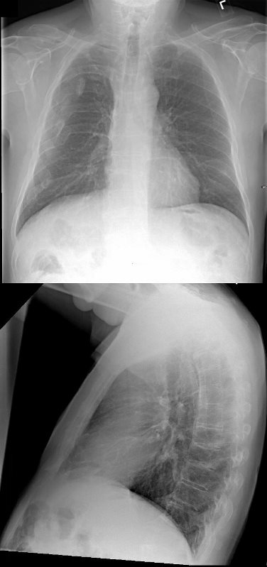
A CXR performed 4 years before, shows no acute cardiopulmonary disease. Healed right sided rib fractures and healed right clavicular fracture were of incidental note
Ashley Davidoff MD
In the ER a non-displaced right hip fracture was identified and uncomplicated hemiarthroplasty was performed.

In the ER a non-displaced right hip fracture was identified and uncomplicated hemiarthroplasty was performed.
Ashley Davidoff MD
CXR at the time showed no acute disease
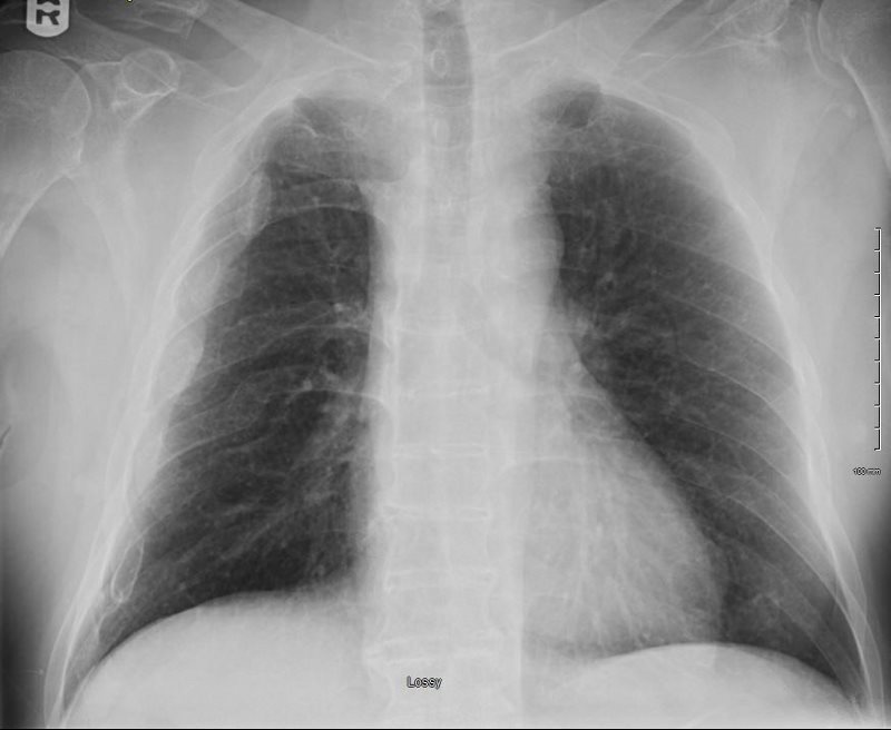
CXR at the time shows no acute disease
Ashley Davidoff MD
He was discharged and in the interim prior to readmission 2 weeks later his wife fell and had to be admitted to the hospital.
At the time of his next readmission he presented in shock. (NYHA Class IV and ASA Class 4)
CXR showed pulmonary edema.
LA? Difficult to see
Fuzzy Vessels
Perihilar Infiltrates
10, 20 or 30?
Wedge pressure >30
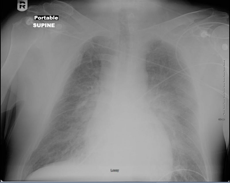
At the time of his next readmission he presented in shock. (NYHA Class IV and ASA Class 4)
CXR showed pulmonary edema.
EKG showed new LBB
Ashley Davidoff MD
EKG showed new LBB
Echo at the bedside in the ICU showed an ejection fraction of 15%, with global hypokinesis sparing the base of the heart. There was moderate MR, PAP 44-65mm Hg, RV was normal.
Preliminary diagnosis of an acute MI was made with acute systolic heart failure and cardiogenic shock
He was transferred to the Cath Lab
Prior to gaining access to the arterial system the patient went into PEA requiring sustained CPR requiring both epinephrine, atropine and urgent intubation
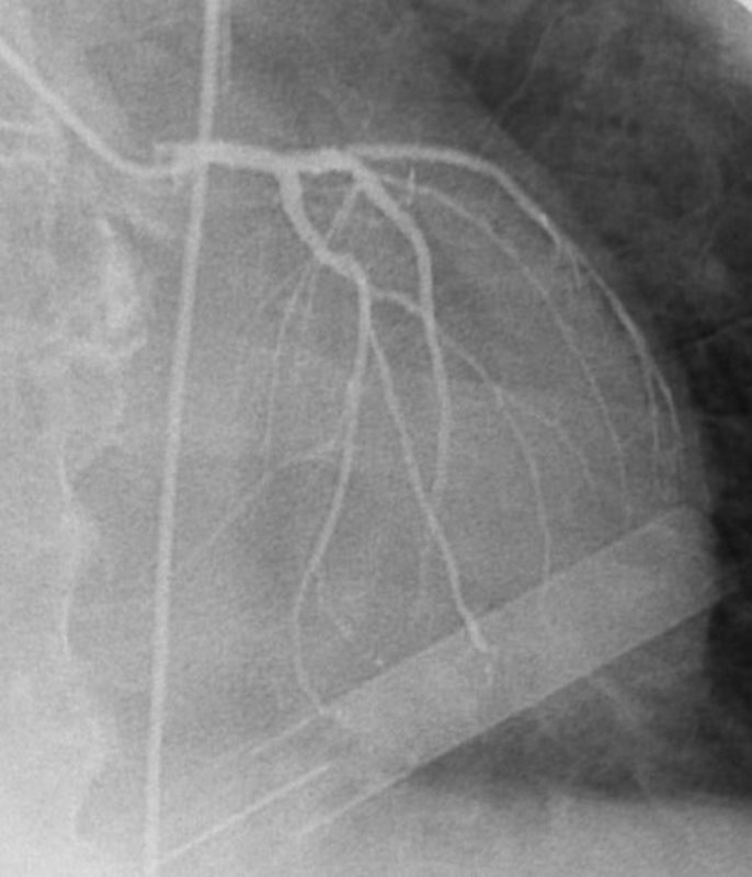
Emergent cardiac catheterization showed an LV pressure of 64/17 and wedge pressure of 41 mmHg. Temporary pacemaker was placed as well as an IABP. No significant CAD was identified.
AP PROJECTION
Which is the LAD? Look for the septal perforators running to the diaphragm
Ashley Davidoff MD
LAD – Now there are some beautiful perforators off the LAD supplying 2/3 of the septum
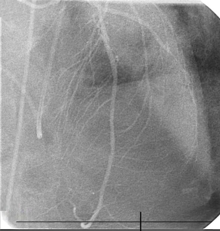
No significant CAD was identified. Ashley Davidoff MD
RCA – SA nodal of RCA (40%) Early Take of of the PDA , RCA Dominant (85%)
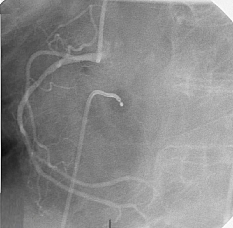
Ashley Davidoff MD
IABP placed
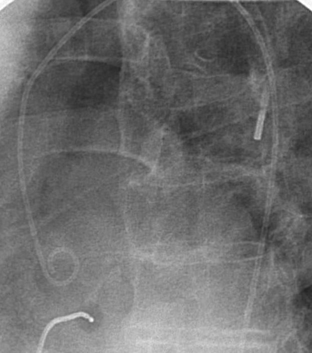
Ashley Davidoff MD
LV gram showed ballooning of the apex of the heart consistent with Takotsubo cardiomyopathy with an estimated ejection fraction of 10%
AKINETIC ANTERIOR WALL AND APEX – TAKOTSUBO
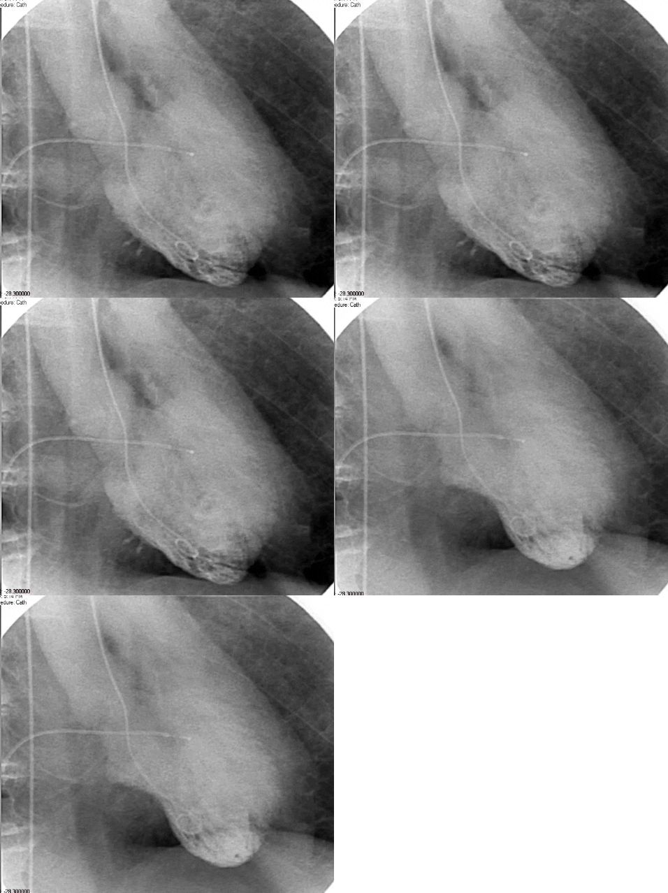
Serial CXR showed ongoing perihilar infiltrates with air bronchograms consistent with cardiogenic and alveolar edema.
Alveolar Edema – Air Bronchograms
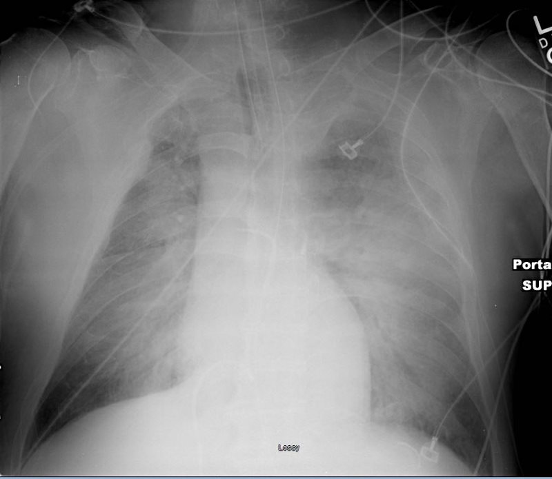
TAKOTSUBO CARDIOMYOPATHY
Ashley Davidoff MD
He passed away 2 days later – Broken Hearted
- Points of Interest
- Acute presentation and death from Takotsubo 1.8%
Two hits personal physical ailment and emotional ailment of his wife’s admission to hospital
- Acute presentation and death from Takotsubo 1.8%


