

Hi Michael
“Sarcoidosis, a Scare, Chest Pain and an Octopus”
This is a story about a 59 year old woman who had sarcoidosis, who had a scare developed chest pain that sounded like acute coronary syndrome and was short of breath
Your part is to teach about Heart Failure on CXR in 4 minutes!
I am close to finishing your part which is now on this page
Let me know about any reservations
Thanks
AD
hi Michael
Hope this is not too late – I exchanged the interstitial edema example for a beautiful example of Kerley B lines and peribronchial cuffing (near the end )
Questions to be asked when looking for CHF
1 Size of the LA?
2 – Ratio branch pulmonary arteries to bronchus?
3 – Fuzziness of the Vessels? (Peribronchial cuffing, Kerley B)
4 Alveolar Edema
Ashley Davidoff MD
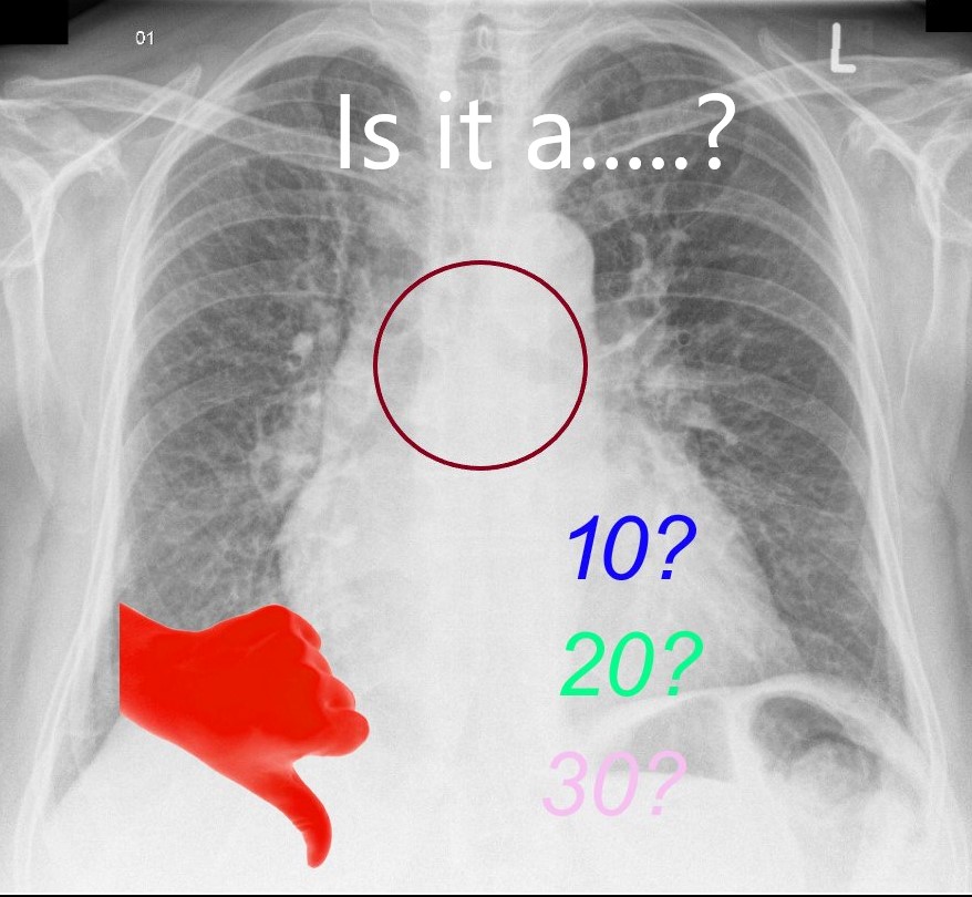
Questions
-
Is the LA Enlarged?
-
Is it an LA with a 10 mmHg, 20 mmHg or 30 mmHg pressure
-
Is the PA diameter>Bronchus in the middle or upper lobe vessels? and or redistribution
-
Is there fuzziness of the vessels? and or peribronchial cuffing, Kerley B, effusions
-
Is there alveolar edema? and or perihilar infiltrates
-
1 Is the LA Enlarged?
Ashley Davidoff MD

Ashley Davidoff MD
Is it an LA with a 10 mmHg, 20 mmHg or 30 mmHg pressure
Ashley Davidoff MD
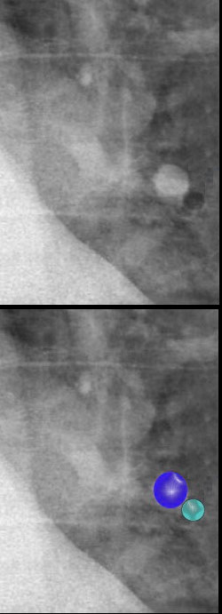
2.1
Is the PA>Bronchus?
and or redistribution
Then it is at least 10 mmHg
Ashley Davidoff MD
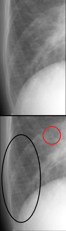
2.2
Is there fuzziness of the vessels? and or peribronchial cuffing, Kerley B, effusions
ie Is there interstitial edema?
Then it is at least 20 mmHg

Ashley Davidoff MD
Kerley B Line and Peribronchial Cuffing
Ashley Davidoff MD
2.3
Are there perihilar batwing infiltrates?
ie Is there alveolar edema?
Then LA pressure is at least 30 mmHg

Ashley Davidoff MD
TOP left image is of normal alveoli
Top Rt is PA slightly greater than bronchus = LVEDP 10-20 mmHg
Bottom left = 20-30 interstitial edema – peribronchial cuffing and Kerley B
Bottom Rt >30 = Alveolar Edema
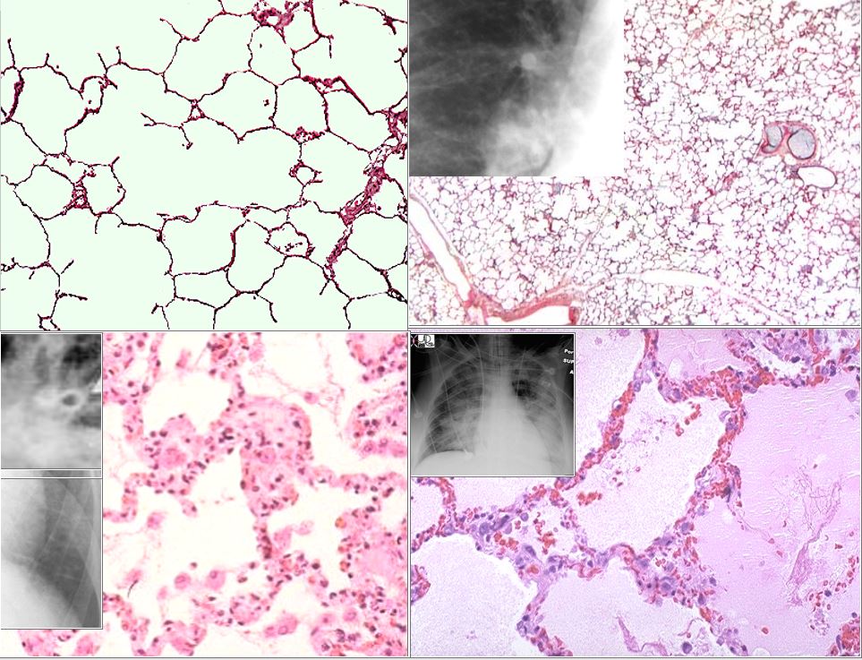
The top left image is the a histological section of normal alveoli and normal wall and interstitium. Heart failure occurs when the left ventricular end diastolic pressure rises. There are 3 basic phases of heart failure. in the first phase (top right) the LVEDP rises above 12 mmHg and on an upright CXR there is equalization of the size of the vessels going to the upper lobes and lower lobes. As the LVEDP goes above about 15-18 mm Hg there is cephalization of the vessels and the upper lobe vessels are larger than the lower lobe vessels.
The second phase of interstitial edema (bottom left) occurs when the intravascular hydrostatic pressure exceeds the intravascular oncotic pressure and this occurs when the LVEDP goes above 25 mm Hg. Fluid accumulates in the alveolar walls and interstitium and the wall becomes thicker with fluid, and the lymphatics and interlobular septa are distended.
The last phase of alveolar edema (lower right) occurs when the pressure exceeds 35 mmHg and the fluid leaks into the alveoli .
Ashley Davidoff MD