Our patient
So we know we have diffuse infiltration of both the LV and RV infiltration based on our nulling attempts Amyloidosis is the classical example of this entity
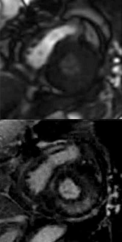
Short axis during first pass while still in RV (above) and then delayed post gadolinium shows diffuse mid myocardial circumferential LGE enhancement consistent with an infiltrative cardiomyopathy
Ashley Davidoff MD
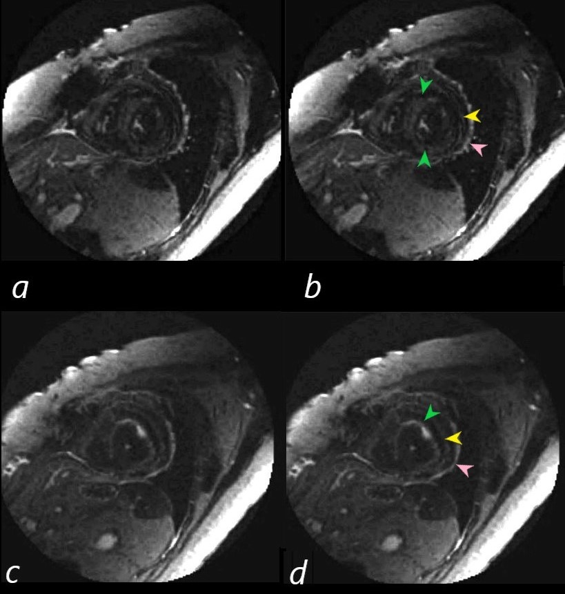
Short axis images on the delayed post Gad images show 3 rings of LGE. Image b (correlate with image a) is through the body of the LV and shows mid myocardial LGE seen as an almost complete ring of diffuse accumulation (green arrowheads), a thin ring of more peripheral mid myocardial LGE (yellow arrowhead) together with probable pericardial LGE (yellow arrow head)
In image d (correlate with image c) near the apex of the heart, there are 2 distinct rings of a linear morphology in the mid myocardium. The inner ring (green arrowhead) has some focal nodularity and an outer mid myocardial ring (yellow arrowhead) . Subepicardial or pericardial enhancement is suggested as well (pink arrowhead).
Ashley Davidoff MD
As we learned
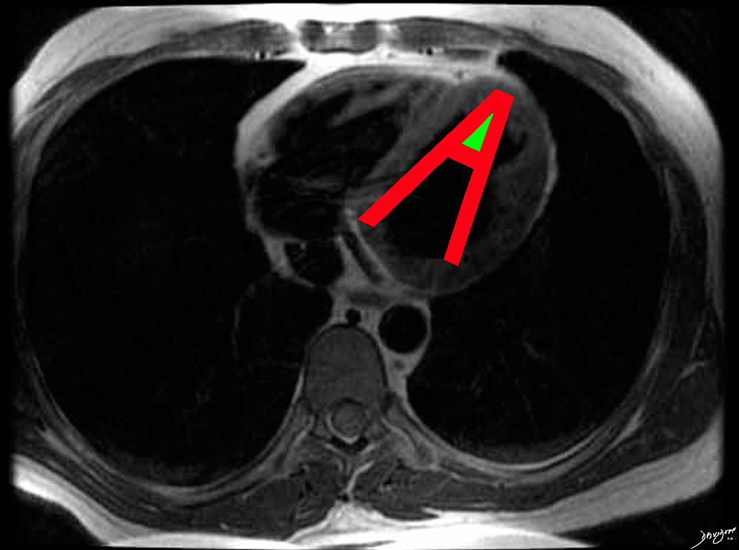
The hallmark of cardiac amyloidosis is
LGE involving subendocardial regions with apical sparing
Ashley Davidoff MD
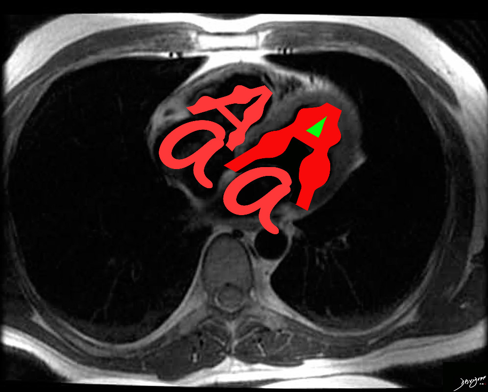
In cardiac amyloidosis increased LV thickness is common , but may involve RV and atrial septum with bilateral atrial enlargement.
Ashley Davidoff MD
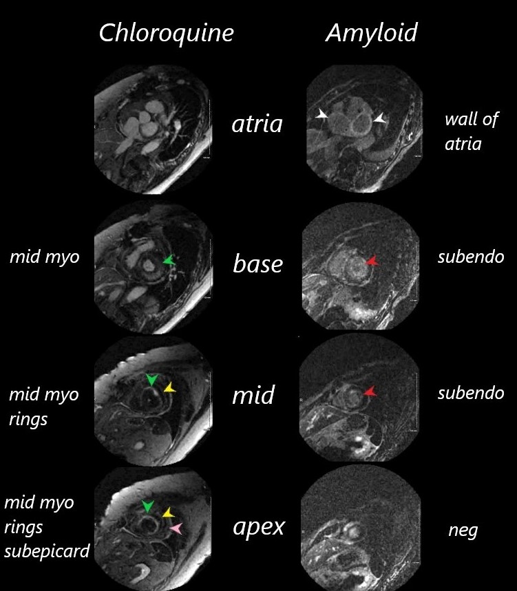
It was difficult to null the myocardium on both these patients.
The images are organized from the atria (top images through the bases, bodies and apices (lowest images) of the left ventricles.
The chloroquine cardiomyopathy shows no LGE of the atria, but progressive linear circumferential mid- ventricular LGE through to the apex
The amyloid cardiomyopathy hase LGE in both atrial walls, circumferential LGE through the base and body o the LV but sparing of the apex.
Ashley Davidoff MD
amyloid images are from image number 131429

