Skull
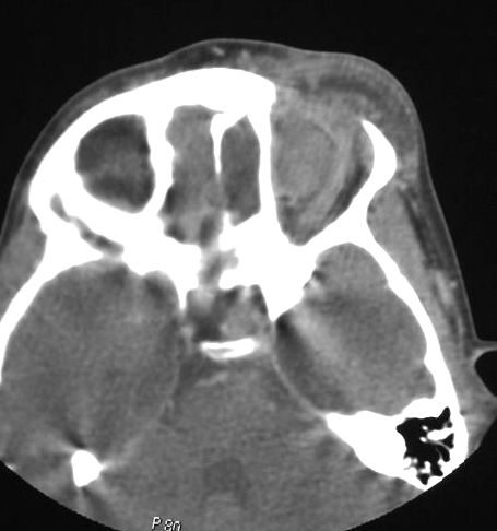
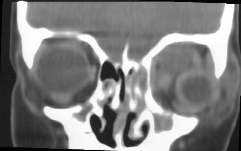
bone, ethmoid sinus, orbit, eye, infection, abscess, pediatric,
Ashley Davidoff MD
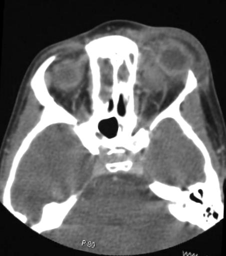
bone, ethmoid sinus, orbit, eye, infection, abscess, pediatric,
Ashley Davidoff MD
Clavicle
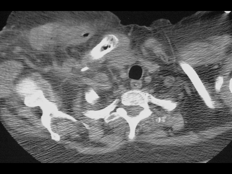
Bone, clavicle, air, osteomyelitis, soft tissue swelling clostridium, CT
Ashley Davidoff MD
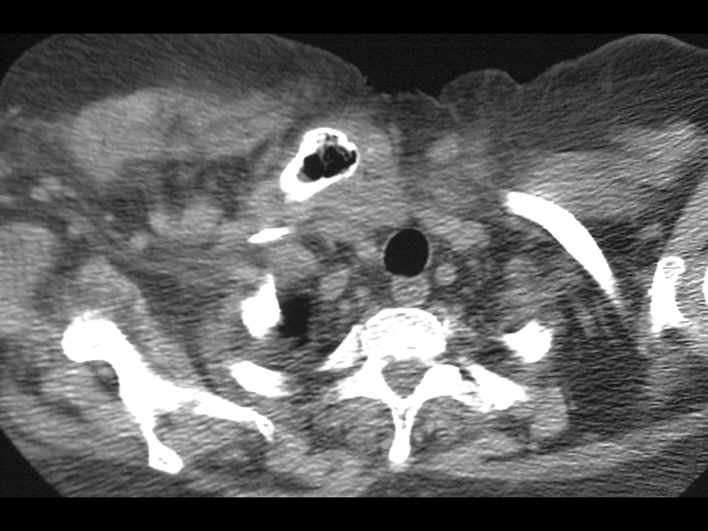
Bone, clavicle, air, osteomyelitis, soft tissue swelling clostridium, CT
Ashley Davidoff MD
Hand
Septic Arthropathy
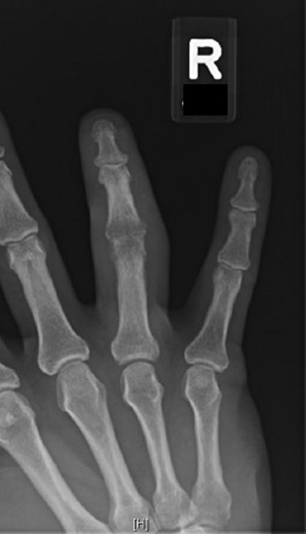
Clinical History: Pain and swelling of the ring finger.
Organ: Finger
Radiologic Finding: Localized osteopenia centered over the PIP joint with marginal joint erosion. Surrounding soft tissue swelling.
Dx: Septic arthropathy with probable osteomyelitis.
Modality: Radiograph
Akira Murakami MD
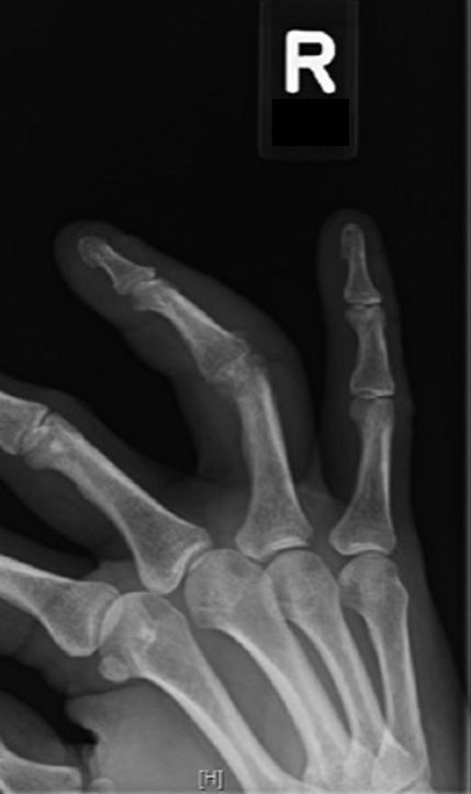
Clinical History: Pain and swelling of the ring finger.
Organ: Finger
Radiologic Finding: Localized osteopenia centered over the PIP joint with marginal joint erosion. Surrounding soft tissue swelling.
Dx: Septic arthropathy with probable osteomyelitis.
Modality: Radiograph
Akira Murakami MD
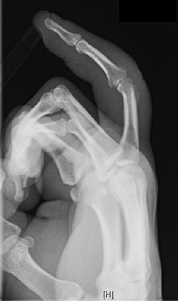
Clinical History: Pain and swelling of the ring finger.
Organ: Finger
Radiologic Finding: Localized osteopenia centered over the PIP joint with marginal joint erosion. Surrounding soft tissue swelling.
Dx: Septic arthropathy with probable osteomyelitis.
Modality: Radiograph
Akira Murakami MD
Lumbar Spine
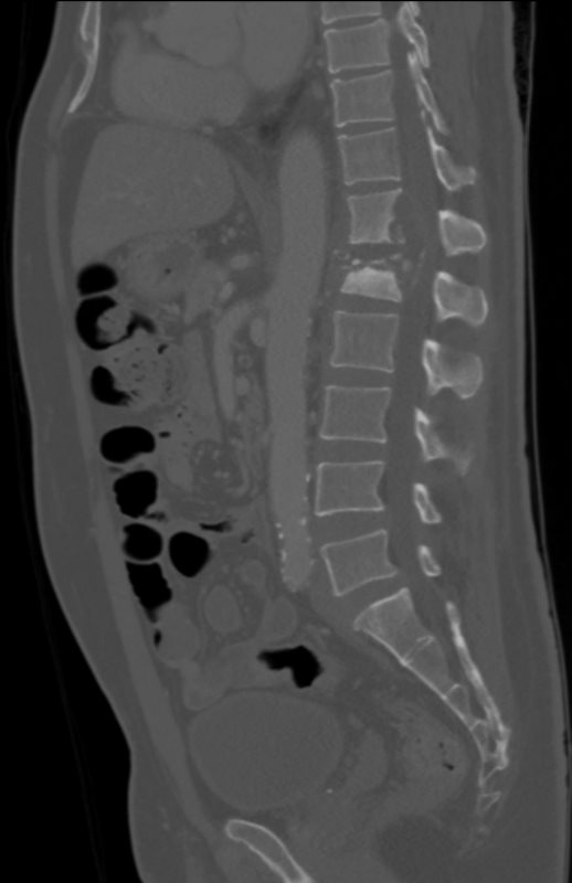
TB KIDNEY, SPINE, EPIDURAL ABSCESS, ADRENAL and PSOAS ABSCESS
58-year-old male with known history of tuberculosis.
There is enlargement of the bilateral adrenal glands, with multiple nodular structures encompassing the left adrenal gland, which may also represent infectious process.
Examination of the lumbar spine on the CT scan shows findings consistent with TB osteomyelitis with destruction of T12 and L1 vertebral bodies with prevertebral and epidural collections extending into the spinal canal.
Contributed by Christina LeBedis MD Case Study
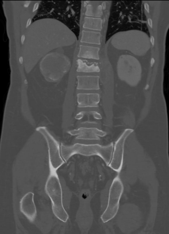
TB KIDNEY, SPINE, EPIDURAL ABSCESS, ADRENAL and PSOAS ABSCESS
58-year-old male with known history of tuberculosis.
Examination of the lumbar spine on the CT scan shows findings consistent with TB osteomyelitis with destruction of T12 and L1 vertebral bodies with prevertebral and epidural collections extending into the spinal canal.
Contributed by Christina LeBedis MD Case Study
Pelvis
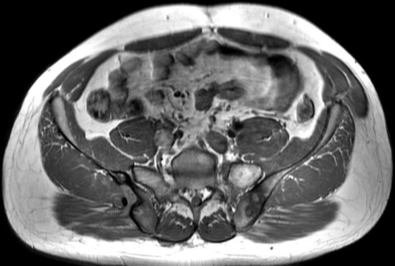
Clinical history: Chronic fever and chills. Weight loss.
Organ: Pelvis
Radiologic Finding: Left Ilium: T2 hyperintense, peripherally enhancing bone lesions with surrounding low T1 signal. Right Ilium: Peripherally enhancing, erosive bone lesion along the posterior cortex with an enhancing sinus tract extending into the gluteus muscle.
Dx: Osteomyelitis and bone abscess secondary to chronic TB.
Modality: Axial MRI T1
Akira Murakami MD
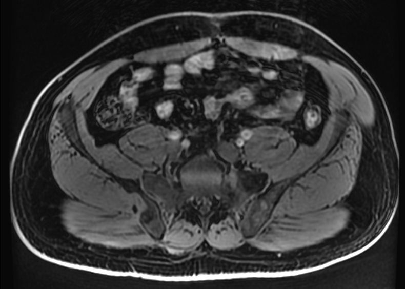
Clinical history: Chronic fever and chills. Weight loss.
Organ: Pelvis
Radiologic Finding: Left Ilium: T2 hyperintense, peripherally enhancing bone lesions with surrounding low T1 signal. Right Ilium: Peripherally enhancing, erosive bone lesion along the posterior cortex with an enhancing sinus tract extending into the gluteus muscle.
Modality: Axial MRI T1 FS pre
Akira Murakami MD
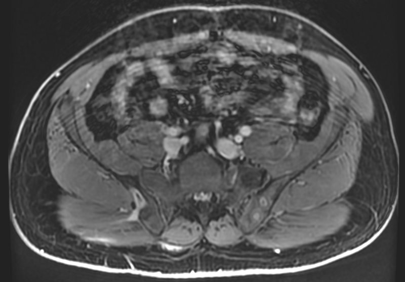
Clinical history: Chronic fever and chills. Weight loss.
Organ: Pelvis
Radiologic Finding: Left Ilium: T2 hyperintense, peripherally enhancing bone lesions with surrounding low T1 signal. Right Ilium: Peripherally enhancing, erosive bone lesion along the posterior cortex with an enhancing sinus tract extending into the gluteus muscle.
Modality: Axial MRI T1 FS post gadolinium
Akira Murakami MD
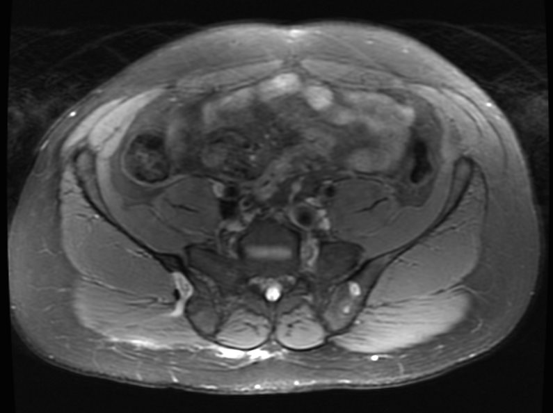
(T2 FS)
Clinical history: Chronic fever and chills. Weight loss.
Organ: Pelvis
Radiologic Finding: Left Ilium: T2 hyperintense, peripherally enhancing bone lesions with surrounding low T1 signal. Right Ilium: Peripherally enhancing, erosive bone lesion along the posterior cortex with an enhancing sinus tract extending into the gluteus muscle.
Modality: Axial MRI T2 Fat Sat
Akira Murakami MD
Pubic Symphysis
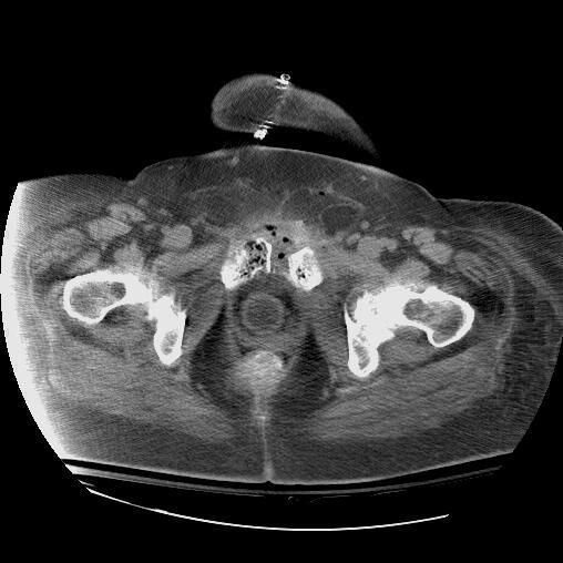
Bone, pubis, pubic symphysis, air, bone destruction, cortical destruction, osteomyelitis, abscess, soft tissue swelling,
drainage,CT
Ashley Davidoff MD
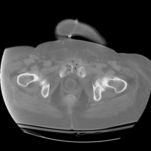
Bone, pubis, pubic symphysis, air, bone destruction, cortical destruction, osteomyelitis, abscess, soft tissue swelling,
drainage,CT
Ashley Davidoff MD
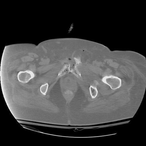
Bone, pubis, pubic symphysis, air, bone destruction, cortical destruction, osteomyelitis, abscess, soft tissue swelling,
drainage,CT
Ashley Davidoff MD
Hip
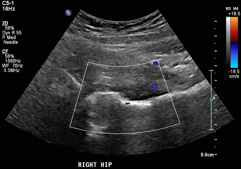
SEPTIC ARTHROPATHY
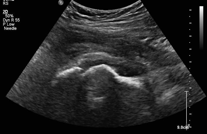
Clinical History: Chronic IV drug use, with new onset fever and chills. Limited range of motion and pain at the hip.
Organ: Hip
Radiologic Finding: MSK Ultrasound image of the anterior hip joint and anterior femoral head neck junction, with the probe held parallel to the femoral neck. Hypoechoic material / joint fluid is distending the anterior joint capsule. Mild hyperemia by color Doppler.
Dx: Acute septic arthropathy of the hip (confirmed by aspiration).
Modality: Ultrasound
Akira Murakami MD
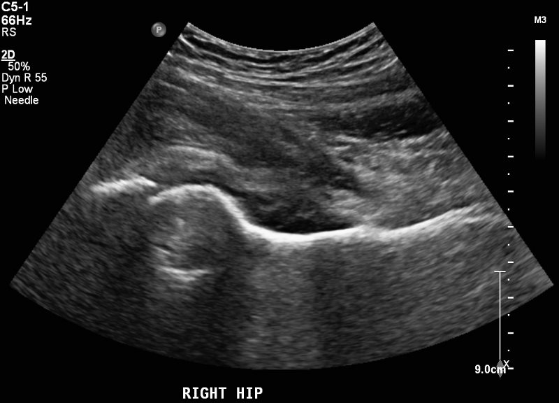
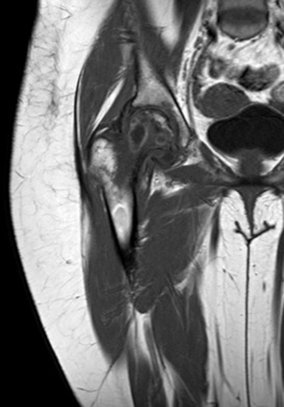
Clinical History: Chronic hip pain with fevers and chills
Organ: Hip
Radiologic Finding: Large hip joint effusion with synovial enhancement. T2 Hyperintense marrow signal / bone marrow edema pattern throughout the femoral neck with corresponding geographic T1 hypointense signal. Cavitary lesion within the center of the femoral neck with peripheral enhancement.
Dx: Septic arthropathy of the hip with Osteomyelitis and bone abscess within the femoral neck.
Modality: MRI T1
Akira Murakami MD
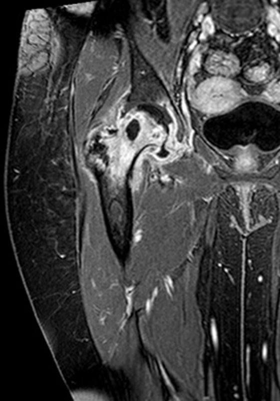
Clinical History: Chronic hip pain with fevers and chills
Organ: Hip
Radiologic Finding: Large hip joint effusion with synovial enhancement. T2 Hyperintense marrow signal / bone marrow edema pattern throughout the femoral neck with corresponding geographic T1 hypointense signal. Cavitary lesion within the center of the femoral neck with peripheral enhancement.
Dx: Septic arthropathy of the hip with Osteomyelitis and bone abscess within the femoral neck.
Modality: MRI T1 Fat Sat
Akira Murakami MD
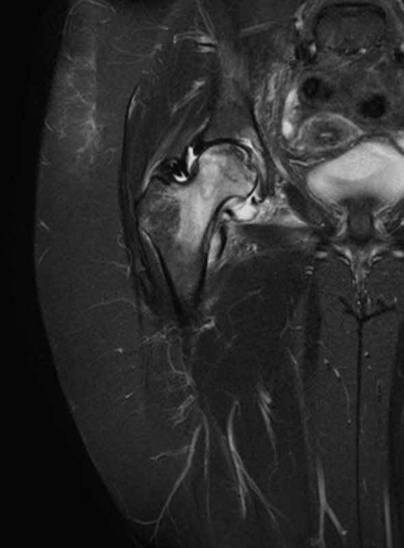
Clinical History: Chronic hip pain with fevers and chills
Organ: Hip
Radiologic Finding: Large hip joint effusion with synovial enhancement. T2 Hyperintense marrow signal / bone marrow edema pattern throughout the femoral neck with corresponding geographic T1 hypointense signal. Cavitary lesion within the center of the femoral neck with peripheral enhancement.
Dx: Septic arthropathy of the hip with Osteomyelitis and bone abscess within the femoral neck.
Modality: MRI T2 Fat Sat
Akira Murakami MD
Femur
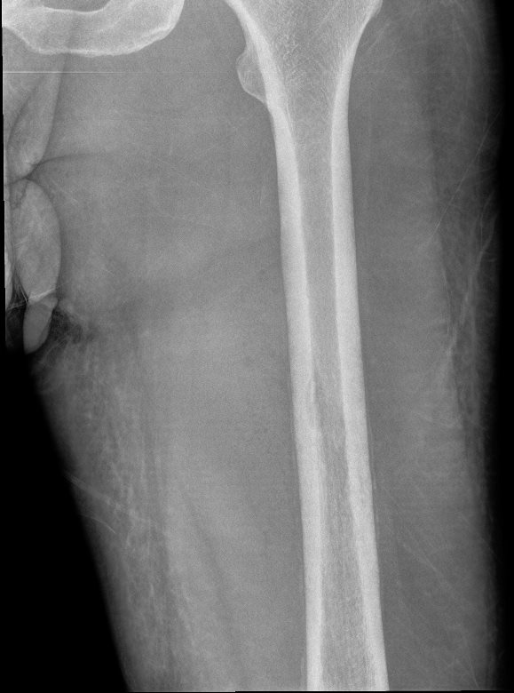
Clinical History: Leg pain with fevers and chills.
Organ: Femur
Radiologic Finding: Periosteal reaction along the femoral diaphysis, surrounding a small lytic lesion within the marrow. Adjacent soft tissue swelling.
Dx: Acute osteomyelitis with periostitis and early bone abscess
Modality: Radiography
Clinical History: Leg pain with fevers and chills.
Akira Murakami MD
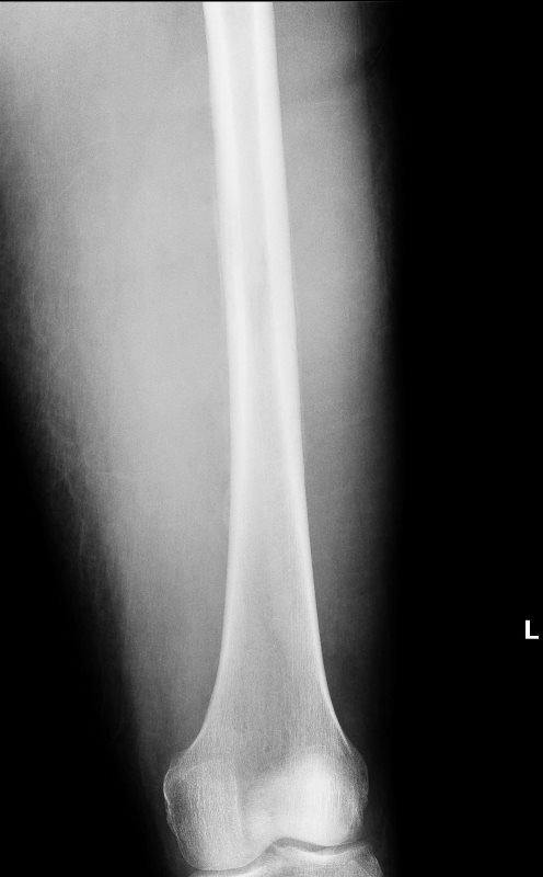
Clinical History: Leg pain with fevers and chills.
Organ: Femur
Radiologic Finding: Periosteal reaction along the femoral diaphysis, surrounding a small lytic lesion within the marrow. Adjacent soft tissue swelling.
Dx: Acute osteomyelitis with periostitis and early bone abscess
Modality: Radiography
Clinical History: Leg pain with fevers and chills.
Akira Murakami MD
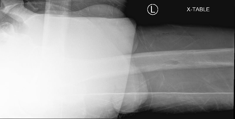
Clinical History: Leg pain with fevers and chills.
Organ: Femur
Radiologic Finding: Periosteal reaction along the femoral diaphysis, surrounding a small lytic lesion within the marrow. Adjacent soft tissue swelling.
Dx: Acute osteomyelitis with periostitis and early bone abscess
Modality: Radiography
Clinical History: Leg pain with fevers and chills.
Akira Murakami MD
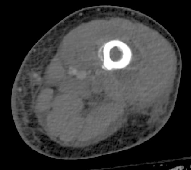
Clinical History: Chronic leg / femur pain with fevers and chills
Organ: Femur
Radiologic Finding: Hypoattenuating material lifting up the periosteum. Surrounding soft tissue attenuation and evolving fluid loculation.
Dx: Soft tissue infection with an adjacent subperiosteal abscess / involucrum of the femur.
Modality: CT
Akira Murakami MD
BONE and SOFT TISSUE ABSCESS
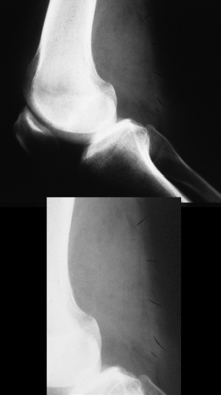
femur, soft tissues, bone, swelling, abscess, infection, X-ray
Ashley Davidoff
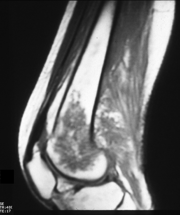
femur, soft tissues, bone, swelling, abscess, infection, MRI, T1 weighted, sagittal
Ashley Davidoff
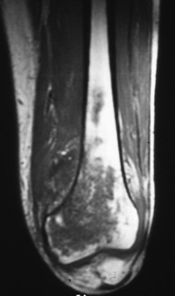
Ashley Davidoff
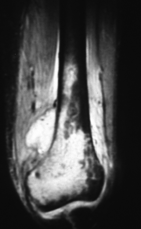
Ashley Davidoff
Tibia
Chronic Osteomyelitis
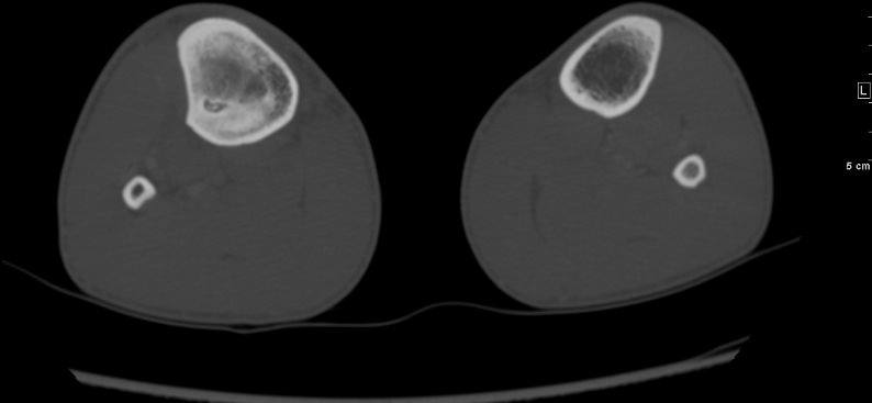
Clinical History: History of fracture, with chronic leg pain and fever.
Organ: Tibia / Lower Leg
Radiologic Finding: Linear, cavitary lesion within the marrow of the tibia diaphysis, with surrounding sclerosis and bony proliferation. Calcified foci within the center of the cavitary lesions. Numerus osseous sinus tracts extending to the soft tissue.
Dx: Chronic osteomyelitis with sequestra.
Modality: CT in the axial planes
Akira Murakami MD
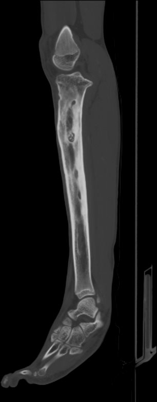
Clinical History: History of fracture, with chronic leg pain and fever.
Organ: Tibia / Lower Leg
Radiologic Finding: Linear, cavitary lesion within the marrow of the tibia diaphysis, with surrounding sclerosis and bony proliferation. Calcified foci within the center of the cavitary lesions. Numerus osseous sinus tracts extending to the soft tissue.
Dx: Chronic osteomyelitis with sequestra.
Modality: CT in the sagittal plane
Akira Murakami MD
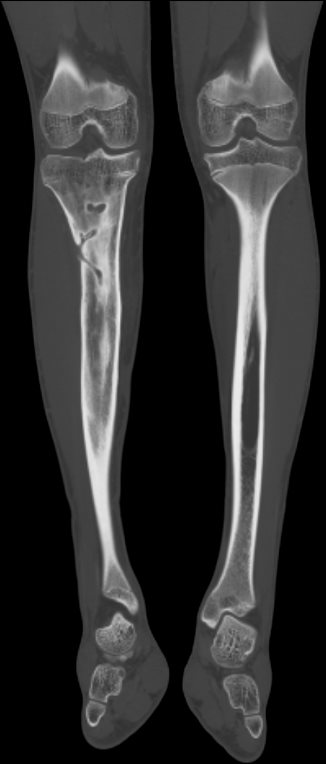
Clinical History: History of fracture, with chronic leg pain and fever.
Organ: Tibia / Lower Leg
Radiologic Finding: Linear, cavitary lesion within the marrow of the tibia diaphysis, with surrounding sclerosis and bony proliferation. Calcified foci within the center of the cavitary lesions. Numerus osseous sinus tracts extending to the soft tissue.
Dx: Chronic osteomyelitis with sequestra.
Modality: CT in the coronal plane
Akira Murakami MD
Brodies Abscess
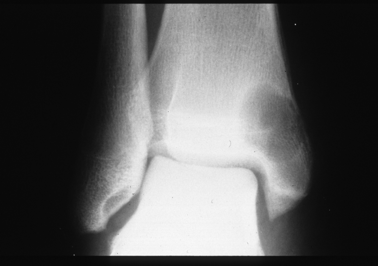
bone, tibia, lucency, lytic lesion, sclerotic margin, infection, Brodies abscess, X-ray, Ashley Davidoff MD
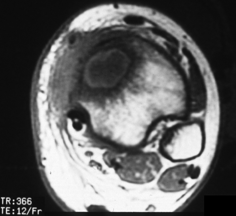
tibia, bone defect, penumbra sign, high signal of rim, subacute infection, Brodie?s abscess, MRI, T1,
Ashley Davidoff MD
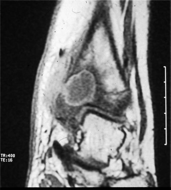
tibia, bone defect, penumbra sign, high signal of rim, subacute infection, Brodie?s abscess, MRI, T1,coronal
Ashley Davidoff MD
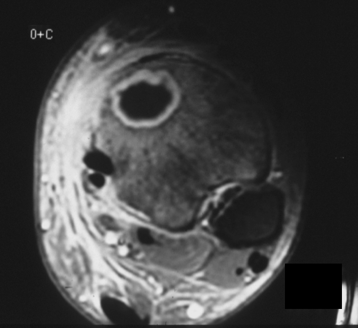
tibia, bone defect, penumbra sign, intense enhancement signal of rim, subacute infection, Brodie?s abscess, MRI, T1, post gadolinium
Ashley Davidoff MD
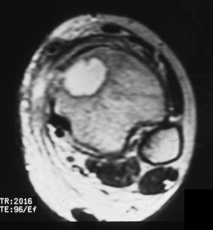
tibia, bone, defect, T2 intense signal, T2 bright, fluid matrix, subacute infection, Brodie?s abscess, MRI, T2
Ashley Davidoff MD
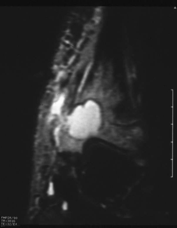
tibia, bone, defect, T2 intense signal, T2 bright, fluid matrix, subacute infection, Brodie?s abscess, MRI, T2, coronal
Ashley Davidoff MD
Foot – Osteomyelitis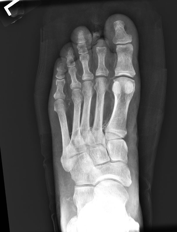
PRIOR X- RAY NORMAL DISTAL PHALANX OF THE GREAT TOE
Clinical History: Diabetes and chronic peripheral vascular disease. Recent swelling over the great toe with a chronic ulcer.
Organ: Foot
Modality: Radiograph X ray
Akira Murakami MD
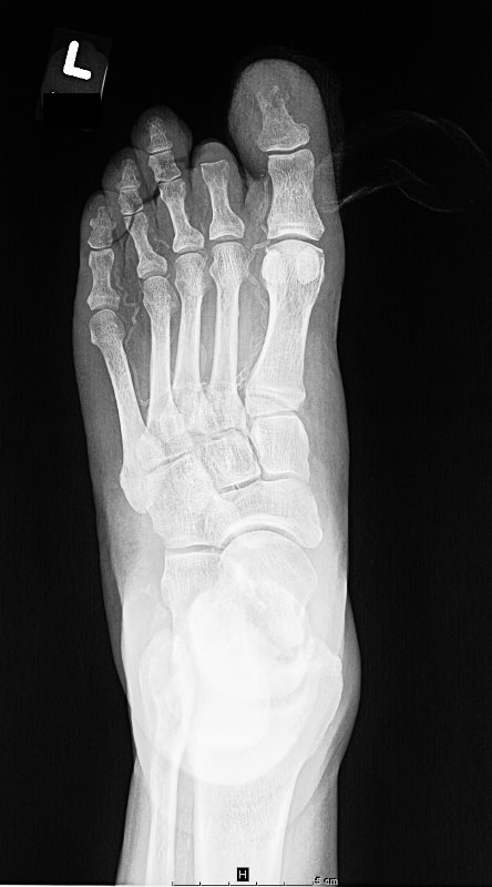
Organ: Foot
Clinical History: Diabetes and chronic peripheral vascular disease. Recent swelling over the great toe with a chronic ulcer.
Radiologic Finding: Erosive bony destruction of the tuft of the great toe distal phalanx with surrounding soft tissue swelling.
Dx: Acute osteomyelitis of the great toe.
Modality: Radiograph X ray
Akira Murakami MD
Foot – Necrotizing Fasciitis
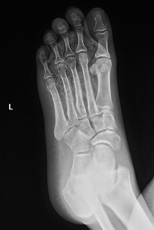
Clinical History: Fever with pain and swelling over the foot.
Organ: Foot
Radiologic Finding: Soft tissue air density throughout the forefoot.
Dx: Necrotizing fasciitis.
Modality: Radiograph X ray
Akira Murakami MD
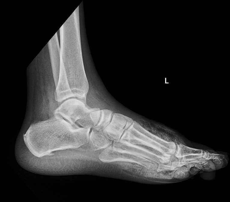
Clinical History: Fever with pain and swelling over the foot.
Organ: Foot
Radiologic Finding: Soft tissue air density throughout the forefoot.
Dx: Necrotizing fasciitis.
Modality: Radiograph X ray
Akira Murakami MD
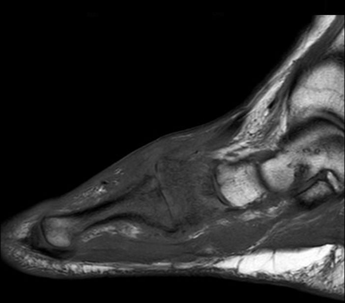
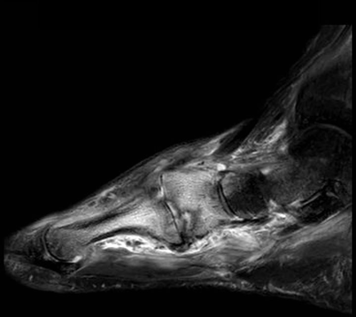
Clinical History: History of poorly controlled diabetes with a chronic foot ulcer over the medial midfoot.
Organ: Foot
Radiologic Finding: Intense T2 signal / bone marrow edema pattern of the medial cuneiform and 1st metatarsal, with corresponding geographic low T1 signal.
Dx: Acute osteomyelitis of the medial cuneiform and 1st metatarsal.
Modality: MRI Sagittal T1 and Sagittal T2 Fat saturated images of the forefoot.
Akira Murakami MD

