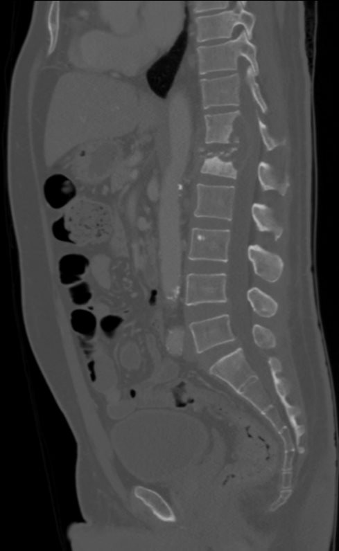
TB KIDNEY, SPINE, EPIDURAL ABSCESS, ADRENAL and PSOAS ABSCESS
58-year-old male with known history of tuberculosis.
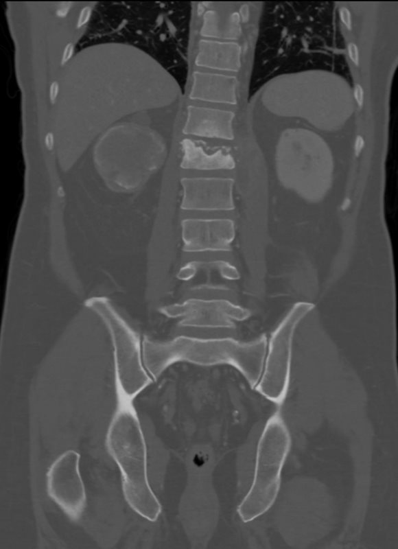
Examination of the lumbar spine on the CT scan shows findings consistent with TB osteomyelitis with destruction of T12 and L1 vertebral bodies with prevertebral and epidural collections extending into the spinal canal.
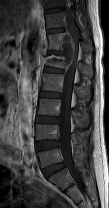
The MRI confirms the presence of an osteomyelitis discitis associated with an epidural abscess with multiple collections in the left psoas muscle
Contributed by Christina LeBedis MD Case Study
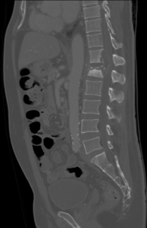
TB KIDNEY, SPINE, EPIDURAL ABSCESS, ADRENAL and PSOAS ABSCESS
58-year-old male with known history of tuberculosis.
The MRI confirms the presence of an osteomyelitis discitis associated with an epidural abscess with multiple collections in the left psoas muscle
Contributed by Christina LeBedis MD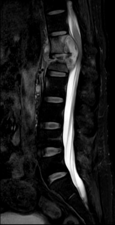
TB KIDNEY, SPINE, EPIDURAL ABSCESS, ADRENAL and PSOAS ABSCESS
58-year-old male with known history of tuberculosis.
The MRI confirms the presence of an osteomyelitis discitis associated with an epidural abscess with multiple collections in the left psoas muscle
Contributed by Christina LeBedis MD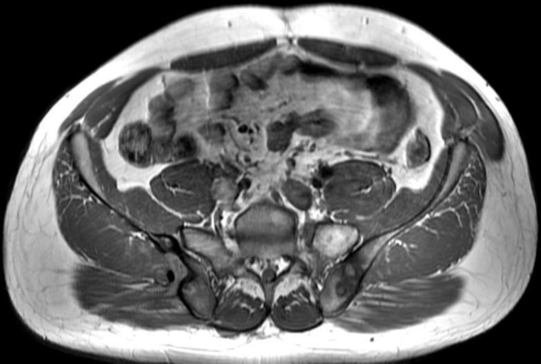
Clinical history: Chronic fever and chills. Weight loss.
Organ: Pelvis
Radiologic Finding: Left Ilium: T2 hyperintense, peripherally enhancing bone lesions with surrounding low T1 signal. Right Ilium: Peripherally enhancing, erosive bone lesion along the posterior cortex with an enhancing sinus tract extending into the gluteus muscle.
Dx: Osteomyelitis and bone abscess secondary to chronic TB.
Modality: Axial MRI T1
Akira Murakami MD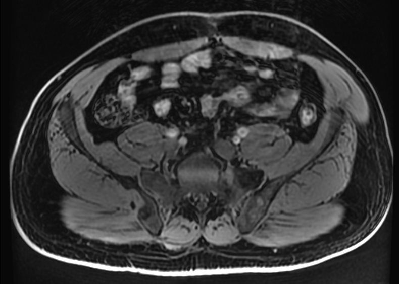
Clinical history: Chronic fever and chills. Weight loss.
Organ: Pelvis
Radiologic Finding: Left Ilium: T2 hyperintense, peripherally enhancing bone lesions with surrounding low T1 signal. Right Ilium: Peripherally enhancing, erosive bone lesion along the posterior cortex with an enhancing sinus tract extending into the gluteus muscle.
Modality: Axial MRI T1 FS pre
Akira Murakami MD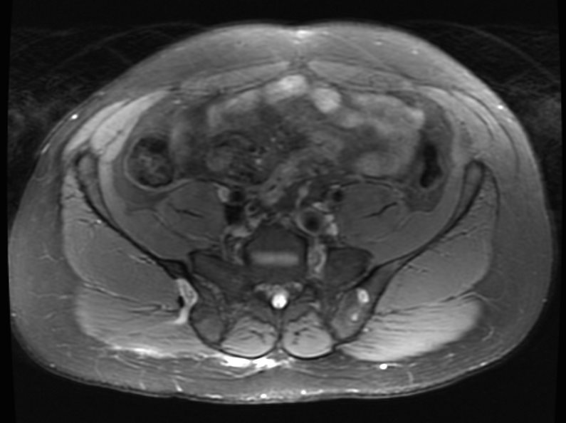
(T2 FS)
Clinical history: Chronic fever and chills. Weight loss.
Organ: Pelvis
Radiologic Finding: Left Ilium: T2 hyperintense, peripherally enhancing bone lesions with surrounding low T1 signal. Right Ilium: Peripherally enhancing, erosive bone lesion along the posterior cortex with an enhancing sinus tract extending into the gluteus muscle.
Modality: Axial MRI T2 Fat Sat
Akira Murakami MD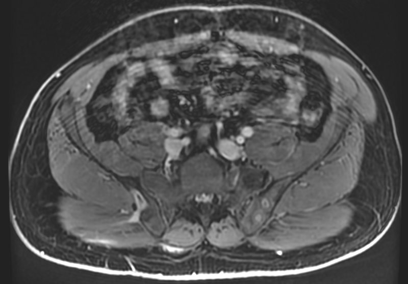
Clinical history: Chronic fever and chills. Weight loss.
Organ: Pelvis
Radiologic Finding: Left Ilium: T2 hyperintense, peripherally enhancing bone lesions with surrounding low T1 signal. Right Ilium: Peripherally enhancing, erosive bone lesion along the posterior cortex with an enhancing sinus tract extending into the gluteus muscle.
Modality: Axial MRI T1 FS post gadolinium
Akira Murakami MD
TCV Case Studies
