Hi Alex
For the sake of time I think we should just do heart/hands in collagen vascular disease – SLE, Scleroderma and RA
You should be able to stay on this page and present it all from here
- The way it is organized
- Disease name
- Heart disease with frequent components bolded
- Hands – an image of the hands in the disease
Our Patient
65 year old female with longstanding history of SLE, Lupus Sjogren?s and Raynaud?s
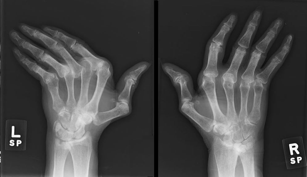
65 year old female with longstanding history of SLE, Lupus Sjogren’s and Raynaud’s Xray shows non erosive arthropathy with ulnar deviation of 2nd through 5th MCP joints
SLE
Heart
- Pancarditis
- pericardium, pericarditis 25% most common
- myocardium, myocarditis is rare and caused by vasculitis
- endocardium ? Libman-Sacks 10% mitral and tricuspid valve
- myocardial infarction 9X increase
- Cardiac complications in about 50% and major cause of death
Scleroderma
-
Heart
-
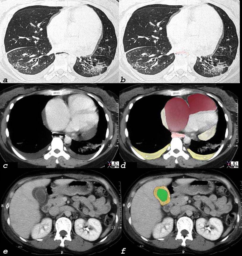
Scleroderma, Pulmonary Hypertension RVF Cor Pulmonale Pericardial effusion
40 year old female with known interstitial lung disease (a and b) shows enlarged right atrium and right ventricle and small pericardial effusion (c and overlaid in maroon in d) and enlarged esophagus (overlay in pink in d) and an edematous gallbladder wall from chronic right heart failure.
Hands
Soft Tissue Calcification Ulnar Deviation
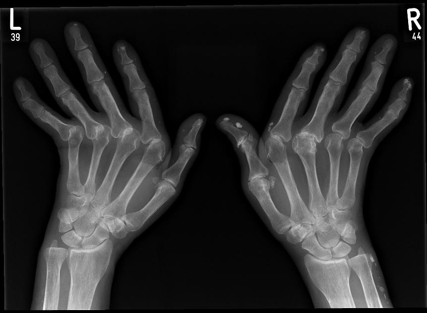
Radiographs of both hands show abnormal alignment of the metacarpophalangeal joints, most marked on the left, in keeping with subluxation. The bone density appears normal. There is joint space loss and evidence of erosive arthropathy particularly evident at the metacarpophalangeal joinft of the right 3rd and 4th MCP’s. Dense soft tissue calcifications are seen in the fingertips and along the ulnar aspect of the right wrist/distal forearm.
Case courtesy of Dr Jan Frank Gerstenmaier,
Radiopaedia.org, rID: 23125
Acroosteolysis
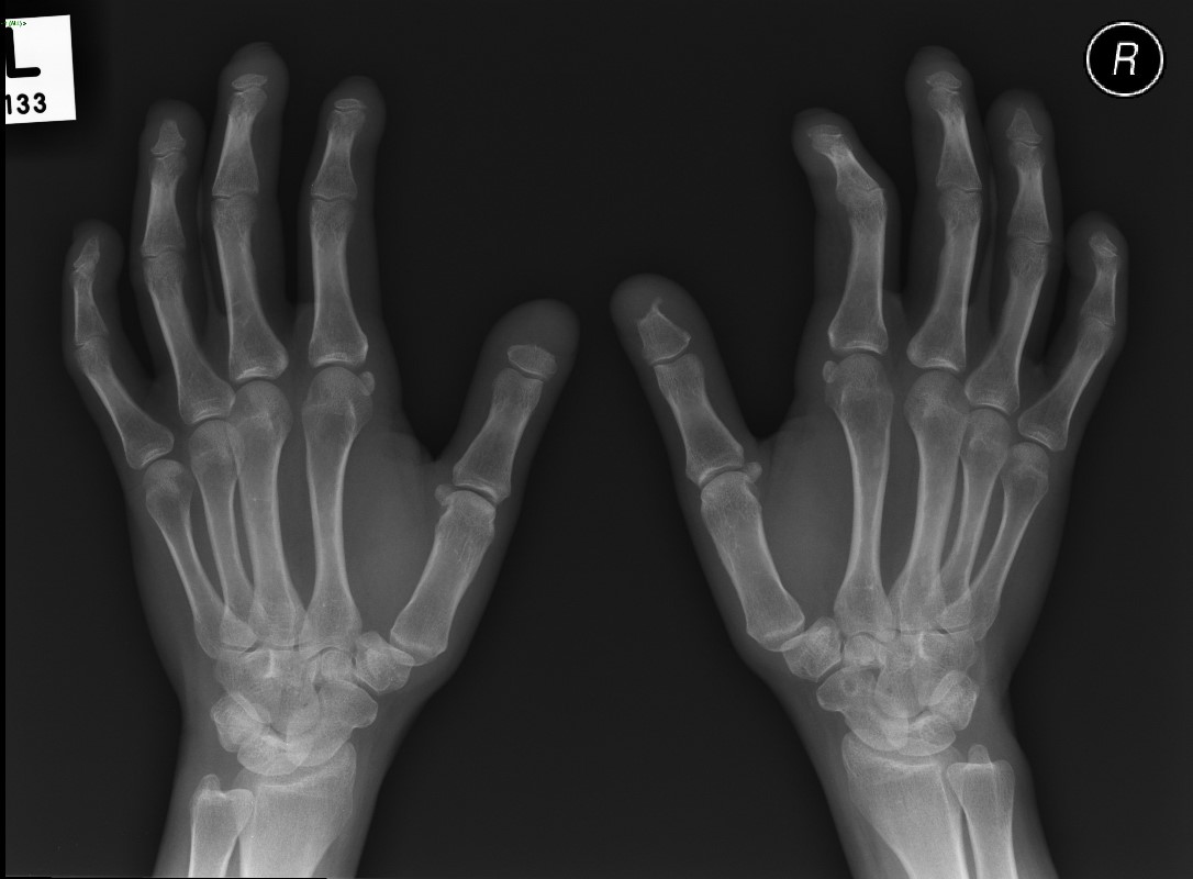
Acroosteolysis in a female patient with scleroderma
Case courtesy of Dr Minh Xuan Truong,
Radiopaedia
TCV –Scleroderma and the Heart
Rheumatoid Arthritis
Heart
- Increased incidence of
- congestive heart failure and
- ischemic heart disease associated with an
- increased mortality
- Pancarditis
Hands
Erosive Osteoarthritis dominant in the MCPs and Carpals
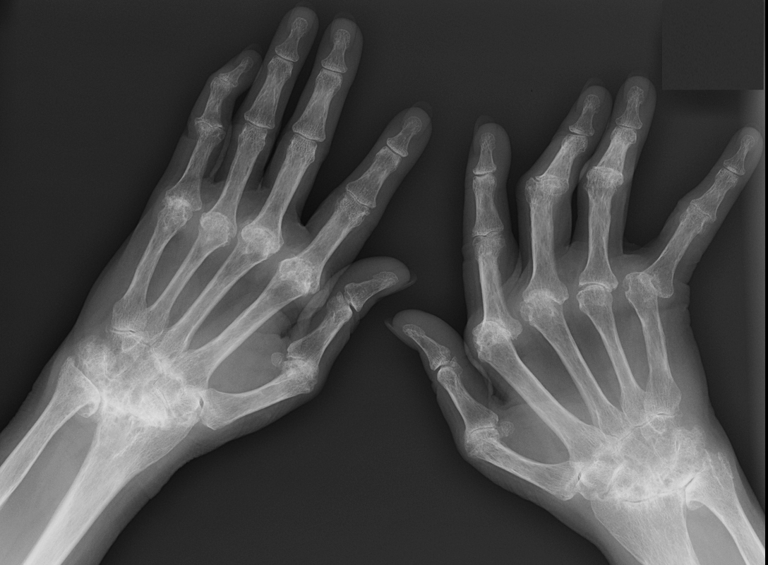
Showing degenerative and erosive changes dominantly at the MCP joints, intercarpal joints, ulnar carpal and radiocarpal joints and to lesser extent the PIP joints. There is ulnar deviation more prominent on the right hand
Ashley Davidoff MD
Rheumatoid Arthritis and Heart Disease TCV
RA and the Hand TCV
For more extensive info in see TCV on Hands and Heart
Next Marc Salomon, 65 F with fatigue and dyspnea

