Vascular Spasm
The Common Vein Copyright 2007
Principles
Inflammation
Vasculitis
Trauma
|
Shock |
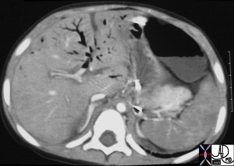
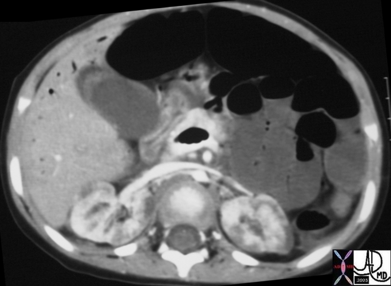
| 20333 20334 hx 3F kidney heterogeneous perfusion terminal failure imaging radiology CTscan C+ liver air dx MVA trauma bowel ischemia Courtesy Ashley Davidoff MD DB |
DOMElement Object
(
    [schemaTypeInfo] =>
    [tagName] => table
    [firstElementChild] => (object value omitted)
    [lastElementChild] => (object value omitted)
    [childElementCount] => 1
    [previousElementSibling] => (object value omitted)
    [nextElementSibling] =>
    [nodeName] => table
    [nodeValue] =>


Shock


20333 20334 hx 3F kidney heterogeneous perfusion terminal failure imaging radiology CTscan C+ liver air dx MVA trauma bowel ischemia Courtesy Ashley Davidoff MD DB


    [nodeType] => 1
    [parentNode] => (object value omitted)
    [childNodes] => (object value omitted)
    [firstChild] => (object value omitted)
    [lastChild] => (object value omitted)
    [previousSibling] => (object value omitted)
    [nextSibling] => (object value omitted)
    [attributes] => (object value omitted)
    [ownerDocument] => (object value omitted)
    [namespaceURI] =>
    [prefix] =>
    [localName] => table
    [baseURI] =>
    [textContent] =>


Shock


20333 20334 hx 3F kidney heterogeneous perfusion terminal failure imaging radiology CTscan C+ liver air dx MVA trauma bowel ischemia Courtesy Ashley Davidoff MD DB


)
DOMElement Object
(
    [schemaTypeInfo] =>
    [tagName] => td
    [firstElementChild] => (object value omitted)
    [lastElementChild] => (object value omitted)
    [childElementCount] => 1
    [previousElementSibling] =>
    [nextElementSibling] =>
    [nodeName] => td
    [nodeValue] => 20333 20334 hx 3F kidney heterogeneous perfusion terminal failure imaging radiology CTscan C+ liver air dx MVA trauma bowel ischemia Courtesy Ashley Davidoff MD DB
    [nodeType] => 1
    [parentNode] => (object value omitted)
    [childNodes] => (object value omitted)
    [firstChild] => (object value omitted)
    [lastChild] => (object value omitted)
    [previousSibling] => (object value omitted)
    [nextSibling] => (object value omitted)
    [attributes] => (object value omitted)
    [ownerDocument] => (object value omitted)
    [namespaceURI] =>
    [prefix] =>
    [localName] => td
    [baseURI] =>
    [textContent] => 20333 20334 hx 3F kidney heterogeneous perfusion terminal failure imaging radiology CTscan C+ liver air dx MVA trauma bowel ischemia Courtesy Ashley Davidoff MD DB
)
DOMElement Object
(
    [schemaTypeInfo] =>
    [tagName] => td
    [firstElementChild] => (object value omitted)
    [lastElementChild] => (object value omitted)
    [childElementCount] => 2
    [previousElementSibling] =>
    [nextElementSibling] =>
    [nodeName] => td
    [nodeValue] =>

Shock

    [nodeType] => 1
    [parentNode] => (object value omitted)
    [childNodes] => (object value omitted)
    [firstChild] => (object value omitted)
    [lastChild] => (object value omitted)
    [previousSibling] => (object value omitted)
    [nextSibling] => (object value omitted)
    [attributes] => (object value omitted)
    [ownerDocument] => (object value omitted)
    [namespaceURI] =>
    [prefix] =>
    [localName] => td
    [baseURI] =>
    [textContent] =>

Shock

)
DOMElement Object
(
    [schemaTypeInfo] =>
    [tagName] => table
    [firstElementChild] => (object value omitted)
    [lastElementChild] => (object value omitted)
    [childElementCount] => 1
    [previousElementSibling] => (object value omitted)
    [nextElementSibling] => (object value omitted)
    [nodeName] => table
    [nodeValue] =>


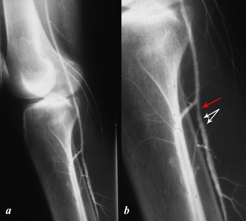
Traumatic Spasm and beading of the Tibioperoneal Trunk


This male patient presented with transient loss of pulses and cyanosis of the right foot following traumatic injury to his knee. This angiogram of the popliteal artery, (a, and magnified in b) show spasm and narrowing (red arrow) of the tibioperoneal trunk as well as beading of the artery (white arrows). No obvious intimal injury is identified.
copyright 2012 Courtesy Ashley Davidoff MD 00008c01.8L


    [nodeType] => 1
    [parentNode] => (object value omitted)
    [childNodes] => (object value omitted)
    [firstChild] => (object value omitted)
    [lastChild] => (object value omitted)
    [previousSibling] => (object value omitted)
    [nextSibling] => (object value omitted)
    [attributes] => (object value omitted)
    [ownerDocument] => (object value omitted)
    [namespaceURI] =>
    [prefix] =>
    [localName] => table
    [baseURI] =>
    [textContent] =>


Traumatic Spasm and beading of the Tibioperoneal Trunk


This male patient presented with transient loss of pulses and cyanosis of the right foot following traumatic injury to his knee. This angiogram of the popliteal artery, (a, and magnified in b) show spasm and narrowing (red arrow) of the tibioperoneal trunk as well as beading of the artery (white arrows). No obvious intimal injury is identified.
copyright 2012 Courtesy Ashley Davidoff MD 00008c01.8L


)
DOMElement Object
(
    [schemaTypeInfo] =>
    [tagName] => td
    [firstElementChild] => (object value omitted)
    [lastElementChild] => (object value omitted)
    [childElementCount] => 1
    [previousElementSibling] =>
    [nextElementSibling] =>
    [nodeName] => td
    [nodeValue] => This male patient presented with transient loss of pulses and cyanosis of the right foot following traumatic injury to his knee. This angiogram of the popliteal artery, (a, and magnified in b) show spasm and narrowing (red arrow) of the tibioperoneal trunk as well as beading of the artery (white arrows). No obvious intimal injury is identified.
copyright 2012 Courtesy Ashley Davidoff MD 00008c01.8L

    [nodeType] => 1
    [parentNode] => (object value omitted)
    [childNodes] => (object value omitted)
    [firstChild] => (object value omitted)
    [lastChild] => (object value omitted)
    [previousSibling] => (object value omitted)
    [nextSibling] => (object value omitted)
    [attributes] => (object value omitted)
    [ownerDocument] => (object value omitted)
    [namespaceURI] =>
    [prefix] =>
    [localName] => td
    [baseURI] =>
    [textContent] => This male patient presented with transient loss of pulses and cyanosis of the right foot following traumatic injury to his knee. This angiogram of the popliteal artery, (a, and magnified in b) show spasm and narrowing (red arrow) of the tibioperoneal trunk as well as beading of the artery (white arrows). No obvious intimal injury is identified.
copyright 2012 Courtesy Ashley Davidoff MD 00008c01.8L

)
DOMElement Object
(
    [schemaTypeInfo] =>
    [tagName] => td
    [firstElementChild] => (object value omitted)
    [lastElementChild] => (object value omitted)
    [childElementCount] => 2
    [previousElementSibling] =>
    [nextElementSibling] =>
    [nodeName] => td
    [nodeValue] =>
Traumatic Spasm and beading of the Tibioperoneal Trunk

    [nodeType] => 1
    [parentNode] => (object value omitted)
    [childNodes] => (object value omitted)
    [firstChild] => (object value omitted)
    [lastChild] => (object value omitted)
    [previousSibling] => (object value omitted)
    [nextSibling] => (object value omitted)
    [attributes] => (object value omitted)
    [ownerDocument] => (object value omitted)
    [namespaceURI] =>
    [prefix] =>
    [localName] => td
    [baseURI] =>
    [textContent] =>
Traumatic Spasm and beading of the Tibioperoneal Trunk

)
DOMElement Object
(
    [schemaTypeInfo] =>
    [tagName] => table
    [firstElementChild] => (object value omitted)
    [lastElementChild] => (object value omitted)
    [childElementCount] => 1
    [previousElementSibling] => (object value omitted)
    [nextElementSibling] => (object value omitted)
    [nodeName] => table
    [nodeValue] =>


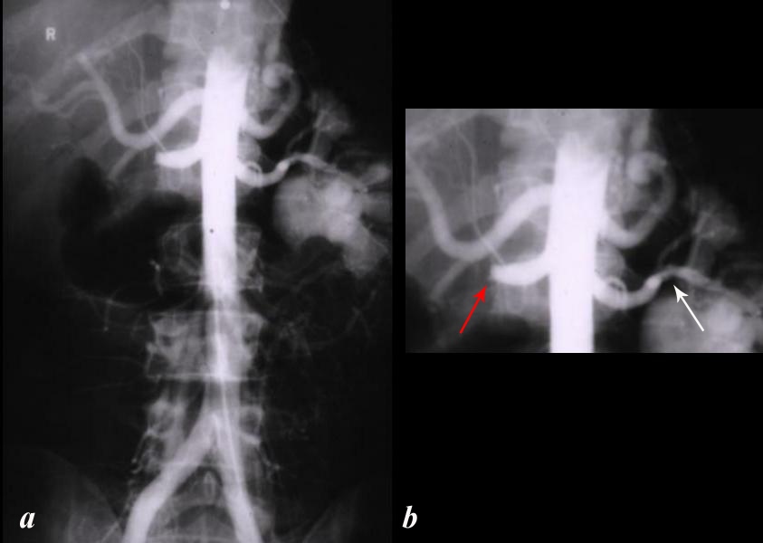
Occlusion and Spasm Following Trauma


The aortogram is from a patient who was admitted to the hospital following a motor vehicle accident and wascomplaining of right flank pain. The aortogram in the AP projection shows an occluded right renal artery (red arrow) and spasm of the left renal artery (white arrow).
Courtesy Ashley Davidoff MD 00086bL


    [nodeType] => 1
    [parentNode] => (object value omitted)
    [childNodes] => (object value omitted)
    [firstChild] => (object value omitted)
    [lastChild] => (object value omitted)
    [previousSibling] => (object value omitted)
    [nextSibling] => (object value omitted)
    [attributes] => (object value omitted)
    [ownerDocument] => (object value omitted)
    [namespaceURI] =>
    [prefix] =>
    [localName] => table
    [baseURI] =>
    [textContent] =>


Occlusion and Spasm Following Trauma


The aortogram is from a patient who was admitted to the hospital following a motor vehicle accident and wascomplaining of right flank pain. The aortogram in the AP projection shows an occluded right renal artery (red arrow) and spasm of the left renal artery (white arrow).
Courtesy Ashley Davidoff MD 00086bL


)
DOMElement Object
(
    [schemaTypeInfo] =>
    [tagName] => td
    [firstElementChild] => (object value omitted)
    [lastElementChild] => (object value omitted)
    [childElementCount] => 2
    [previousElementSibling] =>
    [nextElementSibling] =>
    [nodeName] => td
    [nodeValue] =>
The aortogram is from a patient who was admitted to the hospital following a motor vehicle accident and wascomplaining of right flank pain. The aortogram in the AP projection shows an occluded right renal artery (red arrow) and spasm of the left renal artery (white arrow).
Courtesy Ashley Davidoff MD 00086bL

    [nodeType] => 1
    [parentNode] => (object value omitted)
    [childNodes] => (object value omitted)
    [firstChild] => (object value omitted)
    [lastChild] => (object value omitted)
    [previousSibling] => (object value omitted)
    [nextSibling] => (object value omitted)
    [attributes] => (object value omitted)
    [ownerDocument] => (object value omitted)
    [namespaceURI] =>
    [prefix] =>
    [localName] => td
    [baseURI] =>
    [textContent] =>
The aortogram is from a patient who was admitted to the hospital following a motor vehicle accident and wascomplaining of right flank pain. The aortogram in the AP projection shows an occluded right renal artery (red arrow) and spasm of the left renal artery (white arrow).
Courtesy Ashley Davidoff MD 00086bL

)
DOMElement Object
(
    [schemaTypeInfo] =>
    [tagName] => td
    [firstElementChild] => (object value omitted)
    [lastElementChild] => (object value omitted)
    [childElementCount] => 2
    [previousElementSibling] =>
    [nextElementSibling] =>
    [nodeName] => td
    [nodeValue] =>
Occlusion and Spasm Following Trauma

    [nodeType] => 1
    [parentNode] => (object value omitted)
    [childNodes] => (object value omitted)
    [firstChild] => (object value omitted)
    [lastChild] => (object value omitted)
    [previousSibling] => (object value omitted)
    [nextSibling] => (object value omitted)
    [attributes] => (object value omitted)
    [ownerDocument] => (object value omitted)
    [namespaceURI] =>
    [prefix] =>
    [localName] => td
    [baseURI] =>
    [textContent] =>
Occlusion and Spasm Following Trauma

)
DOMElement Object
(
    [schemaTypeInfo] =>
    [tagName] => table
    [firstElementChild] => (object value omitted)
    [lastElementChild] => (object value omitted)
    [childElementCount] => 1
    [previousElementSibling] => (object value omitted)
    [nextElementSibling] => (object value omitted)
    [nodeName] => table
    [nodeValue] =>


Traumatic Injury to Lumbar Rtaeries Extravasation and Hypotension with Spasm


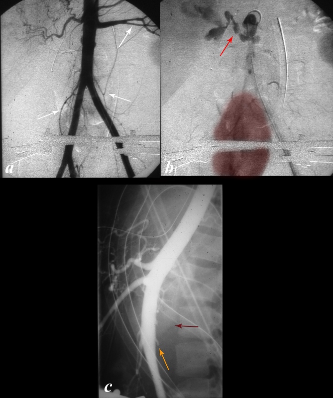
This young male patient fell from high scaffolding and presented in shock. The aortogram shows spasm of the left renal artery, SMA and IMA (white arrows) and diffuse narrowing of the aorta when compared to the common iliacs. The right renal artery is not visualized. Delayed imaging shows active extravasation of contrast (red arrow) and the nephrogram of a pelvic kidney (maroon overlay), explaining the lack of visualization of the right kidney in image (a). The lateral examination (c) shows avulsed lumbar arteries (orange arrows) and an increase in the retro aortic space (maroon arrow).
Copyright 2012 Courtesy Ashley Davidoff MD 00015.1c02.8


    [nodeType] => 1
    [parentNode] => (object value omitted)
    [childNodes] => (object value omitted)
    [firstChild] => (object value omitted)
    [lastChild] => (object value omitted)
    [previousSibling] => (object value omitted)
    [nextSibling] => (object value omitted)
    [attributes] => (object value omitted)
    [ownerDocument] => (object value omitted)
    [namespaceURI] =>
    [prefix] =>
    [localName] => table
    [baseURI] =>
    [textContent] =>


Traumatic Injury to Lumbar Rtaeries Extravasation and Hypotension with Spasm


This young male patient fell from high scaffolding and presented in shock. The aortogram shows spasm of the left renal artery, SMA and IMA (white arrows) and diffuse narrowing of the aorta when compared to the common iliacs. The right renal artery is not visualized. Delayed imaging shows active extravasation of contrast (red arrow) and the nephrogram of a pelvic kidney (maroon overlay), explaining the lack of visualization of the right kidney in image (a). The lateral examination (c) shows avulsed lumbar arteries (orange arrows) and an increase in the retro aortic space (maroon arrow).
Copyright 2012 Courtesy Ashley Davidoff MD 00015.1c02.8


)
DOMElement Object
(
    [schemaTypeInfo] =>
    [tagName] => td
    [firstElementChild] => (object value omitted)
    [lastElementChild] => (object value omitted)
    [childElementCount] => 1
    [previousElementSibling] =>
    [nextElementSibling] =>
    [nodeName] => td
    [nodeValue] => This young male patient fell from high scaffolding and presented in shock. The aortogram shows spasm of the left renal artery, SMA and IMA (white arrows) and diffuse narrowing of the aorta when compared to the common iliacs. The right renal artery is not visualized. Delayed imaging shows active extravasation of contrast (red arrow) and the nephrogram of a pelvic kidney (maroon overlay), explaining the lack of visualization of the right kidney in image (a). The lateral examination (c) shows avulsed lumbar arteries (orange arrows) and an increase in the retro aortic space (maroon arrow).
Copyright 2012 Courtesy Ashley Davidoff MD 00015.1c02.8

    [nodeType] => 1
    [parentNode] => (object value omitted)
    [childNodes] => (object value omitted)
    [firstChild] => (object value omitted)
    [lastChild] => (object value omitted)
    [previousSibling] => (object value omitted)
    [nextSibling] => (object value omitted)
    [attributes] => (object value omitted)
    [ownerDocument] => (object value omitted)
    [namespaceURI] =>
    [prefix] =>
    [localName] => td
    [baseURI] =>
    [textContent] => This young male patient fell from high scaffolding and presented in shock. The aortogram shows spasm of the left renal artery, SMA and IMA (white arrows) and diffuse narrowing of the aorta when compared to the common iliacs. The right renal artery is not visualized. Delayed imaging shows active extravasation of contrast (red arrow) and the nephrogram of a pelvic kidney (maroon overlay), explaining the lack of visualization of the right kidney in image (a). The lateral examination (c) shows avulsed lumbar arteries (orange arrows) and an increase in the retro aortic space (maroon arrow).
Copyright 2012 Courtesy Ashley Davidoff MD 00015.1c02.8

)
DOMElement Object
(
    [schemaTypeInfo] =>
    [tagName] => td
    [firstElementChild] => (object value omitted)
    [lastElementChild] => (object value omitted)
    [childElementCount] => 2
    [previousElementSibling] =>
    [nextElementSibling] =>
    [nodeName] => td
    [nodeValue] =>
Traumatic Injury to Lumbar Rtaeries Extravasation and Hypotension with Spasm

    [nodeType] => 1
    [parentNode] => (object value omitted)
    [childNodes] => (object value omitted)
    [firstChild] => (object value omitted)
    [lastChild] => (object value omitted)
    [previousSibling] => (object value omitted)
    [nextSibling] => (object value omitted)
    [attributes] => (object value omitted)
    [ownerDocument] => (object value omitted)
    [namespaceURI] =>
    [prefix] =>
    [localName] => td
    [baseURI] =>
    [textContent] =>
Traumatic Injury to Lumbar Rtaeries Extravasation and Hypotension with Spasm

)
DOMElement Object
(
    [schemaTypeInfo] =>
    [tagName] => table
    [firstElementChild] => (object value omitted)
    [lastElementChild] => (object value omitted)
    [childElementCount] => 1
    [previousElementSibling] => (object value omitted)
    [nextElementSibling] => (object value omitted)
    [nodeName] => table
    [nodeValue] =>


Question Radiation Induced Arteritis


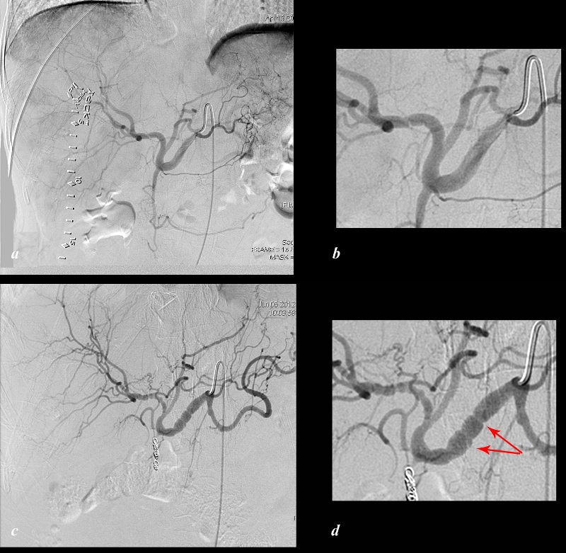
The angiogram of the proper hepatic artery performed in planning for Yttrium therapy (a and magnified in b) in a patient with metastatic carcinoid disease, and shows a normal caliber smooth walled right hepatic artery. Hypervascular metastases are suggested in the image (a). The angiogram of the proper hepatic artery performed 2 months later following Yttrium therapy (c and magnified in d) shows beading of the proper (red arrows) and right hepatic artery. These findings were present without the advance of a wire through the vessel and suggest arteritis post Yttrium therapy.
Copyright 2012 Courtesy Tim Frey MD 113489c01L.8


    [nodeType] => 1
    [parentNode] => (object value omitted)
    [childNodes] => (object value omitted)
    [firstChild] => (object value omitted)
    [lastChild] => (object value omitted)
    [previousSibling] => (object value omitted)
    [nextSibling] => (object value omitted)
    [attributes] => (object value omitted)
    [ownerDocument] => (object value omitted)
    [namespaceURI] =>
    [prefix] =>
    [localName] => table
    [baseURI] =>
    [textContent] =>


Question Radiation Induced Arteritis


The angiogram of the proper hepatic artery performed in planning for Yttrium therapy (a and magnified in b) in a patient with metastatic carcinoid disease, and shows a normal caliber smooth walled right hepatic artery. Hypervascular metastases are suggested in the image (a). The angiogram of the proper hepatic artery performed 2 months later following Yttrium therapy (c and magnified in d) shows beading of the proper (red arrows) and right hepatic artery. These findings were present without the advance of a wire through the vessel and suggest arteritis post Yttrium therapy.
Copyright 2012 Courtesy Tim Frey MD 113489c01L.8


)
DOMElement Object
(
    [schemaTypeInfo] =>
    [tagName] => td
    [firstElementChild] => (object value omitted)
    [lastElementChild] => (object value omitted)
    [childElementCount] => 2
    [previousElementSibling] =>
    [nextElementSibling] =>
    [nodeName] => td
    [nodeValue] =>
The angiogram of the proper hepatic artery performed in planning for Yttrium therapy (a and magnified in b) in a patient with metastatic carcinoid disease, and shows a normal caliber smooth walled right hepatic artery. Hypervascular metastases are suggested in the image (a). The angiogram of the proper hepatic artery performed 2 months later following Yttrium therapy (c and magnified in d) shows beading of the proper (red arrows) and right hepatic artery. These findings were present without the advance of a wire through the vessel and suggest arteritis post Yttrium therapy.
Copyright 2012 Courtesy Tim Frey MD 113489c01L.8

    [nodeType] => 1
    [parentNode] => (object value omitted)
    [childNodes] => (object value omitted)
    [firstChild] => (object value omitted)
    [lastChild] => (object value omitted)
    [previousSibling] => (object value omitted)
    [nextSibling] => (object value omitted)
    [attributes] => (object value omitted)
    [ownerDocument] => (object value omitted)
    [namespaceURI] =>
    [prefix] =>
    [localName] => td
    [baseURI] =>
    [textContent] =>
The angiogram of the proper hepatic artery performed in planning for Yttrium therapy (a and magnified in b) in a patient with metastatic carcinoid disease, and shows a normal caliber smooth walled right hepatic artery. Hypervascular metastases are suggested in the image (a). The angiogram of the proper hepatic artery performed 2 months later following Yttrium therapy (c and magnified in d) shows beading of the proper (red arrows) and right hepatic artery. These findings were present without the advance of a wire through the vessel and suggest arteritis post Yttrium therapy.
Copyright 2012 Courtesy Tim Frey MD 113489c01L.8

)
DOMElement Object
(
    [schemaTypeInfo] =>
    [tagName] => td
    [firstElementChild] => (object value omitted)
    [lastElementChild] => (object value omitted)
    [childElementCount] => 2
    [previousElementSibling] =>
    [nextElementSibling] =>
    [nodeName] => td
    [nodeValue] =>
Question Radiation Induced Arteritis

    [nodeType] => 1
    [parentNode] => (object value omitted)
    [childNodes] => (object value omitted)
    [firstChild] => (object value omitted)
    [lastChild] => (object value omitted)
    [previousSibling] => (object value omitted)
    [nextSibling] => (object value omitted)
    [attributes] => (object value omitted)
    [ownerDocument] => (object value omitted)
    [namespaceURI] =>
    [prefix] =>
    [localName] => td
    [baseURI] =>
    [textContent] =>
Question Radiation Induced Arteritis

)
DOMElement Object
(
    [schemaTypeInfo] =>
    [tagName] => table
    [firstElementChild] => (object value omitted)
    [lastElementChild] => (object value omitted)
    [childElementCount] => 1
    [previousElementSibling] => (object value omitted)
    [nextElementSibling] => (object value omitted)
    [nodeName] => table
    [nodeValue] =>


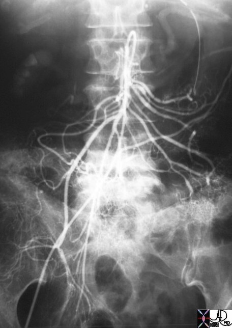
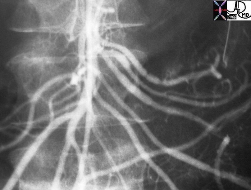
Vasculitis and Spasm


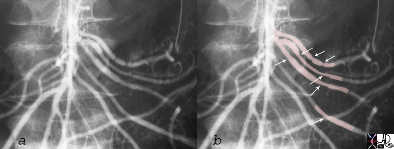
This young woman presented with abdominal pain and bleeding.  She has a known history of henoch Schonlein purpura.  The angiogram shows focal and diffuse areas of vascular spasm (arrows) consistent with a diagnosis of Henoch Schonlein  vasculitis.
28517c03.jpg artery SMA vasculitis Henoch Schonlein arteritis Henoch Schonlein purpura preents with GI bleed fx spasm superior mesenteric artery jejunal branches angiogram angiography Courtesy Ashley DAvidoff MD


    [nodeType] => 1
    [parentNode] => (object value omitted)
    [childNodes] => (object value omitted)
    [firstChild] => (object value omitted)
    [lastChild] => (object value omitted)
    [previousSibling] => (object value omitted)
    [nextSibling] => (object value omitted)
    [attributes] => (object value omitted)
    [ownerDocument] => (object value omitted)
    [namespaceURI] =>
    [prefix] =>
    [localName] => table
    [baseURI] =>
    [textContent] =>


Vasculitis and Spasm


This young woman presented with abdominal pain and bleeding.  She has a known history of henoch Schonlein purpura.  The angiogram shows focal and diffuse areas of vascular spasm (arrows) consistent with a diagnosis of Henoch Schonlein  vasculitis.
28517c03.jpg artery SMA vasculitis Henoch Schonlein arteritis Henoch Schonlein purpura preents with GI bleed fx spasm superior mesenteric artery jejunal branches angiogram angiography Courtesy Ashley DAvidoff MD


)
DOMElement Object
(
    [schemaTypeInfo] =>
    [tagName] => td
    [firstElementChild] => (object value omitted)
    [lastElementChild] => (object value omitted)
    [childElementCount] => 2
    [previousElementSibling] =>
    [nextElementSibling] =>
    [nodeName] => td
    [nodeValue] =>
This young woman presented with abdominal pain and bleeding.  She has a known history of henoch Schonlein purpura.  The angiogram shows focal and diffuse areas of vascular spasm (arrows) consistent with a diagnosis of Henoch Schonlein  vasculitis.
28517c03.jpg artery SMA vasculitis Henoch Schonlein arteritis Henoch Schonlein purpura preents with GI bleed fx spasm superior mesenteric artery jejunal branches angiogram angiography Courtesy Ashley DAvidoff MD
    [nodeType] => 1
    [parentNode] => (object value omitted)
    [childNodes] => (object value omitted)
    [firstChild] => (object value omitted)
    [lastChild] => (object value omitted)
    [previousSibling] => (object value omitted)
    [nextSibling] => (object value omitted)
    [attributes] => (object value omitted)
    [ownerDocument] => (object value omitted)
    [namespaceURI] =>
    [prefix] =>
    [localName] => td
    [baseURI] =>
    [textContent] =>
This young woman presented with abdominal pain and bleeding.  She has a known history of henoch Schonlein purpura.  The angiogram shows focal and diffuse areas of vascular spasm (arrows) consistent with a diagnosis of Henoch Schonlein  vasculitis.
28517c03.jpg artery SMA vasculitis Henoch Schonlein arteritis Henoch Schonlein purpura preents with GI bleed fx spasm superior mesenteric artery jejunal branches angiogram angiography Courtesy Ashley DAvidoff MD
)
DOMElement Object
(
    [schemaTypeInfo] =>
    [tagName] => td
    [firstElementChild] => (object value omitted)
    [lastElementChild] => (object value omitted)
    [childElementCount] => 2
    [previousElementSibling] =>
    [nextElementSibling] =>
    [nodeName] => td
    [nodeValue] =>
Vasculitis and Spasm

    [nodeType] => 1
    [parentNode] => (object value omitted)
    [childNodes] => (object value omitted)
    [firstChild] => (object value omitted)
    [lastChild] => (object value omitted)
    [previousSibling] => (object value omitted)
    [nextSibling] => (object value omitted)
    [attributes] => (object value omitted)
    [ownerDocument] => (object value omitted)
    [namespaceURI] =>
    [prefix] =>
    [localName] => td
    [baseURI] =>
    [textContent] =>
Vasculitis and Spasm

)
DOMElement Object
(
    [schemaTypeInfo] =>
    [tagName] => table
    [firstElementChild] => (object value omitted)
    [lastElementChild] => (object value omitted)
    [childElementCount] => 1
    [previousElementSibling] => (object value omitted)
    [nextElementSibling] => (object value omitted)
    [nodeName] => table
    [nodeValue] =>


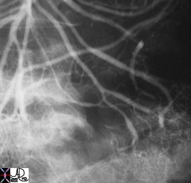
Henoch Schonlein Arteritis Segmental Spasm with Hemorrhage


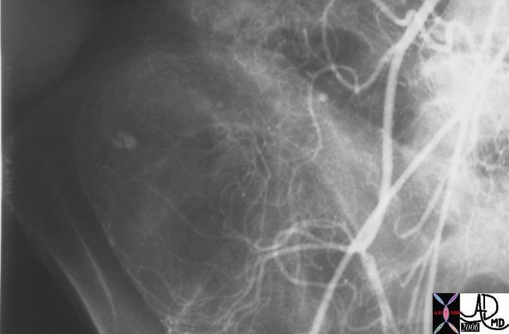
Young female presents with GI bleed hemorrhage bleeding blood small bowel colon SMA superior mesenteric artery jejunal branches ileocolic artery fx arterial spasm fx contrast extravasation RLQ in cecum dx arteritis angiitis vasculitis arteriopathy dx Henoch -Schonlein arteritis angiography angiogram Courtesy Ashley Davidoff MD 28514 28515 28516 28517 surgical specimen showed plaque like ulcers in the bowel consistent with chronic ischemia


    [nodeType] => 1
    [parentNode] => (object value omitted)
    [childNodes] => (object value omitted)
    [firstChild] => (object value omitted)
    [lastChild] => (object value omitted)
    [previousSibling] => (object value omitted)
    [nextSibling] => (object value omitted)
    [attributes] => (object value omitted)
    [ownerDocument] => (object value omitted)
    [namespaceURI] =>
    [prefix] =>
    [localName] => table
    [baseURI] =>
    [textContent] =>


Henoch Schonlein Arteritis Segmental Spasm with Hemorrhage


Young female presents with GI bleed hemorrhage bleeding blood small bowel colon SMA superior mesenteric artery jejunal branches ileocolic artery fx arterial spasm fx contrast extravasation RLQ in cecum dx arteritis angiitis vasculitis arteriopathy dx Henoch -Schonlein arteritis angiography angiogram Courtesy Ashley Davidoff MD 28514 28515 28516 28517 surgical specimen showed plaque like ulcers in the bowel consistent with chronic ischemia


)
DOMElement Object
(
    [schemaTypeInfo] =>
    [tagName] => td
    [firstElementChild] => (object value omitted)
    [lastElementChild] => (object value omitted)
    [childElementCount] => 1
    [previousElementSibling] =>
    [nextElementSibling] =>
    [nodeName] => td
    [nodeValue] => Young female presents with GI bleed hemorrhage bleeding blood small bowel colon SMA superior mesenteric artery jejunal branches ileocolic artery fx arterial spasm fx contrast extravasation RLQ in cecum dx arteritis angiitis vasculitis arteriopathy dx Henoch -Schonlein arteritis angiography angiogram Courtesy Ashley Davidoff MD 28514 28515 28516 28517 surgical specimen showed plaque like ulcers in the bowel consistent with chronic ischemia
    [nodeType] => 1
    [parentNode] => (object value omitted)
    [childNodes] => (object value omitted)
    [firstChild] => (object value omitted)
    [lastChild] => (object value omitted)
    [previousSibling] => (object value omitted)
    [nextSibling] => (object value omitted)
    [attributes] => (object value omitted)
    [ownerDocument] => (object value omitted)
    [namespaceURI] =>
    [prefix] =>
    [localName] => td
    [baseURI] =>
    [textContent] => Young female presents with GI bleed hemorrhage bleeding blood small bowel colon SMA superior mesenteric artery jejunal branches ileocolic artery fx arterial spasm fx contrast extravasation RLQ in cecum dx arteritis angiitis vasculitis arteriopathy dx Henoch -Schonlein arteritis angiography angiogram Courtesy Ashley Davidoff MD 28514 28515 28516 28517 surgical specimen showed plaque like ulcers in the bowel consistent with chronic ischemia
)
DOMElement Object
(
    [schemaTypeInfo] =>
    [tagName] => td
    [firstElementChild] => (object value omitted)
    [lastElementChild] => (object value omitted)
    [childElementCount] => 3
    [previousElementSibling] =>
    [nextElementSibling] =>
    [nodeName] => td
    [nodeValue] =>
Henoch Schonlein Arteritis Segmental Spasm with Hemorrhage

    [nodeType] => 1
    [parentNode] => (object value omitted)
    [childNodes] => (object value omitted)
    [firstChild] => (object value omitted)
    [lastChild] => (object value omitted)
    [previousSibling] => (object value omitted)
    [nextSibling] => (object value omitted)
    [attributes] => (object value omitted)
    [ownerDocument] => (object value omitted)
    [namespaceURI] =>
    [prefix] =>
    [localName] => td
    [baseURI] =>
    [textContent] =>
Henoch Schonlein Arteritis Segmental Spasm with Hemorrhage

)
https://beta.thecommonvein.net/wp-content/uploads/2023/05/28517.jpg https://beta.thecommonvein.net/wp-content/uploads/2023/05/28516.jpg https://beta.thecommonvein.net/wp-content/uploads/2023/05/28515.jpg https://beta.thecommonvein.net/wp-content/uploads/2023/05/28514.jpg
http://thecommonvein.net/media/28516.jpg http://thecommonvein.net/media/28515.jpg
DOMElement Object
(
    [schemaTypeInfo] =>
    [tagName] => table
    [firstElementChild] => (object value omitted)
    [lastElementChild] => (object value omitted)
    [childElementCount] => 1
    [previousElementSibling] => (object value omitted)
    [nextElementSibling] => (object value omitted)
    [nodeName] => table
    [nodeValue] =>


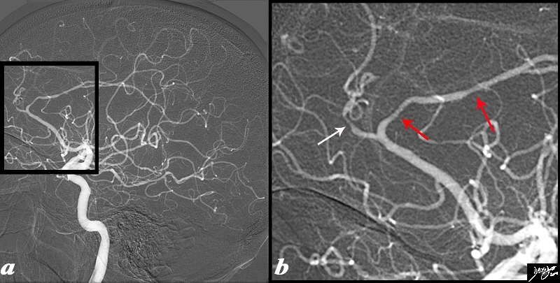
Vasculitis with Multicentric Infarction


The angiogram is from a 62 year old female patient is with acute neurological deficit The intracerebral angiogram shows focal regions of spasm with one area of spasm in the callosomarginal branch (white arrow) and two foci within the pericallosal artery (red arrows) .
Image Ashley Davidoff MD Copyright 2010 89028c04.8s


    [nodeType] => 1
    [parentNode] => (object value omitted)
    [childNodes] => (object value omitted)
    [firstChild] => (object value omitted)
    [lastChild] => (object value omitted)
    [previousSibling] => (object value omitted)
    [nextSibling] => (object value omitted)
    [attributes] => (object value omitted)
    [ownerDocument] => (object value omitted)
    [namespaceURI] =>
    [prefix] =>
    [localName] => table
    [baseURI] =>
    [textContent] =>


Vasculitis with Multicentric Infarction


The angiogram is from a 62 year old female patient is with acute neurological deficit The intracerebral angiogram shows focal regions of spasm with one area of spasm in the callosomarginal branch (white arrow) and two foci within the pericallosal artery (red arrows) .
Image Ashley Davidoff MD Copyright 2010 89028c04.8s


)
DOMElement Object
(
    [schemaTypeInfo] =>
    [tagName] => td
    [firstElementChild] => (object value omitted)
    [lastElementChild] => (object value omitted)
    [childElementCount] => 2
    [previousElementSibling] =>
    [nextElementSibling] =>
    [nodeName] => td
    [nodeValue] =>
The angiogram is from a 62 year old female patient is with acute neurological deficit The intracerebral angiogram shows focal regions of spasm with one area of spasm in the callosomarginal branch (white arrow) and two foci within the pericallosal artery (red arrows) .
Image Ashley Davidoff MD Copyright 2010 89028c04.8s

    [nodeType] => 1
    [parentNode] => (object value omitted)
    [childNodes] => (object value omitted)
    [firstChild] => (object value omitted)
    [lastChild] => (object value omitted)
    [previousSibling] => (object value omitted)
    [nextSibling] => (object value omitted)
    [attributes] => (object value omitted)
    [ownerDocument] => (object value omitted)
    [namespaceURI] =>
    [prefix] =>
    [localName] => td
    [baseURI] =>
    [textContent] =>
The angiogram is from a 62 year old female patient is with acute neurological deficit The intracerebral angiogram shows focal regions of spasm with one area of spasm in the callosomarginal branch (white arrow) and two foci within the pericallosal artery (red arrows) .
Image Ashley Davidoff MD Copyright 2010 89028c04.8s

)
DOMElement Object
(
    [schemaTypeInfo] =>
    [tagName] => td
    [firstElementChild] => (object value omitted)
    [lastElementChild] => (object value omitted)
    [childElementCount] => 2
    [previousElementSibling] =>
    [nextElementSibling] =>
    [nodeName] => td
    [nodeValue] =>
Vasculitis with Multicentric Infarction

    [nodeType] => 1
    [parentNode] => (object value omitted)
    [childNodes] => (object value omitted)
    [firstChild] => (object value omitted)
    [lastChild] => (object value omitted)
    [previousSibling] => (object value omitted)
    [nextSibling] => (object value omitted)
    [attributes] => (object value omitted)
    [ownerDocument] => (object value omitted)
    [namespaceURI] =>
    [prefix] =>
    [localName] => td
    [baseURI] =>
    [textContent] =>
Vasculitis with Multicentric Infarction

)

