The Common Vein Copyright 2009
Definition
Shock is …
characterized by …..
caused by ..etiology or predisposing factors
resulting in a pathological feature (structural change or functional change) or clinical feauture
Sometimes complicated by ….
Diagnosis is suspected clinically by … and confirmed by ….
Imaging includes the use of
Treatment options depend on …. but includes …..
Etymology if available
 
Shock |
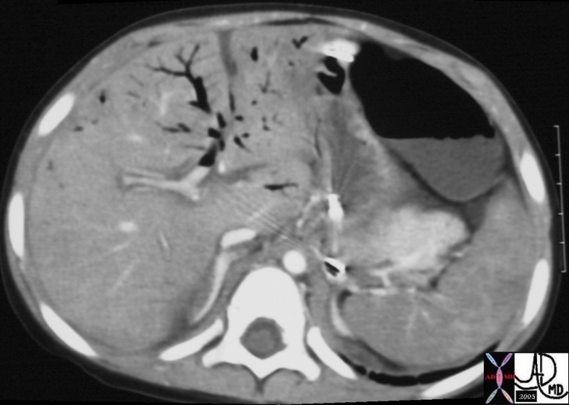
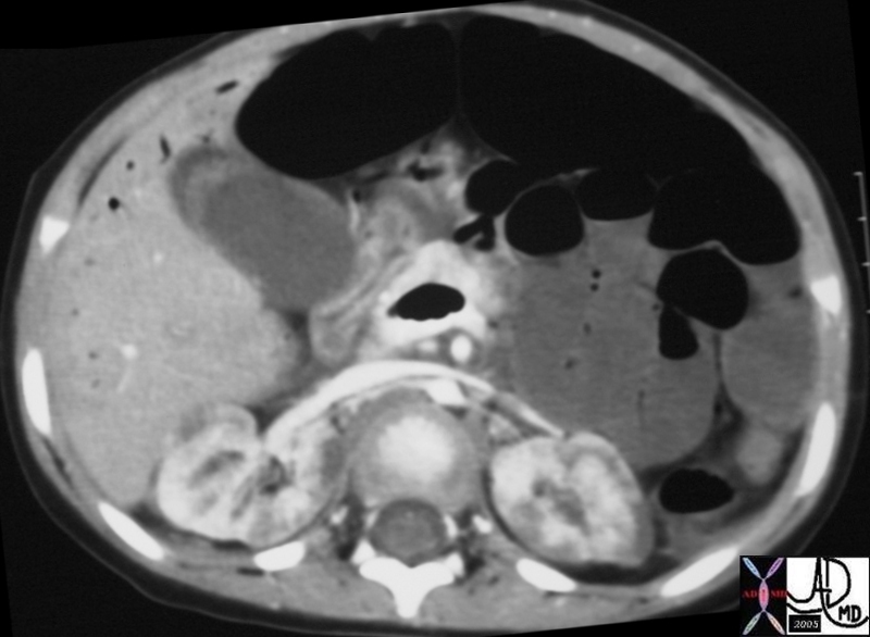
| 20333 20334 hx 3F kidney heterogeneous perfusion terminal failure imaging radiology CTscan C+ liver air dx MVA trauma bowel ischemia Courtesy Ashley Davidoff MD DB |
DOMElement Object
(
    [schemaTypeInfo] =>
    [tagName] => table
    [firstElementChild] => (object value omitted)
    [lastElementChild] => (object value omitted)
    [childElementCount] => 1
    [previousElementSibling] => (object value omitted)
    [nextElementSibling] =>
    [nodeName] => table
    [nodeValue] =>


Shock and the small Aorta


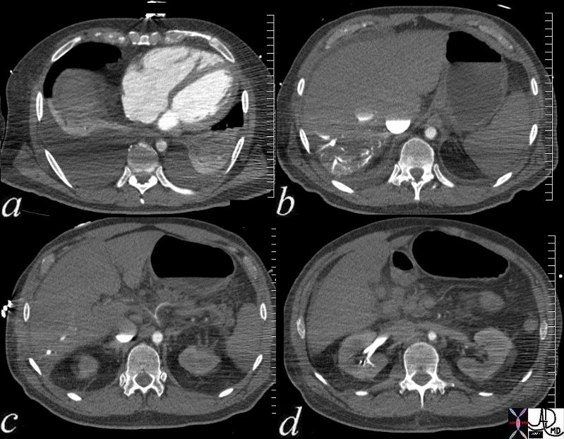
This patient presents with cardiogenic shock In image a, the right ventricle and right atrium are enlarged and thereare bilateral pleura; effusions. Image b shows stasis of contrast into the IVC and the column is relatively stattic due to peripheral constriction and slow return. There is reflux into the hepatic veins due to tricuspid regurgitation and the reflux extnds all the way to the periphery indicating poor forward flow in the hepatic circulaltion again due to operipheral constriction. Note how small the aorta due to contraction of the muscular media in this life threatening situation.. In image c the celiac axis with branches hepatic artery and splenic artery show severe vasoconstriction. In d the reflux of contrast from tricuspid regurgitaion (TR)extends deep into the renal parenchyma for the reasons outlined above.   The patient subsequently developed shocked liver.
73796c01 Courtesy Ashley Davidoff MD


    [nodeType] => 1
    [parentNode] => (object value omitted)
    [childNodes] => (object value omitted)
    [firstChild] => (object value omitted)
    [lastChild] => (object value omitted)
    [previousSibling] => (object value omitted)
    [nextSibling] => (object value omitted)
    [attributes] => (object value omitted)
    [ownerDocument] => (object value omitted)
    [namespaceURI] =>
    [prefix] =>
    [localName] => table
    [baseURI] =>
    [textContent] =>


Shock and the small Aorta


This patient presents with cardiogenic shock In image a, the right ventricle and right atrium are enlarged and thereare bilateral pleura; effusions. Image b shows stasis of contrast into the IVC and the column is relatively stattic due to peripheral constriction and slow return. There is reflux into the hepatic veins due to tricuspid regurgitation and the reflux extnds all the way to the periphery indicating poor forward flow in the hepatic circulaltion again due to operipheral constriction. Note how small the aorta due to contraction of the muscular media in this life threatening situation.. In image c the celiac axis with branches hepatic artery and splenic artery show severe vasoconstriction. In d the reflux of contrast from tricuspid regurgitaion (TR)extends deep into the renal parenchyma for the reasons outlined above.   The patient subsequently developed shocked liver.
73796c01 Courtesy Ashley Davidoff MD


)
DOMElement Object
(
    [schemaTypeInfo] =>
    [tagName] => td
    [firstElementChild] => (object value omitted)
    [lastElementChild] => (object value omitted)
    [childElementCount] => 2
    [previousElementSibling] =>
    [nextElementSibling] =>
    [nodeName] => td
    [nodeValue] => This patient presents with cardiogenic shock In image a, the right ventricle and right atrium are enlarged and thereare bilateral pleura; effusions. Image b shows stasis of contrast into the IVC and the column is relatively stattic due to peripheral constriction and slow return. There is reflux into the hepatic veins due to tricuspid regurgitation and the reflux extnds all the way to the periphery indicating poor forward flow in the hepatic circulaltion again due to operipheral constriction. Note how small the aorta due to contraction of the muscular media in this life threatening situation.. In image c the celiac axis with branches hepatic artery and splenic artery show severe vasoconstriction. In d the reflux of contrast from tricuspid regurgitaion (TR)extends deep into the renal parenchyma for the reasons outlined above.   The patient subsequently developed shocked liver.
73796c01 Courtesy Ashley Davidoff MD
    [nodeType] => 1
    [parentNode] => (object value omitted)
    [childNodes] => (object value omitted)
    [firstChild] => (object value omitted)
    [lastChild] => (object value omitted)
    [previousSibling] => (object value omitted)
    [nextSibling] => (object value omitted)
    [attributes] => (object value omitted)
    [ownerDocument] => (object value omitted)
    [namespaceURI] =>
    [prefix] =>
    [localName] => td
    [baseURI] =>
    [textContent] => This patient presents with cardiogenic shock In image a, the right ventricle and right atrium are enlarged and thereare bilateral pleura; effusions. Image b shows stasis of contrast into the IVC and the column is relatively stattic due to peripheral constriction and slow return. There is reflux into the hepatic veins due to tricuspid regurgitation and the reflux extnds all the way to the periphery indicating poor forward flow in the hepatic circulaltion again due to operipheral constriction. Note how small the aorta due to contraction of the muscular media in this life threatening situation.. In image c the celiac axis with branches hepatic artery and splenic artery show severe vasoconstriction. In d the reflux of contrast from tricuspid regurgitaion (TR)extends deep into the renal parenchyma for the reasons outlined above.   The patient subsequently developed shocked liver.
73796c01 Courtesy Ashley Davidoff MD
)
DOMElement Object
(
    [schemaTypeInfo] =>
    [tagName] => td
    [firstElementChild] => (object value omitted)
    [lastElementChild] => (object value omitted)
    [childElementCount] => 2
    [previousElementSibling] =>
    [nextElementSibling] =>
    [nodeName] => td
    [nodeValue] =>
Shock and the small Aorta

    [nodeType] => 1
    [parentNode] => (object value omitted)
    [childNodes] => (object value omitted)
    [firstChild] => (object value omitted)
    [lastChild] => (object value omitted)
    [previousSibling] => (object value omitted)
    [nextSibling] => (object value omitted)
    [attributes] => (object value omitted)
    [ownerDocument] => (object value omitted)
    [namespaceURI] =>
    [prefix] =>
    [localName] => td
    [baseURI] =>
    [textContent] =>
Shock and the small Aorta

)
DOMElement Object
(
    [schemaTypeInfo] =>
    [tagName] => table
    [firstElementChild] => (object value omitted)
    [lastElementChild] => (object value omitted)
    [childElementCount] => 1
    [previousElementSibling] => (object value omitted)
    [nextElementSibling] => (object value omitted)
    [nodeName] => table
    [nodeValue] =>


Shock


20333 20334 hx 3F kidney heterogeneous perfusion terminal failure imaging radiology CTscan C+ liver air dx MVA trauma bowel ischemia Courtesy Ashley Davidoff MD DB


    [nodeType] => 1
    [parentNode] => (object value omitted)
    [childNodes] => (object value omitted)
    [firstChild] => (object value omitted)
    [lastChild] => (object value omitted)
    [previousSibling] => (object value omitted)
    [nextSibling] => (object value omitted)
    [attributes] => (object value omitted)
    [ownerDocument] => (object value omitted)
    [namespaceURI] =>
    [prefix] =>
    [localName] => table
    [baseURI] =>
    [textContent] =>


Shock


20333 20334 hx 3F kidney heterogeneous perfusion terminal failure imaging radiology CTscan C+ liver air dx MVA trauma bowel ischemia Courtesy Ashley Davidoff MD DB


)
DOMElement Object
(
    [schemaTypeInfo] =>
    [tagName] => td
    [firstElementChild] => (object value omitted)
    [lastElementChild] => (object value omitted)
    [childElementCount] => 1
    [previousElementSibling] =>
    [nextElementSibling] =>
    [nodeName] => td
    [nodeValue] => 20333 20334 hx 3F kidney heterogeneous perfusion terminal failure imaging radiology CTscan C+ liver air dx MVA trauma bowel ischemia Courtesy Ashley Davidoff MD DB
    [nodeType] => 1
    [parentNode] => (object value omitted)
    [childNodes] => (object value omitted)
    [firstChild] => (object value omitted)
    [lastChild] => (object value omitted)
    [previousSibling] => (object value omitted)
    [nextSibling] => (object value omitted)
    [attributes] => (object value omitted)
    [ownerDocument] => (object value omitted)
    [namespaceURI] =>
    [prefix] =>
    [localName] => td
    [baseURI] =>
    [textContent] => 20333 20334 hx 3F kidney heterogeneous perfusion terminal failure imaging radiology CTscan C+ liver air dx MVA trauma bowel ischemia Courtesy Ashley Davidoff MD DB
)
DOMElement Object
(
    [schemaTypeInfo] =>
    [tagName] => td
    [firstElementChild] => (object value omitted)
    [lastElementChild] => (object value omitted)
    [childElementCount] => 3
    [previousElementSibling] =>
    [nextElementSibling] =>
    [nodeName] => td
    [nodeValue] =>
Shock

    [nodeType] => 1
    [parentNode] => (object value omitted)
    [childNodes] => (object value omitted)
    [firstChild] => (object value omitted)
    [lastChild] => (object value omitted)
    [previousSibling] => (object value omitted)
    [nextSibling] => (object value omitted)
    [attributes] => (object value omitted)
    [ownerDocument] => (object value omitted)
    [namespaceURI] =>
    [prefix] =>
    [localName] => td
    [baseURI] =>
    [textContent] =>
Shock

)

