Embolic Disease
The Common Vein Copyright 2009
Author
Definition
Embolic disease is …
characterized by …..
caused by ..etiology or predisposing factors
resulting in a pathological feature (structural change or functional change) or clinical feauture
Sometimes complicated by ….
Diagnosis is suspected clinically by … and confirmed by ….
Imaging includes the use of
Treatment options depend on …. but includes …..
Etymology if available
Principles
Reticuloendothelial System
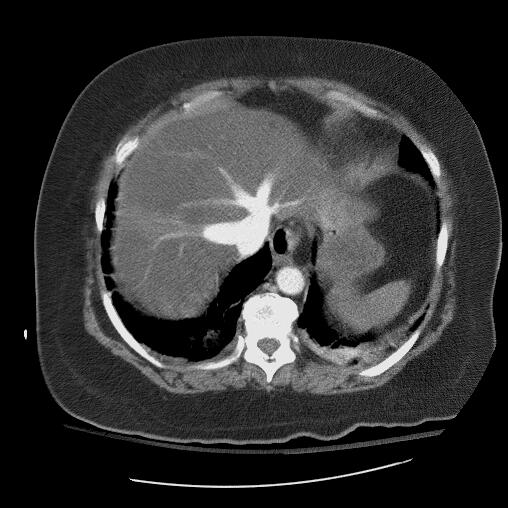
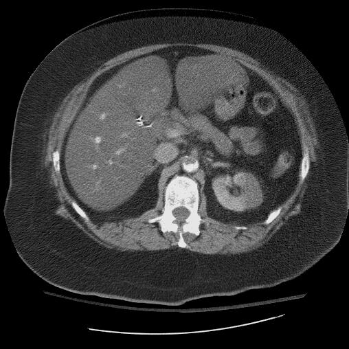
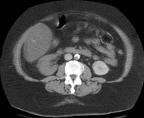
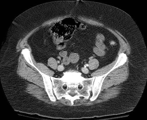
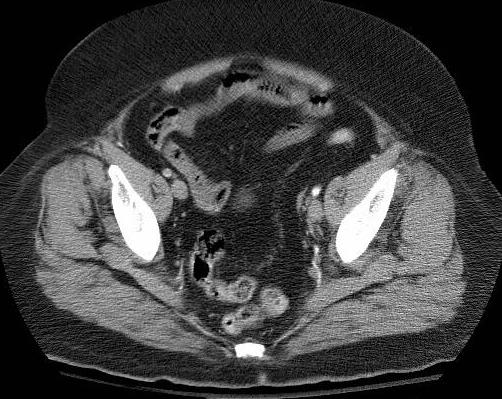
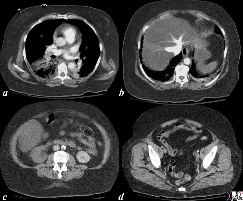
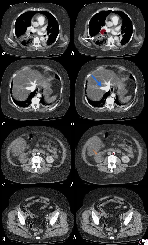
Embolic Infarct of the Splenic Artery |
| 47640 spleen artery fx wedge shaped defect fx occluded artery dx embolism embolic infarction CTscan Davidoff MD |
|
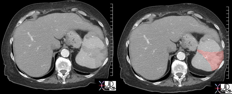
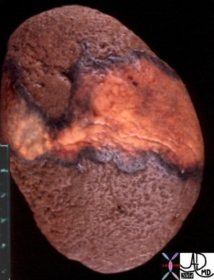
Wedge Shaped Infarct in the Spleen |
| The CTscan with contrast shows a wedge shaped defect in the spleen (overlaid in pink in middle image) characteristic of an acute infarction most commonly caused by emboli. The pathology image from a different patient shows the same wedge shaped process.
46774c01 21321spleen splenic fx wedge shaped defects dx emboli dx systemic embolization dx splenic infarcts CTscan Davidoff MD |
DOMElement Object
(
    [schemaTypeInfo] =>
    [tagName] => table
    [firstElementChild] => (object value omitted)
    [lastElementChild] => (object value omitted)
    [childElementCount] => 1
    [previousElementSibling] => (object value omitted)
    [nextElementSibling] => (object value omitted)
    [nodeName] => table
    [nodeValue] =>


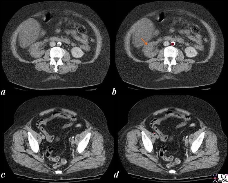
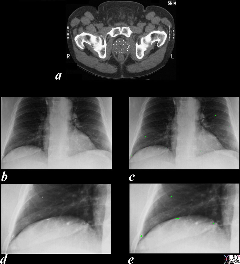
Iatrogenic Embolization of RAdiation Seeds from the Prostate


29676c05.8s 56 year old male with prostate carcinoma has radiation seeds implanted into the prostate gland as seen by the radioopaque pellets in the prostate gland in image a. A chest X-ray following the procedure shows at least 4 of the seeds in the base of the right lung and 2 of the seeds in the mid left lung field overlaid in green (c). Image d is a magnification of the right lower lobe and the seeds are overlaid in green in image e. embolus emboli XRT radiation seeds prostate prostate carcinoma treatment complication iatrogenic Courtesy Ashley Davidoff MD copyright 2009 all rights reserved


    [nodeType] => 1
    [parentNode] => (object value omitted)
    [childNodes] => (object value omitted)
    [firstChild] => (object value omitted)
    [lastChild] => (object value omitted)
    [previousSibling] => (object value omitted)
    [nextSibling] => (object value omitted)
    [attributes] => (object value omitted)
    [ownerDocument] => (object value omitted)
    [namespaceURI] =>
    [prefix] =>
    [localName] => table
    [baseURI] =>
    [textContent] =>


Iatrogenic Embolization of RAdiation Seeds from the Prostate


29676c05.8s 56 year old male with prostate carcinoma has radiation seeds implanted into the prostate gland as seen by the radioopaque pellets in the prostate gland in image a. A chest X-ray following the procedure shows at least 4 of the seeds in the base of the right lung and 2 of the seeds in the mid left lung field overlaid in green (c). Image d is a magnification of the right lower lobe and the seeds are overlaid in green in image e. embolus emboli XRT radiation seeds prostate prostate carcinoma treatment complication iatrogenic Courtesy Ashley Davidoff MD copyright 2009 all rights reserved


)
DOMElement Object
(
    [schemaTypeInfo] =>
    [tagName] => td
    [firstElementChild] => (object value omitted)
    [lastElementChild] => (object value omitted)
    [childElementCount] => 1
    [previousElementSibling] =>
    [nextElementSibling] =>
    [nodeName] => td
    [nodeValue] => 29676c05.8s 56 year old male with prostate carcinoma has radiation seeds implanted into the prostate gland as seen by the radioopaque pellets in the prostate gland in image a. A chest X-ray following the procedure shows at least 4 of the seeds in the base of the right lung and 2 of the seeds in the mid left lung field overlaid in green (c). Image d is a magnification of the right lower lobe and the seeds are overlaid in green in image e. embolus emboli XRT radiation seeds prostate prostate carcinoma treatment complication iatrogenic Courtesy Ashley Davidoff MD copyright 2009 all rights reserved
    [nodeType] => 1
    [parentNode] => (object value omitted)
    [childNodes] => (object value omitted)
    [firstChild] => (object value omitted)
    [lastChild] => (object value omitted)
    [previousSibling] => (object value omitted)
    [nextSibling] => (object value omitted)
    [attributes] => (object value omitted)
    [ownerDocument] => (object value omitted)
    [namespaceURI] =>
    [prefix] =>
    [localName] => td
    [baseURI] =>
    [textContent] => 29676c05.8s 56 year old male with prostate carcinoma has radiation seeds implanted into the prostate gland as seen by the radioopaque pellets in the prostate gland in image a. A chest X-ray following the procedure shows at least 4 of the seeds in the base of the right lung and 2 of the seeds in the mid left lung field overlaid in green (c). Image d is a magnification of the right lower lobe and the seeds are overlaid in green in image e. embolus emboli XRT radiation seeds prostate prostate carcinoma treatment complication iatrogenic Courtesy Ashley Davidoff MD copyright 2009 all rights reserved
)
DOMElement Object
(
    [schemaTypeInfo] =>
    [tagName] => td
    [firstElementChild] => (object value omitted)
    [lastElementChild] => (object value omitted)
    [childElementCount] => 2
    [previousElementSibling] =>
    [nextElementSibling] =>
    [nodeName] => td
    [nodeValue] =>
Iatrogenic Embolization of RAdiation Seeds from the Prostate

    [nodeType] => 1
    [parentNode] => (object value omitted)
    [childNodes] => (object value omitted)
    [firstChild] => (object value omitted)
    [lastChild] => (object value omitted)
    [previousSibling] => (object value omitted)
    [nextSibling] => (object value omitted)
    [attributes] => (object value omitted)
    [ownerDocument] => (object value omitted)
    [namespaceURI] =>
    [prefix] =>
    [localName] => td
    [baseURI] =>
    [textContent] =>
Iatrogenic Embolization of RAdiation Seeds from the Prostate

)
DOMElement Object
(
    [schemaTypeInfo] =>
    [tagName] => table
    [firstElementChild] => (object value omitted)
    [lastElementChild] => (object value omitted)
    [childElementCount] => 1
    [previousElementSibling] => (object value omitted)
    [nextElementSibling] => (object value omitted)
    [nodeName] => table
    [nodeValue] =>


“Hot” Pellet Emboli


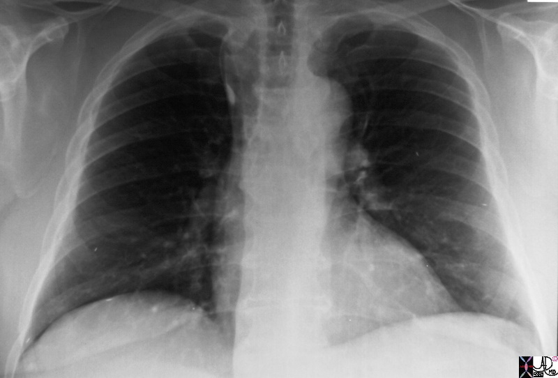
29676b.8s 56 year old male with prostate carcinoma has radiation seeds implanted into the prostate gland A chest X-ray following the procedure shows at least 4 of the seeds in the base of the right lung and 2 of the seeds in the mid left lung field. An azygous lobe is of incidental note. embolus emboli XRT radiation seeds prostate prostate carcinoma treatment complication iatrogenic radiologists and detectives acuity comprehension Courtesy Ashley Davidoff MD copyright 2009 all rights reserved


    [nodeType] => 1
    [parentNode] => (object value omitted)
    [childNodes] => (object value omitted)
    [firstChild] => (object value omitted)
    [lastChild] => (object value omitted)
    [previousSibling] => (object value omitted)
    [nextSibling] => (object value omitted)
    [attributes] => (object value omitted)
    [ownerDocument] => (object value omitted)
    [namespaceURI] =>
    [prefix] =>
    [localName] => table
    [baseURI] =>
    [textContent] =>


“Hot” Pellet Emboli


29676b.8s 56 year old male with prostate carcinoma has radiation seeds implanted into the prostate gland A chest X-ray following the procedure shows at least 4 of the seeds in the base of the right lung and 2 of the seeds in the mid left lung field. An azygous lobe is of incidental note. embolus emboli XRT radiation seeds prostate prostate carcinoma treatment complication iatrogenic radiologists and detectives acuity comprehension Courtesy Ashley Davidoff MD copyright 2009 all rights reserved


)
DOMElement Object
(
    [schemaTypeInfo] =>
    [tagName] => td
    [firstElementChild] => (object value omitted)
    [lastElementChild] => (object value omitted)
    [childElementCount] => 1
    [previousElementSibling] =>
    [nextElementSibling] =>
    [nodeName] => td
    [nodeValue] => 29676b.8s 56 year old male with prostate carcinoma has radiation seeds implanted into the prostate gland A chest X-ray following the procedure shows at least 4 of the seeds in the base of the right lung and 2 of the seeds in the mid left lung field. An azygous lobe is of incidental note. embolus emboli XRT radiation seeds prostate prostate carcinoma treatment complication iatrogenic radiologists and detectives acuity comprehension Courtesy Ashley Davidoff MD copyright 2009 all rights reserved
    [nodeType] => 1
    [parentNode] => (object value omitted)
    [childNodes] => (object value omitted)
    [firstChild] => (object value omitted)
    [lastChild] => (object value omitted)
    [previousSibling] => (object value omitted)
    [nextSibling] => (object value omitted)
    [attributes] => (object value omitted)
    [ownerDocument] => (object value omitted)
    [namespaceURI] =>
    [prefix] =>
    [localName] => td
    [baseURI] =>
    [textContent] => 29676b.8s 56 year old male with prostate carcinoma has radiation seeds implanted into the prostate gland A chest X-ray following the procedure shows at least 4 of the seeds in the base of the right lung and 2 of the seeds in the mid left lung field. An azygous lobe is of incidental note. embolus emboli XRT radiation seeds prostate prostate carcinoma treatment complication iatrogenic radiologists and detectives acuity comprehension Courtesy Ashley Davidoff MD copyright 2009 all rights reserved
)
DOMElement Object
(
    [schemaTypeInfo] =>
    [tagName] => td
    [firstElementChild] => (object value omitted)
    [lastElementChild] => (object value omitted)
    [childElementCount] => 2
    [previousElementSibling] =>
    [nextElementSibling] =>
    [nodeName] => td
    [nodeValue] =>
“Hot” Pellet Emboli

    [nodeType] => 1
    [parentNode] => (object value omitted)
    [childNodes] => (object value omitted)
    [firstChild] => (object value omitted)
    [lastChild] => (object value omitted)
    [previousSibling] => (object value omitted)
    [nextSibling] => (object value omitted)
    [attributes] => (object value omitted)
    [ownerDocument] => (object value omitted)
    [namespaceURI] =>
    [prefix] =>
    [localName] => td
    [baseURI] =>
    [textContent] =>
“Hot” Pellet Emboli

)
DOMElement Object
(
    [schemaTypeInfo] =>
    [tagName] => table
    [firstElementChild] => (object value omitted)
    [lastElementChild] => (object value omitted)
    [childElementCount] => 1
    [previousElementSibling] => (object value omitted)
    [nextElementSibling] => (object value omitted)
    [nodeName] => table
    [nodeValue] =>


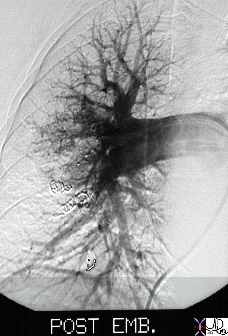
24137


24137 lung pulmonary fx AVM fx arteriovenous malformation dx cirrhosis dx hepatopulmonary syndrome liver hepatic pulmonary artery pulmonary vein MIT post embolization pulmonary angiogram arteriogram angiography Courtesy Ashley Davidoff MD


    [nodeType] => 1
    [parentNode] => (object value omitted)
    [childNodes] => (object value omitted)
    [firstChild] => (object value omitted)
    [lastChild] => (object value omitted)
    [previousSibling] => (object value omitted)
    [nextSibling] => (object value omitted)
    [attributes] => (object value omitted)
    [ownerDocument] => (object value omitted)
    [namespaceURI] =>
    [prefix] =>
    [localName] => table
    [baseURI] =>
    [textContent] =>


24137


24137 lung pulmonary fx AVM fx arteriovenous malformation dx cirrhosis dx hepatopulmonary syndrome liver hepatic pulmonary artery pulmonary vein MIT post embolization pulmonary angiogram arteriogram angiography Courtesy Ashley Davidoff MD


)
DOMElement Object
(
    [schemaTypeInfo] =>
    [tagName] => td
    [firstElementChild] => (object value omitted)
    [lastElementChild] => (object value omitted)
    [childElementCount] => 1
    [previousElementSibling] =>
    [nextElementSibling] =>
    [nodeName] => td
    [nodeValue] => 24137 lung pulmonary fx AVM fx arteriovenous malformation dx cirrhosis dx hepatopulmonary syndrome liver hepatic pulmonary artery pulmonary vein MIT post embolization pulmonary angiogram arteriogram angiography Courtesy Ashley Davidoff MD
    [nodeType] => 1
    [parentNode] => (object value omitted)
    [childNodes] => (object value omitted)
    [firstChild] => (object value omitted)
    [lastChild] => (object value omitted)
    [previousSibling] => (object value omitted)
    [nextSibling] => (object value omitted)
    [attributes] => (object value omitted)
    [ownerDocument] => (object value omitted)
    [namespaceURI] =>
    [prefix] =>
    [localName] => td
    [baseURI] =>
    [textContent] => 24137 lung pulmonary fx AVM fx arteriovenous malformation dx cirrhosis dx hepatopulmonary syndrome liver hepatic pulmonary artery pulmonary vein MIT post embolization pulmonary angiogram arteriogram angiography Courtesy Ashley Davidoff MD
)
DOMElement Object
(
    [schemaTypeInfo] =>
    [tagName] => td
    [firstElementChild] => (object value omitted)
    [lastElementChild] => (object value omitted)
    [childElementCount] => 2
    [previousElementSibling] =>
    [nextElementSibling] =>
    [nodeName] => td
    [nodeValue] =>
24137
    [nodeType] => 1
    [parentNode] => (object value omitted)
    [childNodes] => (object value omitted)
    [firstChild] => (object value omitted)
    [lastChild] => (object value omitted)
    [previousSibling] => (object value omitted)
    [nextSibling] => (object value omitted)
    [attributes] => (object value omitted)
    [ownerDocument] => (object value omitted)
    [namespaceURI] =>
    [prefix] =>
    [localName] => td
    [baseURI] =>
    [textContent] =>
24137
)
https://beta.thecommonvein.net/wp-content/uploads/2023/05/24137.jpg
http://www.thecommonvein.net/media/24137.JPG
DOMElement Object
(
    [schemaTypeInfo] =>
    [tagName] => table
    [firstElementChild] => (object value omitted)
    [lastElementChild] => (object value omitted)
    [childElementCount] => 1
    [previousElementSibling] => (object value omitted)
    [nextElementSibling] => (object value omitted)
    [nodeName] => table
    [nodeValue] =>


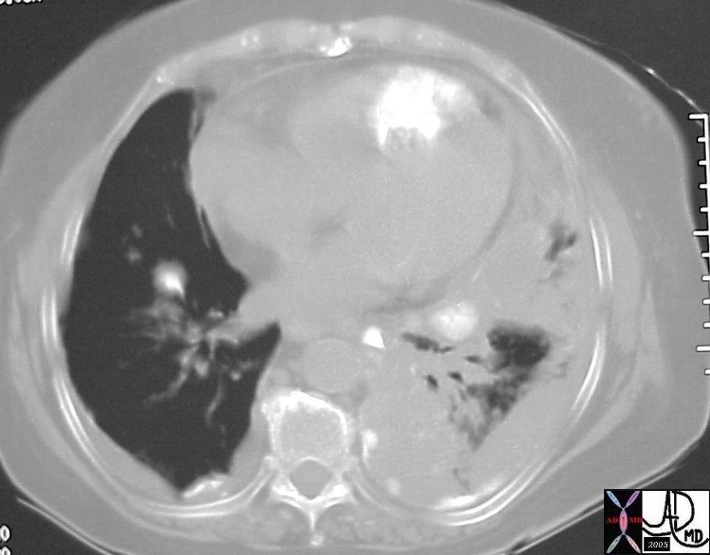
15537b04d


15537b04d hx 75F with left shoulder pain heart lung fx calcification fx parenchymal infiltrate fx pleural effusion dx small pulmonary infarct small Hamptons hump dx metastatic chondrosarcoma metastasis osteogenic sarcomatous malignant pulmonary emboli embolus imaging radiology CTscan Courtesy Ashley Davidoff MD


    [nodeType] => 1
    [parentNode] => (object value omitted)
    [childNodes] => (object value omitted)
    [firstChild] => (object value omitted)
    [lastChild] => (object value omitted)
    [previousSibling] => (object value omitted)
    [nextSibling] => (object value omitted)
    [attributes] => (object value omitted)
    [ownerDocument] => (object value omitted)
    [namespaceURI] =>
    [prefix] =>
    [localName] => table
    [baseURI] =>
    [textContent] =>


15537b04d


15537b04d hx 75F with left shoulder pain heart lung fx calcification fx parenchymal infiltrate fx pleural effusion dx small pulmonary infarct small Hamptons hump dx metastatic chondrosarcoma metastasis osteogenic sarcomatous malignant pulmonary emboli embolus imaging radiology CTscan Courtesy Ashley Davidoff MD


)
DOMElement Object
(
    [schemaTypeInfo] =>
    [tagName] => td
    [firstElementChild] => (object value omitted)
    [lastElementChild] => (object value omitted)
    [childElementCount] => 1
    [previousElementSibling] =>
    [nextElementSibling] =>
    [nodeName] => td
    [nodeValue] => 15537b04d hx 75F with left shoulder pain heart lung fx calcification fx parenchymal infiltrate fx pleural effusion dx small pulmonary infarct small Hamptons hump dx metastatic chondrosarcoma metastasis osteogenic sarcomatous malignant pulmonary emboli embolus imaging radiology CTscan Courtesy Ashley Davidoff MD
    [nodeType] => 1
    [parentNode] => (object value omitted)
    [childNodes] => (object value omitted)
    [firstChild] => (object value omitted)
    [lastChild] => (object value omitted)
    [previousSibling] => (object value omitted)
    [nextSibling] => (object value omitted)
    [attributes] => (object value omitted)
    [ownerDocument] => (object value omitted)
    [namespaceURI] =>
    [prefix] =>
    [localName] => td
    [baseURI] =>
    [textContent] => 15537b04d hx 75F with left shoulder pain heart lung fx calcification fx parenchymal infiltrate fx pleural effusion dx small pulmonary infarct small Hamptons hump dx metastatic chondrosarcoma metastasis osteogenic sarcomatous malignant pulmonary emboli embolus imaging radiology CTscan Courtesy Ashley Davidoff MD
)
DOMElement Object
(
    [schemaTypeInfo] =>
    [tagName] => td
    [firstElementChild] => (object value omitted)
    [lastElementChild] => (object value omitted)
    [childElementCount] => 2
    [previousElementSibling] =>
    [nextElementSibling] =>
    [nodeName] => td
    [nodeValue] =>
15537b04d
    [nodeType] => 1
    [parentNode] => (object value omitted)
    [childNodes] => (object value omitted)
    [firstChild] => (object value omitted)
    [lastChild] => (object value omitted)
    [previousSibling] => (object value omitted)
    [nextSibling] => (object value omitted)
    [attributes] => (object value omitted)
    [ownerDocument] => (object value omitted)
    [namespaceURI] =>
    [prefix] =>
    [localName] => td
    [baseURI] =>
    [textContent] =>
15537b04d
)
DOMElement Object
(
    [schemaTypeInfo] =>
    [tagName] => table
    [firstElementChild] => (object value omitted)
    [lastElementChild] => (object value omitted)
    [childElementCount] => 1
    [previousElementSibling] => (object value omitted)
    [nextElementSibling] => (object value omitted)
    [nodeName] => table
    [nodeValue] =>


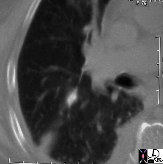
15537b04


15537b04 hx 75F with left shoulder pain heart lung fx calcification fx parenchymal infiltrate fx pleural effusion dx small pulmonary infarct small Hamptons hump dx metastatic chondrosarcoma metastasis osteogenic sarcomatous malignant pulmonary emboli embolus imaging radiology CTscan Courtesy Ashley Davidoff MD


    [nodeType] => 1
    [parentNode] => (object value omitted)
    [childNodes] => (object value omitted)
    [firstChild] => (object value omitted)
    [lastChild] => (object value omitted)
    [previousSibling] => (object value omitted)
    [nextSibling] => (object value omitted)
    [attributes] => (object value omitted)
    [ownerDocument] => (object value omitted)
    [namespaceURI] =>
    [prefix] =>
    [localName] => table
    [baseURI] =>
    [textContent] =>


15537b04


15537b04 hx 75F with left shoulder pain heart lung fx calcification fx parenchymal infiltrate fx pleural effusion dx small pulmonary infarct small Hamptons hump dx metastatic chondrosarcoma metastasis osteogenic sarcomatous malignant pulmonary emboli embolus imaging radiology CTscan Courtesy Ashley Davidoff MD


)
DOMElement Object
(
    [schemaTypeInfo] =>
    [tagName] => td
    [firstElementChild] => (object value omitted)
    [lastElementChild] => (object value omitted)
    [childElementCount] => 1
    [previousElementSibling] =>
    [nextElementSibling] =>
    [nodeName] => td
    [nodeValue] => 15537b04 hx 75F with left shoulder pain heart lung fx calcification fx parenchymal infiltrate fx pleural effusion dx small pulmonary infarct small Hamptons hump dx metastatic chondrosarcoma metastasis osteogenic sarcomatous malignant pulmonary emboli embolus imaging radiology CTscan Courtesy Ashley Davidoff MD
    [nodeType] => 1
    [parentNode] => (object value omitted)
    [childNodes] => (object value omitted)
    [firstChild] => (object value omitted)
    [lastChild] => (object value omitted)
    [previousSibling] => (object value omitted)
    [nextSibling] => (object value omitted)
    [attributes] => (object value omitted)
    [ownerDocument] => (object value omitted)
    [namespaceURI] =>
    [prefix] =>
    [localName] => td
    [baseURI] =>
    [textContent] => 15537b04 hx 75F with left shoulder pain heart lung fx calcification fx parenchymal infiltrate fx pleural effusion dx small pulmonary infarct small Hamptons hump dx metastatic chondrosarcoma metastasis osteogenic sarcomatous malignant pulmonary emboli embolus imaging radiology CTscan Courtesy Ashley Davidoff MD
)
DOMElement Object
(
    [schemaTypeInfo] =>
    [tagName] => td
    [firstElementChild] => (object value omitted)
    [lastElementChild] => (object value omitted)
    [childElementCount] => 2
    [previousElementSibling] =>
    [nextElementSibling] =>
    [nodeName] => td
    [nodeValue] =>
15537b04
    [nodeType] => 1
    [parentNode] => (object value omitted)
    [childNodes] => (object value omitted)
    [firstChild] => (object value omitted)
    [lastChild] => (object value omitted)
    [previousSibling] => (object value omitted)
    [nextSibling] => (object value omitted)
    [attributes] => (object value omitted)
    [ownerDocument] => (object value omitted)
    [namespaceURI] =>
    [prefix] =>
    [localName] => td
    [baseURI] =>
    [textContent] =>
15537b04
)
DOMElement Object
(
    [schemaTypeInfo] =>
    [tagName] => table
    [firstElementChild] => (object value omitted)
    [lastElementChild] => (object value omitted)
    [childElementCount] => 1
    [previousElementSibling] => (object value omitted)
    [nextElementSibling] => (object value omitted)
    [nodeName] => table
    [nodeValue] =>


15551b01


15551b01 bone heart cardiac pulmonary artery tumor pulmonary emboli 15537 T2 intensity over the heart T2 hyperintensity over left humerus shoulder MRIscan DxChondrosarcoma of the humerus metatstattic to the heart lungs Davidoff MD


    [nodeType] => 1
    [parentNode] => (object value omitted)
    [childNodes] => (object value omitted)
    [firstChild] => (object value omitted)
    [lastChild] => (object value omitted)
    [previousSibling] => (object value omitted)
    [nextSibling] => (object value omitted)
    [attributes] => (object value omitted)
    [ownerDocument] => (object value omitted)
    [namespaceURI] =>
    [prefix] =>
    [localName] => table
    [baseURI] =>
    [textContent] =>


15551b01


15551b01 bone heart cardiac pulmonary artery tumor pulmonary emboli 15537 T2 intensity over the heart T2 hyperintensity over left humerus shoulder MRIscan DxChondrosarcoma of the humerus metatstattic to the heart lungs Davidoff MD


)
DOMElement Object
(
    [schemaTypeInfo] =>
    [tagName] => td
    [firstElementChild] => (object value omitted)
    [lastElementChild] => (object value omitted)
    [childElementCount] => 1
    [previousElementSibling] =>
    [nextElementSibling] =>
    [nodeName] => td
    [nodeValue] => 15551b01 bone heart cardiac pulmonary artery tumor pulmonary emboli 15537 T2 intensity over the heart T2 hyperintensity over left humerus shoulder MRIscan DxChondrosarcoma of the humerus metatstattic to the heart lungs Davidoff MD
    [nodeType] => 1
    [parentNode] => (object value omitted)
    [childNodes] => (object value omitted)
    [firstChild] => (object value omitted)
    [lastChild] => (object value omitted)
    [previousSibling] => (object value omitted)
    [nextSibling] => (object value omitted)
    [attributes] => (object value omitted)
    [ownerDocument] => (object value omitted)
    [namespaceURI] =>
    [prefix] =>
    [localName] => td
    [baseURI] =>
    [textContent] => 15551b01 bone heart cardiac pulmonary artery tumor pulmonary emboli 15537 T2 intensity over the heart T2 hyperintensity over left humerus shoulder MRIscan DxChondrosarcoma of the humerus metatstattic to the heart lungs Davidoff MD
)
DOMElement Object
(
    [schemaTypeInfo] =>
    [tagName] => td
    [firstElementChild] => (object value omitted)
    [lastElementChild] => (object value omitted)
    [childElementCount] => 2
    [previousElementSibling] =>
    [nextElementSibling] =>
    [nodeName] => td
    [nodeValue] =>
15551b01
    [nodeType] => 1
    [parentNode] => (object value omitted)
    [childNodes] => (object value omitted)
    [firstChild] => (object value omitted)
    [lastChild] => (object value omitted)
    [previousSibling] => (object value omitted)
    [nextSibling] => (object value omitted)
    [attributes] => (object value omitted)
    [ownerDocument] => (object value omitted)
    [namespaceURI] =>
    [prefix] =>
    [localName] => td
    [baseURI] =>
    [textContent] =>
15551b01
)
DOMElement Object
(
    [schemaTypeInfo] =>
    [tagName] => table
    [firstElementChild] => (object value omitted)
    [lastElementChild] => (object value omitted)
    [childElementCount] => 1
    [previousElementSibling] => (object value omitted)
    [nextElementSibling] => (object value omitted)
    [nodeName] => table
    [nodeValue] =>


The Other Circulation


43684c05b01.8s  The series of CTscans are from an elderly lady who presents with chest pain and right leg pain lung fx infiltrate artery lung pulmonary embolus pulmonary atelectasis vs infarction PE PFO heart patent foramen ovale tricuspid regurgitation acute pulmonary hypertension TR kidney embolic infarction aorta embolus embolic disease right to left shunt imaging radiology CTscan Courtesy Ashley Davidoff MD copyright 2009 all rights reserved 43666 43670 43672 43675 43680 43683 5star


    [nodeType] => 1
    [parentNode] => (object value omitted)
    [childNodes] => (object value omitted)
    [firstChild] => (object value omitted)
    [lastChild] => (object value omitted)
    [previousSibling] => (object value omitted)
    [nextSibling] => (object value omitted)
    [attributes] => (object value omitted)
    [ownerDocument] => (object value omitted)
    [namespaceURI] =>
    [prefix] =>
    [localName] => table
    [baseURI] =>
    [textContent] =>


The Other Circulation


43684c05b01.8s  The series of CTscans are from an elderly lady who presents with chest pain and right leg pain lung fx infiltrate artery lung pulmonary embolus pulmonary atelectasis vs infarction PE PFO heart patent foramen ovale tricuspid regurgitation acute pulmonary hypertension TR kidney embolic infarction aorta embolus embolic disease right to left shunt imaging radiology CTscan Courtesy Ashley Davidoff MD copyright 2009 all rights reserved 43666 43670 43672 43675 43680 43683 5star


)
DOMElement Object
(
    [schemaTypeInfo] =>
    [tagName] => td
    [firstElementChild] => (object value omitted)
    [lastElementChild] => (object value omitted)
    [childElementCount] => 1
    [previousElementSibling] =>
    [nextElementSibling] =>
    [nodeName] => td
    [nodeValue] => 43684c05b01.8s  The series of CTscans are from an elderly lady who presents with chest pain and right leg pain lung fx infiltrate artery lung pulmonary embolus pulmonary atelectasis vs infarction PE PFO heart patent foramen ovale tricuspid regurgitation acute pulmonary hypertension TR kidney embolic infarction aorta embolus embolic disease right to left shunt imaging radiology CTscan Courtesy Ashley Davidoff MD copyright 2009 all rights reserved 43666 43670 43672 43675 43680 43683 5star
    [nodeType] => 1
    [parentNode] => (object value omitted)
    [childNodes] => (object value omitted)
    [firstChild] => (object value omitted)
    [lastChild] => (object value omitted)
    [previousSibling] => (object value omitted)
    [nextSibling] => (object value omitted)
    [attributes] => (object value omitted)
    [ownerDocument] => (object value omitted)
    [namespaceURI] =>
    [prefix] =>
    [localName] => td
    [baseURI] =>
    [textContent] => 43684c05b01.8s  The series of CTscans are from an elderly lady who presents with chest pain and right leg pain lung fx infiltrate artery lung pulmonary embolus pulmonary atelectasis vs infarction PE PFO heart patent foramen ovale tricuspid regurgitation acute pulmonary hypertension TR kidney embolic infarction aorta embolus embolic disease right to left shunt imaging radiology CTscan Courtesy Ashley Davidoff MD copyright 2009 all rights reserved 43666 43670 43672 43675 43680 43683 5star
)
DOMElement Object
(
    [schemaTypeInfo] =>
    [tagName] => td
    [firstElementChild] => (object value omitted)
    [lastElementChild] => (object value omitted)
    [childElementCount] => 2
    [previousElementSibling] =>
    [nextElementSibling] =>
    [nodeName] => td
    [nodeValue] =>
The Other Circulation

    [nodeType] => 1
    [parentNode] => (object value omitted)
    [childNodes] => (object value omitted)
    [firstChild] => (object value omitted)
    [lastChild] => (object value omitted)
    [previousSibling] => (object value omitted)
    [nextSibling] => (object value omitted)
    [attributes] => (object value omitted)
    [ownerDocument] => (object value omitted)
    [namespaceURI] =>
    [prefix] =>
    [localName] => td
    [baseURI] =>
    [textContent] =>
The Other Circulation

)
https://beta.thecommonvein.net/wp-content/uploads/2023/06/43684c05b01.8s.jpg https://beta.thecommonvein.net/wp-content/uploads/2023/06/43683.jpg https://beta.thecommonvein.net/wp-content/uploads/2023/06/43680.jpg https://beta.thecommonvein.net/wp-content/uploads/2023/06/43675.jpg https://beta.thecommonvein.net/wp-content/uploads/2023/06/43672.jpg https://beta.thecommonvein.net/wp-content/uploads/2023/06/43670.jpg https://beta.thecommonvein.net/wp-content/uploads/2023/06/43666.jpg
http://www.thecommonvein.net/search/assets/43684c05b01.8s.jpg
DOMElement Object
(
    [schemaTypeInfo] =>
    [tagName] => table
    [firstElementChild] => (object value omitted)
    [lastElementChild] => (object value omitted)
    [childElementCount] => 1
    [previousElementSibling] => (object value omitted)
    [nextElementSibling] => (object value omitted)
    [nodeName] => table
    [nodeValue] =>


One Circulation


43684c06b02.8s The series of CTscans are from an elderly lady who presents with chest pain and right leg pain lung fx infiltrate artery lung pulmonary embolus pulmonary atelectasis vs infarction PE PFO heart patent foramen ovale tricuspid regurgitation acute pulmonary hypertension TR kidney embolic infarction aorta embolus embolic disease right to left shunt imaging radiology CTscan Courtesy Ashley Davidoff MD copyright 2009 all rights reserved 43666 43670 43672 43675 43680 43683 5star


    [nodeType] => 1
    [parentNode] => (object value omitted)
    [childNodes] => (object value omitted)
    [firstChild] => (object value omitted)
    [lastChild] => (object value omitted)
    [previousSibling] => (object value omitted)
    [nextSibling] => (object value omitted)
    [attributes] => (object value omitted)
    [ownerDocument] => (object value omitted)
    [namespaceURI] =>
    [prefix] =>
    [localName] => table
    [baseURI] =>
    [textContent] =>


One Circulation


43684c06b02.8s The series of CTscans are from an elderly lady who presents with chest pain and right leg pain lung fx infiltrate artery lung pulmonary embolus pulmonary atelectasis vs infarction PE PFO heart patent foramen ovale tricuspid regurgitation acute pulmonary hypertension TR kidney embolic infarction aorta embolus embolic disease right to left shunt imaging radiology CTscan Courtesy Ashley Davidoff MD copyright 2009 all rights reserved 43666 43670 43672 43675 43680 43683 5star


)
DOMElement Object
(
    [schemaTypeInfo] =>
    [tagName] => td
    [firstElementChild] => (object value omitted)
    [lastElementChild] => (object value omitted)
    [childElementCount] => 1
    [previousElementSibling] =>
    [nextElementSibling] =>
    [nodeName] => td
    [nodeValue] => 43684c06b02.8s The series of CTscans are from an elderly lady who presents with chest pain and right leg pain lung fx infiltrate artery lung pulmonary embolus pulmonary atelectasis vs infarction PE PFO heart patent foramen ovale tricuspid regurgitation acute pulmonary hypertension TR kidney embolic infarction aorta embolus embolic disease right to left shunt imaging radiology CTscan Courtesy Ashley Davidoff MD copyright 2009 all rights reserved 43666 43670 43672 43675 43680 43683 5star
    [nodeType] => 1
    [parentNode] => (object value omitted)
    [childNodes] => (object value omitted)
    [firstChild] => (object value omitted)
    [lastChild] => (object value omitted)
    [previousSibling] => (object value omitted)
    [nextSibling] => (object value omitted)
    [attributes] => (object value omitted)
    [ownerDocument] => (object value omitted)
    [namespaceURI] =>
    [prefix] =>
    [localName] => td
    [baseURI] =>
    [textContent] => 43684c06b02.8s The series of CTscans are from an elderly lady who presents with chest pain and right leg pain lung fx infiltrate artery lung pulmonary embolus pulmonary atelectasis vs infarction PE PFO heart patent foramen ovale tricuspid regurgitation acute pulmonary hypertension TR kidney embolic infarction aorta embolus embolic disease right to left shunt imaging radiology CTscan Courtesy Ashley Davidoff MD copyright 2009 all rights reserved 43666 43670 43672 43675 43680 43683 5star
)
DOMElement Object
(
    [schemaTypeInfo] =>
    [tagName] => td
    [firstElementChild] => (object value omitted)
    [lastElementChild] => (object value omitted)
    [childElementCount] => 2
    [previousElementSibling] =>
    [nextElementSibling] =>
    [nodeName] => td
    [nodeValue] =>
One Circulation

    [nodeType] => 1
    [parentNode] => (object value omitted)
    [childNodes] => (object value omitted)
    [firstChild] => (object value omitted)
    [lastChild] => (object value omitted)
    [previousSibling] => (object value omitted)
    [nextSibling] => (object value omitted)
    [attributes] => (object value omitted)
    [ownerDocument] => (object value omitted)
    [namespaceURI] =>
    [prefix] =>
    [localName] => td
    [baseURI] =>
    [textContent] =>
One Circulation

)
https://beta.thecommonvein.net/wp-content/uploads/2023/06/43684c06b02.8s.jpg https://beta.thecommonvein.net/wp-content/uploads/2023/06/43683.jpg https://beta.thecommonvein.net/wp-content/uploads/2023/06/43680.jpg https://beta.thecommonvein.net/wp-content/uploads/2023/06/43675.jpg https://beta.thecommonvein.net/wp-content/uploads/2023/06/43672.jpg https://beta.thecommonvein.net/wp-content/uploads/2023/06/43670.jpg https://beta.thecommonvein.net/wp-content/uploads/2023/06/43666.jpg
http://www.thecommonvein.net/search/assets/43684c06b02.8s.jpg
DOMElement Object
(
    [schemaTypeInfo] =>
    [tagName] => table
    [firstElementChild] => (object value omitted)
    [lastElementChild] => (object value omitted)
    [childElementCount] => 1
    [previousElementSibling] => (object value omitted)
    [nextElementSibling] => (object value omitted)
    [nodeName] => table
    [nodeValue] =>


The Findings


43684c06b.8s The series of CTscans are from an elderly lady who presents with chest pain and right leg pain lung fx infiltrate artery lung pulmonary embolus pulmonary atelectasis vs infarction PE PFO heart patent foramen ovale tricuspid regurgitation acute pulmonary hypertension TR kidney embolic infarction aorta embolus embolic disease right to left shunt imaging radiology CTscan Courtesy Ashley Davidoff MD copyright 2009 all rights reserved 43666 43670 43672 43675 43680 43683 5star


    [nodeType] => 1
    [parentNode] => (object value omitted)
    [childNodes] => (object value omitted)
    [firstChild] => (object value omitted)
    [lastChild] => (object value omitted)
    [previousSibling] => (object value omitted)
    [nextSibling] => (object value omitted)
    [attributes] => (object value omitted)
    [ownerDocument] => (object value omitted)
    [namespaceURI] =>
    [prefix] =>
    [localName] => table
    [baseURI] =>
    [textContent] =>


The Findings


43684c06b.8s The series of CTscans are from an elderly lady who presents with chest pain and right leg pain lung fx infiltrate artery lung pulmonary embolus pulmonary atelectasis vs infarction PE PFO heart patent foramen ovale tricuspid regurgitation acute pulmonary hypertension TR kidney embolic infarction aorta embolus embolic disease right to left shunt imaging radiology CTscan Courtesy Ashley Davidoff MD copyright 2009 all rights reserved 43666 43670 43672 43675 43680 43683 5star


)
DOMElement Object
(
    [schemaTypeInfo] =>
    [tagName] => td
    [firstElementChild] => (object value omitted)
    [lastElementChild] => (object value omitted)
    [childElementCount] => 1
    [previousElementSibling] =>
    [nextElementSibling] =>
    [nodeName] => td
    [nodeValue] => 43684c06b.8s The series of CTscans are from an elderly lady who presents with chest pain and right leg pain lung fx infiltrate artery lung pulmonary embolus pulmonary atelectasis vs infarction PE PFO heart patent foramen ovale tricuspid regurgitation acute pulmonary hypertension TR kidney embolic infarction aorta embolus embolic disease right to left shunt imaging radiology CTscan Courtesy Ashley Davidoff MD copyright 2009 all rights reserved 43666 43670 43672 43675 43680 43683 5star
    [nodeType] => 1
    [parentNode] => (object value omitted)
    [childNodes] => (object value omitted)
    [firstChild] => (object value omitted)
    [lastChild] => (object value omitted)
    [previousSibling] => (object value omitted)
    [nextSibling] => (object value omitted)
    [attributes] => (object value omitted)
    [ownerDocument] => (object value omitted)
    [namespaceURI] =>
    [prefix] =>
    [localName] => td
    [baseURI] =>
    [textContent] => 43684c06b.8s The series of CTscans are from an elderly lady who presents with chest pain and right leg pain lung fx infiltrate artery lung pulmonary embolus pulmonary atelectasis vs infarction PE PFO heart patent foramen ovale tricuspid regurgitation acute pulmonary hypertension TR kidney embolic infarction aorta embolus embolic disease right to left shunt imaging radiology CTscan Courtesy Ashley Davidoff MD copyright 2009 all rights reserved 43666 43670 43672 43675 43680 43683 5star
)
DOMElement Object
(
    [schemaTypeInfo] =>
    [tagName] => td
    [firstElementChild] => (object value omitted)
    [lastElementChild] => (object value omitted)
    [childElementCount] => 2
    [previousElementSibling] =>
    [nextElementSibling] =>
    [nodeName] => td
    [nodeValue] =>
The Findings

    [nodeType] => 1
    [parentNode] => (object value omitted)
    [childNodes] => (object value omitted)
    [firstChild] => (object value omitted)
    [lastChild] => (object value omitted)
    [previousSibling] => (object value omitted)
    [nextSibling] => (object value omitted)
    [attributes] => (object value omitted)
    [ownerDocument] => (object value omitted)
    [namespaceURI] =>
    [prefix] =>
    [localName] => td
    [baseURI] =>
    [textContent] =>
The Findings

)
https://beta.thecommonvein.net/wp-content/uploads/2023/06/43684c06b.8s.jpg https://beta.thecommonvein.net/wp-content/uploads/2023/06/43683.jpg https://beta.thecommonvein.net/wp-content/uploads/2023/06/43680.jpg https://beta.thecommonvein.net/wp-content/uploads/2023/06/43675.jpg https://beta.thecommonvein.net/wp-content/uploads/2023/06/43672.jpg https://beta.thecommonvein.net/wp-content/uploads/2023/06/43670.jpg https://beta.thecommonvein.net/wp-content/uploads/2023/06/43666.jpg
http://www.thecommonvein.net/search/assets/43684c06b.8s.jpg
DOMElement Object
(
    [schemaTypeInfo] =>
    [tagName] => table
    [firstElementChild] => (object value omitted)
    [lastElementChild] => (object value omitted)
    [childElementCount] => 1
    [previousElementSibling] => (object value omitted)
    [nextElementSibling] => (object value omitted)
    [nodeName] => table
    [nodeValue] =>


Tell the Story


43684c01.8s The series of CTscans are from an elderly lady who presents with chest pain and right leg pain lung fx infiltrate artery lung pulmonary embolus pulmonary atelectasis vs infarction PE PFO heart patent foramen ovale tricuspid regurgitation acute pulmonary hypertension TR kidney embolic infarction aorta embolus embolic disease right to left shunt imaging radiology CTscan Courtesy Ashley Davidoff MD copyright 2009 all rights reserved 43666 43670 43672 43675 43680 43683 5star


    [nodeType] => 1
    [parentNode] => (object value omitted)
    [childNodes] => (object value omitted)
    [firstChild] => (object value omitted)
    [lastChild] => (object value omitted)
    [previousSibling] => (object value omitted)
    [nextSibling] => (object value omitted)
    [attributes] => (object value omitted)
    [ownerDocument] => (object value omitted)
    [namespaceURI] =>
    [prefix] =>
    [localName] => table
    [baseURI] =>
    [textContent] =>


Tell the Story


43684c01.8s The series of CTscans are from an elderly lady who presents with chest pain and right leg pain lung fx infiltrate artery lung pulmonary embolus pulmonary atelectasis vs infarction PE PFO heart patent foramen ovale tricuspid regurgitation acute pulmonary hypertension TR kidney embolic infarction aorta embolus embolic disease right to left shunt imaging radiology CTscan Courtesy Ashley Davidoff MD copyright 2009 all rights reserved 43666 43670 43672 43675 43680 43683 5star


)
DOMElement Object
(
    [schemaTypeInfo] =>
    [tagName] => td
    [firstElementChild] => (object value omitted)
    [lastElementChild] => (object value omitted)
    [childElementCount] => 1
    [previousElementSibling] =>
    [nextElementSibling] =>
    [nodeName] => td
    [nodeValue] => 43684c01.8s The series of CTscans are from an elderly lady who presents with chest pain and right leg pain lung fx infiltrate artery lung pulmonary embolus pulmonary atelectasis vs infarction PE PFO heart patent foramen ovale tricuspid regurgitation acute pulmonary hypertension TR kidney embolic infarction aorta embolus embolic disease right to left shunt imaging radiology CTscan Courtesy Ashley Davidoff MD copyright 2009 all rights reserved 43666 43670 43672 43675 43680 43683 5star
    [nodeType] => 1
    [parentNode] => (object value omitted)
    [childNodes] => (object value omitted)
    [firstChild] => (object value omitted)
    [lastChild] => (object value omitted)
    [previousSibling] => (object value omitted)
    [nextSibling] => (object value omitted)
    [attributes] => (object value omitted)
    [ownerDocument] => (object value omitted)
    [namespaceURI] =>
    [prefix] =>
    [localName] => td
    [baseURI] =>
    [textContent] => 43684c01.8s The series of CTscans are from an elderly lady who presents with chest pain and right leg pain lung fx infiltrate artery lung pulmonary embolus pulmonary atelectasis vs infarction PE PFO heart patent foramen ovale tricuspid regurgitation acute pulmonary hypertension TR kidney embolic infarction aorta embolus embolic disease right to left shunt imaging radiology CTscan Courtesy Ashley Davidoff MD copyright 2009 all rights reserved 43666 43670 43672 43675 43680 43683 5star
)
DOMElement Object
(
    [schemaTypeInfo] =>
    [tagName] => td
    [firstElementChild] => (object value omitted)
    [lastElementChild] => (object value omitted)
    [childElementCount] => 2
    [previousElementSibling] =>
    [nextElementSibling] =>
    [nodeName] => td
    [nodeValue] =>
Tell the Story

    [nodeType] => 1
    [parentNode] => (object value omitted)
    [childNodes] => (object value omitted)
    [firstChild] => (object value omitted)
    [lastChild] => (object value omitted)
    [previousSibling] => (object value omitted)
    [nextSibling] => (object value omitted)
    [attributes] => (object value omitted)
    [ownerDocument] => (object value omitted)
    [namespaceURI] =>
    [prefix] =>
    [localName] => td
    [baseURI] =>
    [textContent] =>
Tell the Story

)
https://beta.thecommonvein.net/wp-content/uploads/2023/06/43684c01.8s.jpg https://beta.thecommonvein.net/wp-content/uploads/2023/06/43683.jpg https://beta.thecommonvein.net/wp-content/uploads/2023/06/43680.jpg https://beta.thecommonvein.net/wp-content/uploads/2023/06/43675.jpg https://beta.thecommonvein.net/wp-content/uploads/2023/06/43672.jpg https://beta.thecommonvein.net/wp-content/uploads/2023/06/43670.jpg https://beta.thecommonvein.net/wp-content/uploads/2023/06/43666.jpg
http://www.thecommonvein.net/search/assets/43684c01.8s.jpg
DOMElement Object
(
    [schemaTypeInfo] =>
    [tagName] => table
    [firstElementChild] => (object value omitted)
    [lastElementChild] => (object value omitted)
    [childElementCount] => 1
    [previousElementSibling] => (object value omitted)
    [nextElementSibling] => (object value omitted)
    [nodeName] => table
    [nodeValue] =>


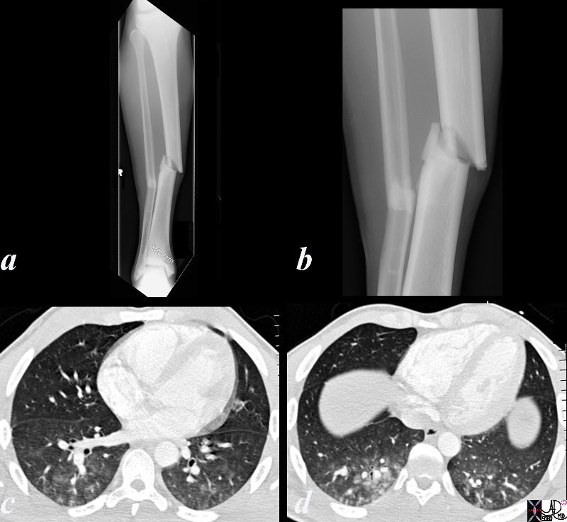
Football Player with a Comminuted Fracture and Fat Embolism


21 year old college student and football player who sustained a compound fracture of his right tibia and fibula. While awaiting surgery he became short of breasth, tachpneic and tachycardic and his saturations dropped to 70’s from 90’s on room air. He had a fever to 103, heart rate of 100, respiratory rate of 30, and a systolic pressure of 140. He required intubation for 5 days but did recover thereafter.
code lung bone fracture fat embolus respiratory distress Courtesy Ashley Davidoff MD copyright 2009 all rights reserved 86255c.8s


    [nodeType] => 1
    [parentNode] => (object value omitted)
    [childNodes] => (object value omitted)
    [firstChild] => (object value omitted)
    [lastChild] => (object value omitted)
    [previousSibling] => (object value omitted)
    [nextSibling] => (object value omitted)
    [attributes] => (object value omitted)
    [ownerDocument] => (object value omitted)
    [namespaceURI] =>
    [prefix] =>
    [localName] => table
    [baseURI] =>
    [textContent] =>


Football Player with a Comminuted Fracture and Fat Embolism


21 year old college student and football player who sustained a compound fracture of his right tibia and fibula. While awaiting surgery he became short of breasth, tachpneic and tachycardic and his saturations dropped to 70’s from 90’s on room air. He had a fever to 103, heart rate of 100, respiratory rate of 30, and a systolic pressure of 140. He required intubation for 5 days but did recover thereafter.
code lung bone fracture fat embolus respiratory distress Courtesy Ashley Davidoff MD copyright 2009 all rights reserved 86255c.8s


)
DOMElement Object
(
    [schemaTypeInfo] =>
    [tagName] => td
    [firstElementChild] => (object value omitted)
    [lastElementChild] => (object value omitted)
    [childElementCount] => 2
    [previousElementSibling] =>
    [nextElementSibling] =>
    [nodeName] => td
    [nodeValue] => 21 year old college student and football player who sustained a compound fracture of his right tibia and fibula. While awaiting surgery he became short of breasth, tachpneic and tachycardic and his saturations dropped to 70’s from 90’s on room air. He had a fever to 103, heart rate of 100, respiratory rate of 30, and a systolic pressure of 140. He required intubation for 5 days but did recover thereafter.
code lung bone fracture fat embolus respiratory distress Courtesy Ashley Davidoff MD copyright 2009 all rights reserved 86255c.8s
    [nodeType] => 1
    [parentNode] => (object value omitted)
    [childNodes] => (object value omitted)
    [firstChild] => (object value omitted)
    [lastChild] => (object value omitted)
    [previousSibling] => (object value omitted)
    [nextSibling] => (object value omitted)
    [attributes] => (object value omitted)
    [ownerDocument] => (object value omitted)
    [namespaceURI] =>
    [prefix] =>
    [localName] => td
    [baseURI] =>
    [textContent] => 21 year old college student and football player who sustained a compound fracture of his right tibia and fibula. While awaiting surgery he became short of breasth, tachpneic and tachycardic and his saturations dropped to 70’s from 90’s on room air. He had a fever to 103, heart rate of 100, respiratory rate of 30, and a systolic pressure of 140. He required intubation for 5 days but did recover thereafter.
code lung bone fracture fat embolus respiratory distress Courtesy Ashley Davidoff MD copyright 2009 all rights reserved 86255c.8s
)
DOMElement Object
(
    [schemaTypeInfo] =>
    [tagName] => td
    [firstElementChild] => (object value omitted)
    [lastElementChild] => (object value omitted)
    [childElementCount] => 2
    [previousElementSibling] =>
    [nextElementSibling] =>
    [nodeName] => td
    [nodeValue] =>
Football Player with a Comminuted Fracture and Fat Embolism

    [nodeType] => 1
    [parentNode] => (object value omitted)
    [childNodes] => (object value omitted)
    [firstChild] => (object value omitted)
    [lastChild] => (object value omitted)
    [previousSibling] => (object value omitted)
    [nextSibling] => (object value omitted)
    [attributes] => (object value omitted)
    [ownerDocument] => (object value omitted)
    [namespaceURI] =>
    [prefix] =>
    [localName] => td
    [baseURI] =>
    [textContent] =>
Football Player with a Comminuted Fracture and Fat Embolism

)
DOMElement Object
(
    [schemaTypeInfo] =>
    [tagName] => table
    [firstElementChild] => (object value omitted)
    [lastElementChild] => (object value omitted)
    [childElementCount] => 1
    [previousElementSibling] => (object value omitted)
    [nextElementSibling] => (object value omitted)
    [nodeName] => table
    [nodeValue] =>


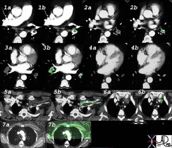
PE from Upper Limb Thrombosis


This elderly woman presented with acute respiratory difficulty with swelling of her left upper limb. CT shows extensive pulmonary embolic disease to the lower lobes from second order branches to the tertiary branches (1-4 with green overlay) and evidence of thrombosis of the left subclavian vein. (5,6). Swelling over the anterior chest including the pectoralis muscles is evident in 7. Courtesy Priscilla Slanetz MD. 32538cLW


    [nodeType] => 1
    [parentNode] => (object value omitted)
    [childNodes] => (object value omitted)
    [firstChild] => (object value omitted)
    [lastChild] => (object value omitted)
    [previousSibling] => (object value omitted)
    [nextSibling] => (object value omitted)
    [attributes] => (object value omitted)
    [ownerDocument] => (object value omitted)
    [namespaceURI] =>
    [prefix] =>
    [localName] => table
    [baseURI] =>
    [textContent] =>


PE from Upper Limb Thrombosis


This elderly woman presented with acute respiratory difficulty with swelling of her left upper limb. CT shows extensive pulmonary embolic disease to the lower lobes from second order branches to the tertiary branches (1-4 with green overlay) and evidence of thrombosis of the left subclavian vein. (5,6). Swelling over the anterior chest including the pectoralis muscles is evident in 7. Courtesy Priscilla Slanetz MD. 32538cLW


)
DOMElement Object
(
    [schemaTypeInfo] =>
    [tagName] => td
    [firstElementChild] => (object value omitted)
    [lastElementChild] => (object value omitted)
    [childElementCount] => 1
    [previousElementSibling] =>
    [nextElementSibling] =>
    [nodeName] => td
    [nodeValue] => This elderly woman presented with acute respiratory difficulty with swelling of her left upper limb. CT shows extensive pulmonary embolic disease to the lower lobes from second order branches to the tertiary branches (1-4 with green overlay) and evidence of thrombosis of the left subclavian vein. (5,6). Swelling over the anterior chest including the pectoralis muscles is evident in 7. Courtesy Priscilla Slanetz MD. 32538cLW
    [nodeType] => 1
    [parentNode] => (object value omitted)
    [childNodes] => (object value omitted)
    [firstChild] => (object value omitted)
    [lastChild] => (object value omitted)
    [previousSibling] => (object value omitted)
    [nextSibling] => (object value omitted)
    [attributes] => (object value omitted)
    [ownerDocument] => (object value omitted)
    [namespaceURI] =>
    [prefix] =>
    [localName] => td
    [baseURI] =>
    [textContent] => This elderly woman presented with acute respiratory difficulty with swelling of her left upper limb. CT shows extensive pulmonary embolic disease to the lower lobes from second order branches to the tertiary branches (1-4 with green overlay) and evidence of thrombosis of the left subclavian vein. (5,6). Swelling over the anterior chest including the pectoralis muscles is evident in 7. Courtesy Priscilla Slanetz MD. 32538cLW
)
DOMElement Object
(
    [schemaTypeInfo] =>
    [tagName] => td
    [firstElementChild] => (object value omitted)
    [lastElementChild] => (object value omitted)
    [childElementCount] => 2
    [previousElementSibling] =>
    [nextElementSibling] =>
    [nodeName] => td
    [nodeValue] =>
PE from Upper Limb Thrombosis

    [nodeType] => 1
    [parentNode] => (object value omitted)
    [childNodes] => (object value omitted)
    [firstChild] => (object value omitted)
    [lastChild] => (object value omitted)
    [previousSibling] => (object value omitted)
    [nextSibling] => (object value omitted)
    [attributes] => (object value omitted)
    [ownerDocument] => (object value omitted)
    [namespaceURI] =>
    [prefix] =>
    [localName] => td
    [baseURI] =>
    [textContent] =>
PE from Upper Limb Thrombosis

)
DOMElement Object
(
    [schemaTypeInfo] =>
    [tagName] => table
    [firstElementChild] => (object value omitted)
    [lastElementChild] => (object value omitted)
    [childElementCount] => 1
    [previousElementSibling] => (object value omitted)
    [nextElementSibling] => (object value omitted)
    [nodeName] => table
    [nodeValue] =>


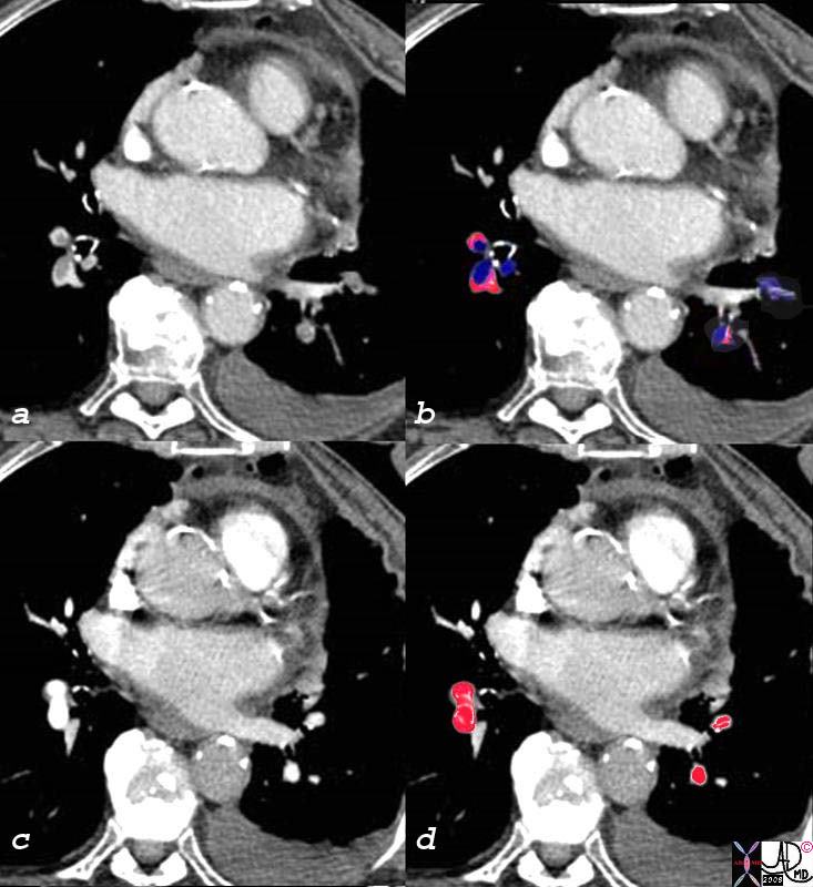
Pulmonary Emboli Before and After Treatment


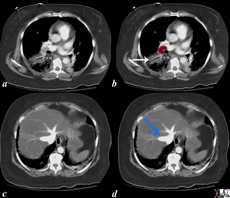
The patient has breast carcinoma and was shown in the set of images above.  She presented with shortness of breath and pulmonary emboli were diagnosed as filling defects (overlay in royal blue c), in all the visualized vessels.  After 2 weeks of anticiacougulant therapy the emboli were lysed and the vessels were completely patent (overlaid in red in d)
78540c02s elderly lady wih breast carcinoma presents with SOB lung artery filling defects pulmonary emboli 2 weeks following treatment emboli resolved Courtesy Ashley Davidoff MD copyright 2008


    [nodeType] => 1
    [parentNode] => (object value omitted)
    [childNodes] => (object value omitted)
    [firstChild] => (object value omitted)
    [lastChild] => (object value omitted)
    [previousSibling] => (object value omitted)
    [nextSibling] => (object value omitted)
    [attributes] => (object value omitted)
    [ownerDocument] => (object value omitted)
    [namespaceURI] =>
    [prefix] =>
    [localName] => table
    [baseURI] =>
    [textContent] =>


Pulmonary Emboli Before and After Treatment


The patient has breast carcinoma and was shown in the set of images above.  She presented with shortness of breath and pulmonary emboli were diagnosed as filling defects (overlay in royal blue c), in all the visualized vessels.  After 2 weeks of anticiacougulant therapy the emboli were lysed and the vessels were completely patent (overlaid in red in d)
78540c02s elderly lady wih breast carcinoma presents with SOB lung artery filling defects pulmonary emboli 2 weeks following treatment emboli resolved Courtesy Ashley Davidoff MD copyright 2008


)
DOMElement Object
(
    [schemaTypeInfo] =>
    [tagName] => td
    [firstElementChild] => (object value omitted)
    [lastElementChild] => (object value omitted)
    [childElementCount] => 2
    [previousElementSibling] =>
    [nextElementSibling] =>
    [nodeName] => td
    [nodeValue] => The patient has breast carcinoma and was shown in the set of images above.  She presented with shortness of breath and pulmonary emboli were diagnosed as filling defects (overlay in royal blue c), in all the visualized vessels.  After 2 weeks of anticiacougulant therapy the emboli were lysed and the vessels were completely patent (overlaid in red in d)
78540c02s elderly lady wih breast carcinoma presents with SOB lung artery filling defects pulmonary emboli 2 weeks following treatment emboli resolved Courtesy Ashley Davidoff MD copyright 2008
    [nodeType] => 1
    [parentNode] => (object value omitted)
    [childNodes] => (object value omitted)
    [firstChild] => (object value omitted)
    [lastChild] => (object value omitted)
    [previousSibling] => (object value omitted)
    [nextSibling] => (object value omitted)
    [attributes] => (object value omitted)
    [ownerDocument] => (object value omitted)
    [namespaceURI] =>
    [prefix] =>
    [localName] => td
    [baseURI] =>
    [textContent] => The patient has breast carcinoma and was shown in the set of images above.  She presented with shortness of breath and pulmonary emboli were diagnosed as filling defects (overlay in royal blue c), in all the visualized vessels.  After 2 weeks of anticiacougulant therapy the emboli were lysed and the vessels were completely patent (overlaid in red in d)
78540c02s elderly lady wih breast carcinoma presents with SOB lung artery filling defects pulmonary emboli 2 weeks following treatment emboli resolved Courtesy Ashley Davidoff MD copyright 2008
)
DOMElement Object
(
    [schemaTypeInfo] =>
    [tagName] => td
    [firstElementChild] => (object value omitted)
    [lastElementChild] => (object value omitted)
    [childElementCount] => 2
    [previousElementSibling] =>
    [nextElementSibling] =>
    [nodeName] => td
    [nodeValue] =>
Pulmonary Emboli Before and After Treatment

    [nodeType] => 1
    [parentNode] => (object value omitted)
    [childNodes] => (object value omitted)
    [firstChild] => (object value omitted)
    [lastChild] => (object value omitted)
    [previousSibling] => (object value omitted)
    [nextSibling] => (object value omitted)
    [attributes] => (object value omitted)
    [ownerDocument] => (object value omitted)
    [namespaceURI] =>
    [prefix] =>
    [localName] => td
    [baseURI] =>
    [textContent] =>
Pulmonary Emboli Before and After Treatment

)
DOMElement Object
(
    [schemaTypeInfo] =>
    [tagName] => table
    [firstElementChild] => (object value omitted)
    [lastElementChild] => (object value omitted)
    [childElementCount] => 1
    [previousElementSibling] => (object value omitted)
    [nextElementSibling] => (object value omitted)
    [nodeName] => table
    [nodeValue] =>


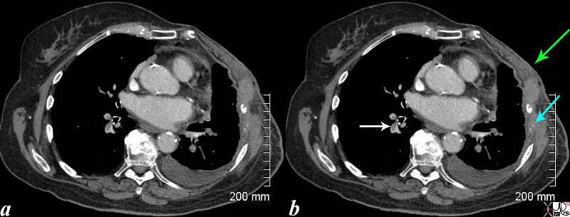
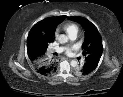
Breast Carcinoma and Pulmonary Emboli


This CT is from an elderly lady wih breast carcinoma, who presents with SOB .  Noted are filling defects in the pulmonary arteries (white arrow). The left sided pulmonary arteries also have filling defects. (see next set of images) She has had a left mastectomy, (green arrow) and has lytic metastases of the left ribs with destruction of the bone which remain as two white calcific specks (teal blue arrow). A small pleural effusion on the left may be due to the malignancy (likely) or due to the pulmonary embolus.
lung artery filling defects pulmonary emboli 2 weeks following treatment emboli resolved Courtesy Ashley Davidoff MD copyright 2008 78540c03.8s


    [nodeType] => 1
    [parentNode] => (object value omitted)
    [childNodes] => (object value omitted)
    [firstChild] => (object value omitted)
    [lastChild] => (object value omitted)
    [previousSibling] => (object value omitted)
    [nextSibling] => (object value omitted)
    [attributes] => (object value omitted)
    [ownerDocument] => (object value omitted)
    [namespaceURI] =>
    [prefix] =>
    [localName] => table
    [baseURI] =>
    [textContent] =>


Breast Carcinoma and Pulmonary Emboli


This CT is from an elderly lady wih breast carcinoma, who presents with SOB .  Noted are filling defects in the pulmonary arteries (white arrow). The left sided pulmonary arteries also have filling defects. (see next set of images) She has had a left mastectomy, (green arrow) and has lytic metastases of the left ribs with destruction of the bone which remain as two white calcific specks (teal blue arrow). A small pleural effusion on the left may be due to the malignancy (likely) or due to the pulmonary embolus.
lung artery filling defects pulmonary emboli 2 weeks following treatment emboli resolved Courtesy Ashley Davidoff MD copyright 2008 78540c03.8s


)
DOMElement Object
(
    [schemaTypeInfo] =>
    [tagName] => td
    [firstElementChild] => (object value omitted)
    [lastElementChild] => (object value omitted)
    [childElementCount] => 2
    [previousElementSibling] =>
    [nextElementSibling] =>
    [nodeName] => td
    [nodeValue] => This CT is from an elderly lady wih breast carcinoma, who presents with SOB .  Noted are filling defects in the pulmonary arteries (white arrow). The left sided pulmonary arteries also have filling defects. (see next set of images) She has had a left mastectomy, (green arrow) and has lytic metastases of the left ribs with destruction of the bone which remain as two white calcific specks (teal blue arrow). A small pleural effusion on the left may be due to the malignancy (likely) or due to the pulmonary embolus.
lung artery filling defects pulmonary emboli 2 weeks following treatment emboli resolved Courtesy Ashley Davidoff MD copyright 2008 78540c03.8s
    [nodeType] => 1
    [parentNode] => (object value omitted)
    [childNodes] => (object value omitted)
    [firstChild] => (object value omitted)
    [lastChild] => (object value omitted)
    [previousSibling] => (object value omitted)
    [nextSibling] => (object value omitted)
    [attributes] => (object value omitted)
    [ownerDocument] => (object value omitted)
    [namespaceURI] =>
    [prefix] =>
    [localName] => td
    [baseURI] =>
    [textContent] => This CT is from an elderly lady wih breast carcinoma, who presents with SOB .  Noted are filling defects in the pulmonary arteries (white arrow). The left sided pulmonary arteries also have filling defects. (see next set of images) She has had a left mastectomy, (green arrow) and has lytic metastases of the left ribs with destruction of the bone which remain as two white calcific specks (teal blue arrow). A small pleural effusion on the left may be due to the malignancy (likely) or due to the pulmonary embolus.
lung artery filling defects pulmonary emboli 2 weeks following treatment emboli resolved Courtesy Ashley Davidoff MD copyright 2008 78540c03.8s
)
DOMElement Object
(
    [schemaTypeInfo] =>
    [tagName] => td
    [firstElementChild] => (object value omitted)
    [lastElementChild] => (object value omitted)
    [childElementCount] => 2
    [previousElementSibling] =>
    [nextElementSibling] =>
    [nodeName] => td
    [nodeValue] =>
Breast Carcinoma and Pulmonary Emboli

    [nodeType] => 1
    [parentNode] => (object value omitted)
    [childNodes] => (object value omitted)
    [firstChild] => (object value omitted)
    [lastChild] => (object value omitted)
    [previousSibling] => (object value omitted)
    [nextSibling] => (object value omitted)
    [attributes] => (object value omitted)
    [ownerDocument] => (object value omitted)
    [namespaceURI] =>
    [prefix] =>
    [localName] => td
    [baseURI] =>
    [textContent] =>
Breast Carcinoma and Pulmonary Emboli

)
DOMElement Object
(
    [schemaTypeInfo] =>
    [tagName] => table
    [firstElementChild] => (object value omitted)
    [lastElementChild] => (object value omitted)
    [childElementCount] => 1
    [previousElementSibling] => (object value omitted)
    [nextElementSibling] => (object value omitted)
    [nodeName] => table
    [nodeValue] =>


Wedge Shaped Infarct in the Spleen


The CTscan with contrast shows a wedge shaped defect in the spleen (overlaid in pink in middle image) characteristic of an acute infarction most commonly caused by emboli.  The pathology image from a different patient shows the same wedge shaped process.
46774c01 21321spleen splenic fx wedge shaped defects dx emboli dx systemic embolization dx splenic infarcts CTscan Davidoff MD


    [nodeType] => 1
    [parentNode] => (object value omitted)
    [childNodes] => (object value omitted)
    [firstChild] => (object value omitted)
    [lastChild] => (object value omitted)
    [previousSibling] => (object value omitted)
    [nextSibling] => (object value omitted)
    [attributes] => (object value omitted)
    [ownerDocument] => (object value omitted)
    [namespaceURI] =>
    [prefix] =>
    [localName] => table
    [baseURI] =>
    [textContent] =>


Wedge Shaped Infarct in the Spleen


The CTscan with contrast shows a wedge shaped defect in the spleen (overlaid in pink in middle image) characteristic of an acute infarction most commonly caused by emboli.  The pathology image from a different patient shows the same wedge shaped process.
46774c01 21321spleen splenic fx wedge shaped defects dx emboli dx systemic embolization dx splenic infarcts CTscan Davidoff MD


)
DOMElement Object
(
    [schemaTypeInfo] =>
    [tagName] => td
    [firstElementChild] => (object value omitted)
    [lastElementChild] => (object value omitted)
    [childElementCount] => 2
    [previousElementSibling] =>
    [nextElementSibling] =>
    [nodeName] => td
    [nodeValue] => The CTscan with contrast shows a wedge shaped defect in the spleen (overlaid in pink in middle image) characteristic of an acute infarction most commonly caused by emboli.  The pathology image from a different patient shows the same wedge shaped process.
46774c01 21321spleen splenic fx wedge shaped defects dx emboli dx systemic embolization dx splenic infarcts CTscan Davidoff MD
    [nodeType] => 1
    [parentNode] => (object value omitted)
    [childNodes] => (object value omitted)
    [firstChild] => (object value omitted)
    [lastChild] => (object value omitted)
    [previousSibling] => (object value omitted)
    [nextSibling] => (object value omitted)
    [attributes] => (object value omitted)
    [ownerDocument] => (object value omitted)
    [namespaceURI] =>
    [prefix] =>
    [localName] => td
    [baseURI] =>
    [textContent] => The CTscan with contrast shows a wedge shaped defect in the spleen (overlaid in pink in middle image) characteristic of an acute infarction most commonly caused by emboli.  The pathology image from a different patient shows the same wedge shaped process.
46774c01 21321spleen splenic fx wedge shaped defects dx emboli dx systemic embolization dx splenic infarcts CTscan Davidoff MD
)
DOMElement Object
(
    [schemaTypeInfo] =>
    [tagName] => td
    [firstElementChild] => (object value omitted)
    [lastElementChild] => (object value omitted)
    [childElementCount] => 2
    [previousElementSibling] =>
    [nextElementSibling] =>
    [nodeName] => td
    [nodeValue] =>

Wedge Shaped Infarct in the Spleen

    [nodeType] => 1
    [parentNode] => (object value omitted)
    [childNodes] => (object value omitted)
    [firstChild] => (object value omitted)
    [lastChild] => (object value omitted)
    [previousSibling] => (object value omitted)
    [nextSibling] => (object value omitted)
    [attributes] => (object value omitted)
    [ownerDocument] => (object value omitted)
    [namespaceURI] =>
    [prefix] =>
    [localName] => td
    [baseURI] =>
    [textContent] =>

Wedge Shaped Infarct in the Spleen

)
DOMElement Object
(
    [schemaTypeInfo] =>
    [tagName] => table
    [firstElementChild] => (object value omitted)
    [lastElementChild] => (object value omitted)
    [childElementCount] => 1
    [previousElementSibling] => (object value omitted)
    [nextElementSibling] => (object value omitted)
    [nodeName] => table
    [nodeValue] =>


Embolic Infarct of the Splenic Artery


47640 spleen artery fx wedge shaped defect fx occluded artery dx embolism embolic infarction CTscan Davidoff MD


    [nodeType] => 1
    [parentNode] => (object value omitted)
    [childNodes] => (object value omitted)
    [firstChild] => (object value omitted)
    [lastChild] => (object value omitted)
    [previousSibling] => (object value omitted)
    [nextSibling] => (object value omitted)
    [attributes] => (object value omitted)
    [ownerDocument] => (object value omitted)
    [namespaceURI] =>
    [prefix] =>
    [localName] => table
    [baseURI] =>
    [textContent] =>


Embolic Infarct of the Splenic Artery


47640 spleen artery fx wedge shaped defect fx occluded artery dx embolism embolic infarction CTscan Davidoff MD


)
DOMElement Object
(
    [schemaTypeInfo] =>
    [tagName] => td
    [firstElementChild] => (object value omitted)
    [lastElementChild] => (object value omitted)
    [childElementCount] => 1
    [previousElementSibling] =>
    [nextElementSibling] =>
    [nodeName] => td
    [nodeValue] => 47640 spleen artery fx wedge shaped defect fx occluded artery dx embolism embolic infarction CTscan Davidoff MD
    [nodeType] => 1
    [parentNode] => (object value omitted)
    [childNodes] => (object value omitted)
    [firstChild] => (object value omitted)
    [lastChild] => (object value omitted)
    [previousSibling] => (object value omitted)
    [nextSibling] => (object value omitted)
    [attributes] => (object value omitted)
    [ownerDocument] => (object value omitted)
    [namespaceURI] =>
    [prefix] =>
    [localName] => td
    [baseURI] =>
    [textContent] => 47640 spleen artery fx wedge shaped defect fx occluded artery dx embolism embolic infarction CTscan Davidoff MD
)
DOMElement Object
(
    [schemaTypeInfo] =>
    [tagName] => td
    [firstElementChild] => (object value omitted)
    [lastElementChild] => (object value omitted)
    [childElementCount] => 2
    [previousElementSibling] =>
    [nextElementSibling] =>
    [nodeName] => td
    [nodeValue] =>
Embolic Infarct of the Splenic Artery
    [nodeType] => 1
    [parentNode] => (object value omitted)
    [childNodes] => (object value omitted)
    [firstChild] => (object value omitted)
    [lastChild] => (object value omitted)
    [previousSibling] => (object value omitted)
    [nextSibling] => (object value omitted)
    [attributes] => (object value omitted)
    [ownerDocument] => (object value omitted)
    [namespaceURI] =>
    [prefix] =>
    [localName] => td
    [baseURI] =>
    [textContent] =>
Embolic Infarct of the Splenic Artery
)
DOMElement Object
(
    [schemaTypeInfo] =>
    [tagName] => table
    [firstElementChild] => (object value omitted)
    [lastElementChild] => (object value omitted)
    [childElementCount] => 1
    [previousElementSibling] => (object value omitted)
    [nextElementSibling] => (object value omitted)
    [nodeName] => table
    [nodeValue] =>


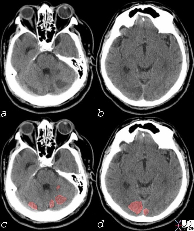
Emboli to the Posterior Circulation Involving the Cerbellum and the Occipital Cortex


74923c01 brain emboli low density lesions posterior cerebral circulation cerebellum occipital lobe occipital infarcts cerebellar infarcts embolic disease CTscan Courtesy Ashley DAvidoff MD


    [nodeType] => 1
    [parentNode] => (object value omitted)
    [childNodes] => (object value omitted)
    [firstChild] => (object value omitted)
    [lastChild] => (object value omitted)
    [previousSibling] => (object value omitted)
    [nextSibling] => (object value omitted)
    [attributes] => (object value omitted)
    [ownerDocument] => (object value omitted)
    [namespaceURI] =>
    [prefix] =>
    [localName] => table
    [baseURI] =>
    [textContent] =>


Emboli to the Posterior Circulation Involving the Cerbellum and the Occipital Cortex


74923c01 brain emboli low density lesions posterior cerebral circulation cerebellum occipital lobe occipital infarcts cerebellar infarcts embolic disease CTscan Courtesy Ashley DAvidoff MD


)
DOMElement Object
(
    [schemaTypeInfo] =>
    [tagName] => td
    [firstElementChild] => (object value omitted)
    [lastElementChild] => (object value omitted)
    [childElementCount] => 1
    [previousElementSibling] =>
    [nextElementSibling] =>
    [nodeName] => td
    [nodeValue] => 74923c01 brain emboli low density lesions posterior cerebral circulation cerebellum occipital lobe occipital infarcts cerebellar infarcts embolic disease CTscan Courtesy Ashley DAvidoff MD
    [nodeType] => 1
    [parentNode] => (object value omitted)
    [childNodes] => (object value omitted)
    [firstChild] => (object value omitted)
    [lastChild] => (object value omitted)
    [previousSibling] => (object value omitted)
    [nextSibling] => (object value omitted)
    [attributes] => (object value omitted)
    [ownerDocument] => (object value omitted)
    [namespaceURI] =>
    [prefix] =>
    [localName] => td
    [baseURI] =>
    [textContent] => 74923c01 brain emboli low density lesions posterior cerebral circulation cerebellum occipital lobe occipital infarcts cerebellar infarcts embolic disease CTscan Courtesy Ashley DAvidoff MD
)
DOMElement Object
(
    [schemaTypeInfo] =>
    [tagName] => td
    [firstElementChild] => (object value omitted)
    [lastElementChild] => (object value omitted)
    [childElementCount] => 2
    [previousElementSibling] =>
    [nextElementSibling] =>
    [nodeName] => td
    [nodeValue] =>
Emboli to the Posterior Circulation Involving the Cerbellum and the Occipital Cortex

    [nodeType] => 1
    [parentNode] => (object value omitted)
    [childNodes] => (object value omitted)
    [firstChild] => (object value omitted)
    [lastChild] => (object value omitted)
    [previousSibling] => (object value omitted)
    [nextSibling] => (object value omitted)
    [attributes] => (object value omitted)
    [ownerDocument] => (object value omitted)
    [namespaceURI] =>
    [prefix] =>
    [localName] => td
    [baseURI] =>
    [textContent] =>
Emboli to the Posterior Circulation Involving the Cerbellum and the Occipital Cortex

)
DOMElement Object
(
    [schemaTypeInfo] =>
    [tagName] => table
    [firstElementChild] => (object value omitted)
    [lastElementChild] => (object value omitted)
    [childElementCount] => 1
    [previousElementSibling] => (object value omitted)
    [nextElementSibling] => (object value omitted)
    [nodeName] => table
    [nodeValue] =>


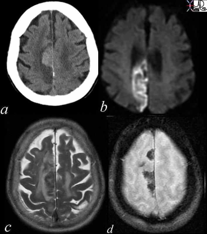
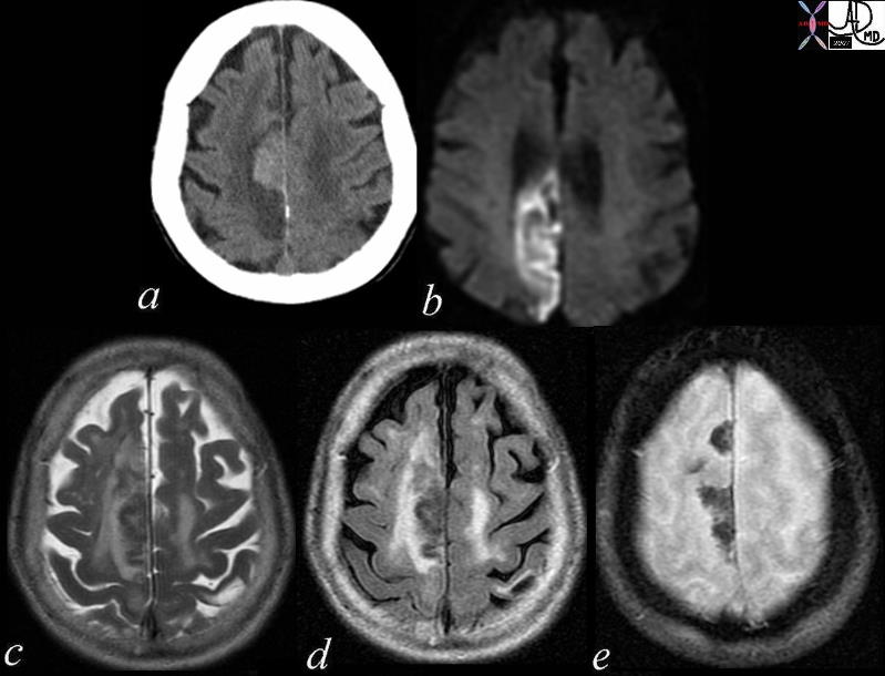
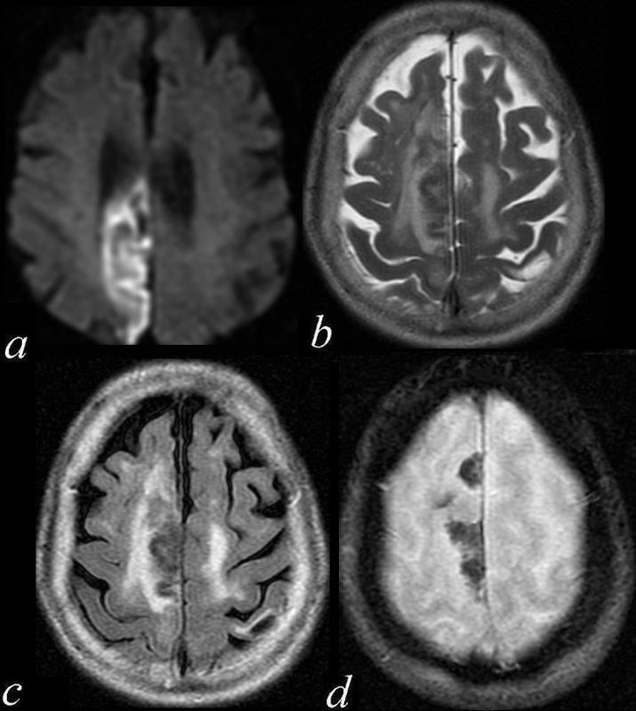
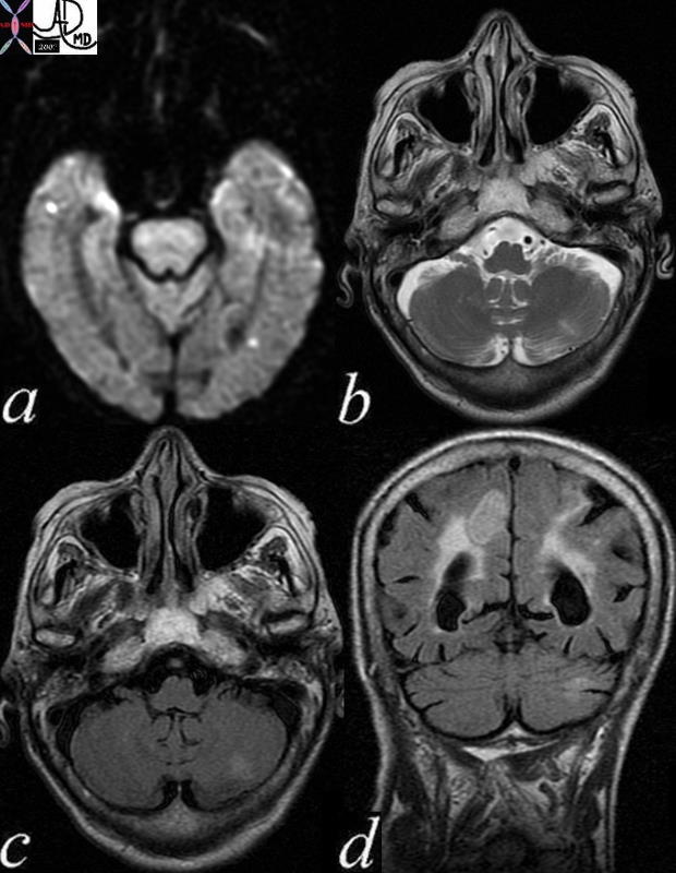
Cerebellar and Parietal Lobe Involvement


71239c04 patient with atrial fibrillation brain cerebrum cerebelum cerebellar hemisphere multicentric parietal lobe parasagittal hemorrhagic infarct paracentral DWII bright T2 bright FLAIR bright GRE mixed blood products dx acute hemorrhagic infarction secondary to embolic event the patient disd have other non hemorrhagic infrctions in other parts of the brain MRI Davidoff MD 71239c0171239c02 71239c03 71239c04


    [nodeType] => 1
    [parentNode] => (object value omitted)
    [childNodes] => (object value omitted)
    [firstChild] => (object value omitted)
    [lastChild] => (object value omitted)
    [previousSibling] => (object value omitted)
    [nextSibling] => (object value omitted)
    [attributes] => (object value omitted)
    [ownerDocument] => (object value omitted)
    [namespaceURI] =>
    [prefix] =>
    [localName] => table
    [baseURI] =>
    [textContent] =>


Cerebellar and Parietal Lobe Involvement


71239c04 patient with atrial fibrillation brain cerebrum cerebelum cerebellar hemisphere multicentric parietal lobe parasagittal hemorrhagic infarct paracentral DWII bright T2 bright FLAIR bright GRE mixed blood products dx acute hemorrhagic infarction secondary to embolic event the patient disd have other non hemorrhagic infrctions in other parts of the brain MRI Davidoff MD 71239c0171239c02 71239c03 71239c04


)
DOMElement Object
(
    [schemaTypeInfo] =>
    [tagName] => td
    [firstElementChild] => (object value omitted)
    [lastElementChild] => (object value omitted)
    [childElementCount] => 1
    [previousElementSibling] =>
    [nextElementSibling] =>
    [nodeName] => td
    [nodeValue] => 71239c04 patient with atrial fibrillation brain cerebrum cerebelum cerebellar hemisphere multicentric parietal lobe parasagittal hemorrhagic infarct paracentral DWII bright T2 bright FLAIR bright GRE mixed blood products dx acute hemorrhagic infarction secondary to embolic event the patient disd have other non hemorrhagic infrctions in other parts of the brain MRI Davidoff MD 71239c0171239c02 71239c03 71239c04
    [nodeType] => 1
    [parentNode] => (object value omitted)
    [childNodes] => (object value omitted)
    [firstChild] => (object value omitted)
    [lastChild] => (object value omitted)
    [previousSibling] => (object value omitted)
    [nextSibling] => (object value omitted)
    [attributes] => (object value omitted)
    [ownerDocument] => (object value omitted)
    [namespaceURI] =>
    [prefix] =>
    [localName] => td
    [baseURI] =>
    [textContent] => 71239c04 patient with atrial fibrillation brain cerebrum cerebelum cerebellar hemisphere multicentric parietal lobe parasagittal hemorrhagic infarct paracentral DWII bright T2 bright FLAIR bright GRE mixed blood products dx acute hemorrhagic infarction secondary to embolic event the patient disd have other non hemorrhagic infrctions in other parts of the brain MRI Davidoff MD 71239c0171239c02 71239c03 71239c04
)
DOMElement Object
(
    [schemaTypeInfo] =>
    [tagName] => td
    [firstElementChild] => (object value omitted)
    [lastElementChild] => (object value omitted)
    [childElementCount] => 2
    [previousElementSibling] =>
    [nextElementSibling] =>
    [nodeName] => td
    [nodeValue] =>
Cerebellar and Parietal Lobe Involvement

    [nodeType] => 1
    [parentNode] => (object value omitted)
    [childNodes] => (object value omitted)
    [firstChild] => (object value omitted)
    [lastChild] => (object value omitted)
    [previousSibling] => (object value omitted)
    [nextSibling] => (object value omitted)
    [attributes] => (object value omitted)
    [ownerDocument] => (object value omitted)
    [namespaceURI] =>
    [prefix] =>
    [localName] => td
    [baseURI] =>
    [textContent] =>
Cerebellar and Parietal Lobe Involvement

)
https://beta.thecommonvein.net/wp-content/uploads/2023/06/71239c04.jpg https://beta.thecommonvein.net/wp-content/uploads/2023/06/71239c03.jpg https://beta.thecommonvein.net/wp-content/uploads/2023/06/71239c02.jpg https://beta.thecommonvein.net/wp-content/uploads/2023/06/71239c01.jpg
http://thecommonvein.net/media/71239c04.jpg

