Arterial System
Ashley Davidoff MD
Copyright 2012
Introduction
DOMElement Object
(
    [schemaTypeInfo] =>
    [tagName] => table
    [firstElementChild] => (object value omitted)
    [lastElementChild] => (object value omitted)
    [childElementCount] => 1
    [previousElementSibling] => (object value omitted)
    [nextElementSibling] =>
    [nodeName] => table
    [nodeValue] =>


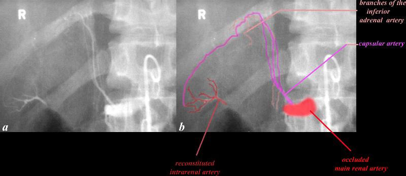
Traumatic occlusion of the Renal Artery
Collateral Supply via the Capsular Artery


The angiogram of the right renal artery following traumatic injury shows an occluded artery (bright red) with an intact capsular artery (bright pink) that gives rise to a capsular artery (bright pink) that gives rise to branches of the inferior adrenal but also serves to collateralize the intrarenal arterial system (dull red).
Copyright 2012 Courtesy Ashley Davidoff MD 26495c01L.8


    [nodeType] => 1
    [parentNode] => (object value omitted)
    [childNodes] => (object value omitted)
    [firstChild] => (object value omitted)
    [lastChild] => (object value omitted)
    [previousSibling] => (object value omitted)
    [nextSibling] => (object value omitted)
    [attributes] => (object value omitted)
    [ownerDocument] => (object value omitted)
    [namespaceURI] =>
    [prefix] =>
    [localName] => table
    [baseURI] =>
    [textContent] =>


Traumatic occlusion of the Renal Artery
Collateral Supply via the Capsular Artery


The angiogram of the right renal artery following traumatic injury shows an occluded artery (bright red) with an intact capsular artery (bright pink) that gives rise to a capsular artery (bright pink) that gives rise to branches of the inferior adrenal but also serves to collateralize the intrarenal arterial system (dull red).
Copyright 2012 Courtesy Ashley Davidoff MD 26495c01L.8


)
DOMElement Object
(
    [schemaTypeInfo] =>
    [tagName] => td
    [firstElementChild] => (object value omitted)
    [lastElementChild] => (object value omitted)
    [childElementCount] => 2
    [previousElementSibling] =>
    [nextElementSibling] =>
    [nodeName] => td
    [nodeValue] =>
The angiogram of the right renal artery following traumatic injury shows an occluded artery (bright red) with an intact capsular artery (bright pink) that gives rise to a capsular artery (bright pink) that gives rise to branches of the inferior adrenal but also serves to collateralize the intrarenal arterial system (dull red).
Copyright 2012 Courtesy Ashley Davidoff MD 26495c01L.8

    [nodeType] => 1
    [parentNode] => (object value omitted)
    [childNodes] => (object value omitted)
    [firstChild] => (object value omitted)
    [lastChild] => (object value omitted)
    [previousSibling] => (object value omitted)
    [nextSibling] => (object value omitted)
    [attributes] => (object value omitted)
    [ownerDocument] => (object value omitted)
    [namespaceURI] =>
    [prefix] =>
    [localName] => td
    [baseURI] =>
    [textContent] =>
The angiogram of the right renal artery following traumatic injury shows an occluded artery (bright red) with an intact capsular artery (bright pink) that gives rise to a capsular artery (bright pink) that gives rise to branches of the inferior adrenal but also serves to collateralize the intrarenal arterial system (dull red).
Copyright 2012 Courtesy Ashley Davidoff MD 26495c01L.8

)
DOMElement Object
(
    [schemaTypeInfo] =>
    [tagName] => td
    [firstElementChild] => (object value omitted)
    [lastElementChild] => (object value omitted)
    [childElementCount] => 3
    [previousElementSibling] =>
    [nextElementSibling] =>
    [nodeName] => td
    [nodeValue] =>
Traumatic occlusion of the Renal Artery
Collateral Supply via the Capsular Artery

    [nodeType] => 1
    [parentNode] => (object value omitted)
    [childNodes] => (object value omitted)
    [firstChild] => (object value omitted)
    [lastChild] => (object value omitted)
    [previousSibling] => (object value omitted)
    [nextSibling] => (object value omitted)
    [attributes] => (object value omitted)
    [ownerDocument] => (object value omitted)
    [namespaceURI] =>
    [prefix] =>
    [localName] => td
    [baseURI] =>
    [textContent] =>
Traumatic occlusion of the Renal Artery
Collateral Supply via the Capsular Artery

)
DOMElement Object
(
    [schemaTypeInfo] =>
    [tagName] => table
    [firstElementChild] => (object value omitted)
    [lastElementChild] => (object value omitted)
    [childElementCount] => 1
    [previousElementSibling] => (object value omitted)
    [nextElementSibling] => (object value omitted)
    [nodeName] => table
    [nodeValue] =>


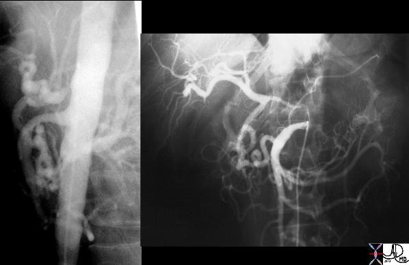
Long Segment Celiac Axis Stenosis with Collaterals via the Pancreaticoduodenal Arcade


Angiogram of the SMA shows retrograde filling of the celiac axis which has a significant long segment stenosis. This stenosis has an unusual appearance in that it is relatively long but the most likely cause is atherosclerosis.
Courtesy Laura Feldman MD 35076c


    [nodeType] => 1
    [parentNode] => (object value omitted)
    [childNodes] => (object value omitted)
    [firstChild] => (object value omitted)
    [lastChild] => (object value omitted)
    [previousSibling] => (object value omitted)
    [nextSibling] => (object value omitted)
    [attributes] => (object value omitted)
    [ownerDocument] => (object value omitted)
    [namespaceURI] =>
    [prefix] =>
    [localName] => table
    [baseURI] =>
    [textContent] =>


Long Segment Celiac Axis Stenosis with Collaterals via the Pancreaticoduodenal Arcade


Angiogram of the SMA shows retrograde filling of the celiac axis which has a significant long segment stenosis. This stenosis has an unusual appearance in that it is relatively long but the most likely cause is atherosclerosis.
Courtesy Laura Feldman MD 35076c


)
DOMElement Object
(
    [schemaTypeInfo] =>
    [tagName] => td
    [firstElementChild] => (object value omitted)
    [lastElementChild] => (object value omitted)
    [childElementCount] => 2
    [previousElementSibling] =>
    [nextElementSibling] =>
    [nodeName] => td
    [nodeValue] =>
Angiogram of the SMA shows retrograde filling of the celiac axis which has a significant long segment stenosis. This stenosis has an unusual appearance in that it is relatively long but the most likely cause is atherosclerosis.
Courtesy Laura Feldman MD 35076c

    [nodeType] => 1
    [parentNode] => (object value omitted)
    [childNodes] => (object value omitted)
    [firstChild] => (object value omitted)
    [lastChild] => (object value omitted)
    [previousSibling] => (object value omitted)
    [nextSibling] => (object value omitted)
    [attributes] => (object value omitted)
    [ownerDocument] => (object value omitted)
    [namespaceURI] =>
    [prefix] =>
    [localName] => td
    [baseURI] =>
    [textContent] =>
Angiogram of the SMA shows retrograde filling of the celiac axis which has a significant long segment stenosis. This stenosis has an unusual appearance in that it is relatively long but the most likely cause is atherosclerosis.
Courtesy Laura Feldman MD 35076c

)
DOMElement Object
(
    [schemaTypeInfo] =>
    [tagName] => td
    [firstElementChild] => (object value omitted)
    [lastElementChild] => (object value omitted)
    [childElementCount] => 2
    [previousElementSibling] =>
    [nextElementSibling] =>
    [nodeName] => td
    [nodeValue] =>
Long Segment Celiac Axis Stenosis with Collaterals via the Pancreaticoduodenal Arcade

    [nodeType] => 1
    [parentNode] => (object value omitted)
    [childNodes] => (object value omitted)
    [firstChild] => (object value omitted)
    [lastChild] => (object value omitted)
    [previousSibling] => (object value omitted)
    [nextSibling] => (object value omitted)
    [attributes] => (object value omitted)
    [ownerDocument] => (object value omitted)
    [namespaceURI] =>
    [prefix] =>
    [localName] => td
    [baseURI] =>
    [textContent] =>
Long Segment Celiac Axis Stenosis with Collaterals via the Pancreaticoduodenal Arcade

)
DOMElement Object
(
    [schemaTypeInfo] =>
    [tagName] => table
    [firstElementChild] => (object value omitted)
    [lastElementChild] => (object value omitted)
    [childElementCount] => 1
    [previousElementSibling] => (object value omitted)
    [nextElementSibling] => (object value omitted)
    [nodeName] => table
    [nodeValue] =>


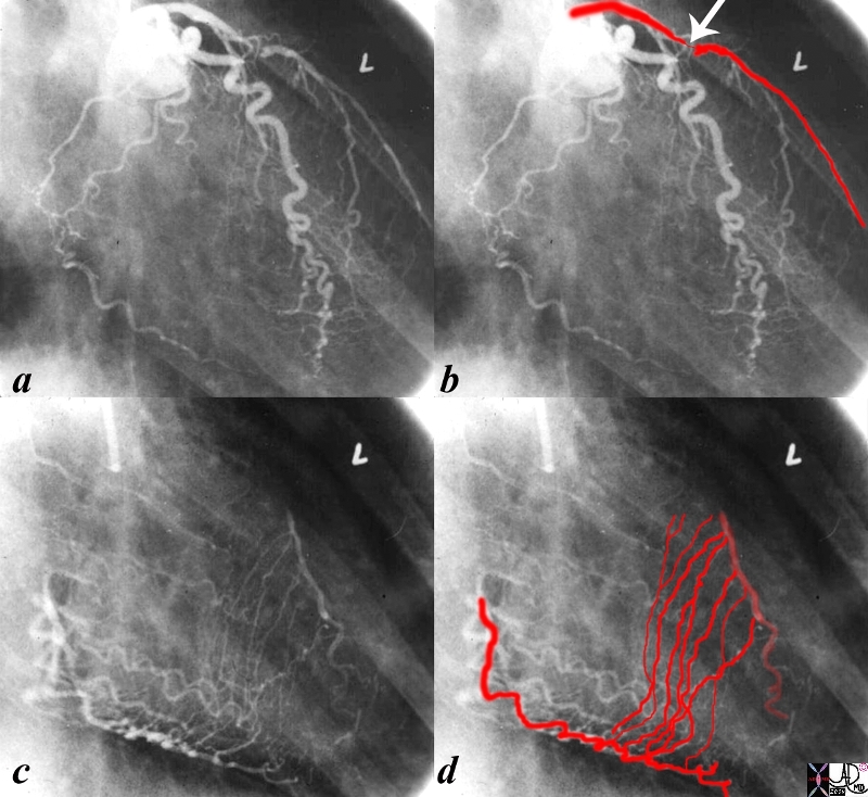
High Grade Stenosis of the LAD and Collateral Circulation theough the PDA


The first angiogram is from a an injection of the left coronary artery in the RAO projection. A subtotal stenosis is seen in the proximal LAD (arrow). There is an implied high pressure gradient between the proximal and distal LAD and a predicted low pressure in the distal vessel. The second injection was into a normal right coronary artery with delayed iaging when most of the contrast had disappeared from the vessel and shows septal collateral vessels supplying the downstrem low pressure LAD from a high pressure RCA. code heart cardiac stenosis atherosclerosis collaterals septal collaterals coronary artery septal perforators angiogram angiography
Courtesy Ashley Davidoff MD copyright 2012 all rights reserved 15042c02.8s


    [nodeType] => 1
    [parentNode] => (object value omitted)
    [childNodes] => (object value omitted)
    [firstChild] => (object value omitted)
    [lastChild] => (object value omitted)
    [previousSibling] => (object value omitted)
    [nextSibling] => (object value omitted)
    [attributes] => (object value omitted)
    [ownerDocument] => (object value omitted)
    [namespaceURI] =>
    [prefix] =>
    [localName] => table
    [baseURI] =>
    [textContent] =>


High Grade Stenosis of the LAD and Collateral Circulation theough the PDA


The first angiogram is from a an injection of the left coronary artery in the RAO projection. A subtotal stenosis is seen in the proximal LAD (arrow). There is an implied high pressure gradient between the proximal and distal LAD and a predicted low pressure in the distal vessel. The second injection was into a normal right coronary artery with delayed iaging when most of the contrast had disappeared from the vessel and shows septal collateral vessels supplying the downstrem low pressure LAD from a high pressure RCA. code heart cardiac stenosis atherosclerosis collaterals septal collaterals coronary artery septal perforators angiogram angiography
Courtesy Ashley Davidoff MD copyright 2012 all rights reserved 15042c02.8s


)
DOMElement Object
(
    [schemaTypeInfo] =>
    [tagName] => td
    [firstElementChild] => (object value omitted)
    [lastElementChild] => (object value omitted)
    [childElementCount] => 2
    [previousElementSibling] =>
    [nextElementSibling] =>
    [nodeName] => td
    [nodeValue] =>
The first angiogram is from a an injection of the left coronary artery in the RAO projection. A subtotal stenosis is seen in the proximal LAD (arrow). There is an implied high pressure gradient between the proximal and distal LAD and a predicted low pressure in the distal vessel. The second injection was into a normal right coronary artery with delayed iaging when most of the contrast had disappeared from the vessel and shows septal collateral vessels supplying the downstrem low pressure LAD from a high pressure RCA. code heart cardiac stenosis atherosclerosis collaterals septal collaterals coronary artery septal perforators angiogram angiography
Courtesy Ashley Davidoff MD copyright 2012 all rights reserved 15042c02.8s

    [nodeType] => 1
    [parentNode] => (object value omitted)
    [childNodes] => (object value omitted)
    [firstChild] => (object value omitted)
    [lastChild] => (object value omitted)
    [previousSibling] => (object value omitted)
    [nextSibling] => (object value omitted)
    [attributes] => (object value omitted)
    [ownerDocument] => (object value omitted)
    [namespaceURI] =>
    [prefix] =>
    [localName] => td
    [baseURI] =>
    [textContent] =>
The first angiogram is from a an injection of the left coronary artery in the RAO projection. A subtotal stenosis is seen in the proximal LAD (arrow). There is an implied high pressure gradient between the proximal and distal LAD and a predicted low pressure in the distal vessel. The second injection was into a normal right coronary artery with delayed iaging when most of the contrast had disappeared from the vessel and shows septal collateral vessels supplying the downstrem low pressure LAD from a high pressure RCA. code heart cardiac stenosis atherosclerosis collaterals septal collaterals coronary artery septal perforators angiogram angiography
Courtesy Ashley Davidoff MD copyright 2012 all rights reserved 15042c02.8s

)
DOMElement Object
(
    [schemaTypeInfo] =>
    [tagName] => td
    [firstElementChild] => (object value omitted)
    [lastElementChild] => (object value omitted)
    [childElementCount] => 2
    [previousElementSibling] =>
    [nextElementSibling] =>
    [nodeName] => td
    [nodeValue] =>
High Grade Stenosis of the LAD and Collateral Circulation theough the PDA

    [nodeType] => 1
    [parentNode] => (object value omitted)
    [childNodes] => (object value omitted)
    [firstChild] => (object value omitted)
    [lastChild] => (object value omitted)
    [previousSibling] => (object value omitted)
    [nextSibling] => (object value omitted)
    [attributes] => (object value omitted)
    [ownerDocument] => (object value omitted)
    [namespaceURI] =>
    [prefix] =>
    [localName] => td
    [baseURI] =>
    [textContent] =>
High Grade Stenosis of the LAD and Collateral Circulation theough the PDA

)
DOMElement Object
(
    [schemaTypeInfo] =>
    [tagName] => table
    [firstElementChild] => (object value omitted)
    [lastElementChild] => (object value omitted)
    [childElementCount] => 1
    [previousElementSibling] => (object value omitted)
    [nextElementSibling] => (object value omitted)
    [nodeName] => table
    [nodeValue] =>


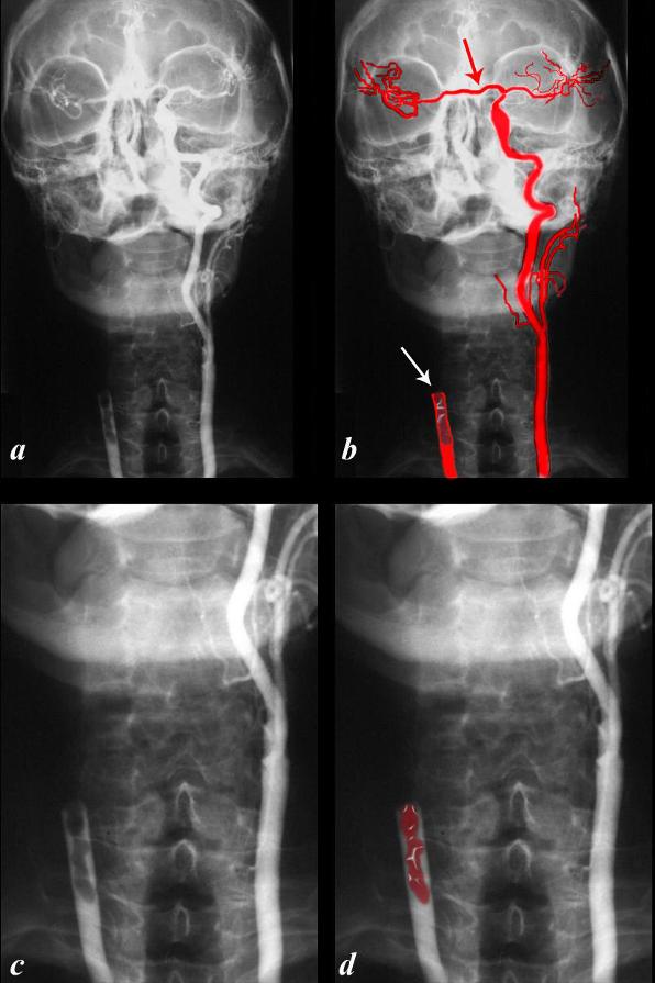
Occluded Right Common Carotid
Caollateral via the Circle of Willis


This angiogram of the carotid arteries, (a,b) show an occluded right common carotid artery (white arrow)with serpiginous thrombus in the artery behind the occlusion (maroon overlay in b and d). The left common carotid artery is almost a normal artery and fills the right system via the circle of Willis (red arrow) The occluded left common carotid and thrombus is magnified in c and d.
Davidoff art copyright 2012 Courtesy Ashley Davidoff MD 113403c02L04.9


    [nodeType] => 1
    [parentNode] => (object value omitted)
    [childNodes] => (object value omitted)
    [firstChild] => (object value omitted)
    [lastChild] => (object value omitted)
    [previousSibling] => (object value omitted)
    [nextSibling] => (object value omitted)
    [attributes] => (object value omitted)
    [ownerDocument] => (object value omitted)
    [namespaceURI] =>
    [prefix] =>
    [localName] => table
    [baseURI] =>
    [textContent] =>


Occluded Right Common Carotid
Caollateral via the Circle of Willis


This angiogram of the carotid arteries, (a,b) show an occluded right common carotid artery (white arrow)with serpiginous thrombus in the artery behind the occlusion (maroon overlay in b and d). The left common carotid artery is almost a normal artery and fills the right system via the circle of Willis (red arrow) The occluded left common carotid and thrombus is magnified in c and d.
Davidoff art copyright 2012 Courtesy Ashley Davidoff MD 113403c02L04.9


)
DOMElement Object
(
    [schemaTypeInfo] =>
    [tagName] => td
    [firstElementChild] => (object value omitted)
    [lastElementChild] => (object value omitted)
    [childElementCount] => 1
    [previousElementSibling] =>
    [nextElementSibling] =>
    [nodeName] => td
    [nodeValue] => This angiogram of the carotid arteries, (a,b) show an occluded right common carotid artery (white arrow)with serpiginous thrombus in the artery behind the occlusion (maroon overlay in b and d). The left common carotid artery is almost a normal artery and fills the right system via the circle of Willis (red arrow) The occluded left common carotid and thrombus is magnified in c and d.
Davidoff art copyright 2012 Courtesy Ashley Davidoff MD 113403c02L04.9

    [nodeType] => 1
    [parentNode] => (object value omitted)
    [childNodes] => (object value omitted)
    [firstChild] => (object value omitted)
    [lastChild] => (object value omitted)
    [previousSibling] => (object value omitted)
    [nextSibling] => (object value omitted)
    [attributes] => (object value omitted)
    [ownerDocument] => (object value omitted)
    [namespaceURI] =>
    [prefix] =>
    [localName] => td
    [baseURI] =>
    [textContent] => This angiogram of the carotid arteries, (a,b) show an occluded right common carotid artery (white arrow)with serpiginous thrombus in the artery behind the occlusion (maroon overlay in b and d). The left common carotid artery is almost a normal artery and fills the right system via the circle of Willis (red arrow) The occluded left common carotid and thrombus is magnified in c and d.
Davidoff art copyright 2012 Courtesy Ashley Davidoff MD 113403c02L04.9

)
DOMElement Object
(
    [schemaTypeInfo] =>
    [tagName] => td
    [firstElementChild] => (object value omitted)
    [lastElementChild] => (object value omitted)
    [childElementCount] => 3
    [previousElementSibling] =>
    [nextElementSibling] =>
    [nodeName] => td
    [nodeValue] =>
Occluded Right Common Carotid
Caollateral via the Circle of Willis

    [nodeType] => 1
    [parentNode] => (object value omitted)
    [childNodes] => (object value omitted)
    [firstChild] => (object value omitted)
    [lastChild] => (object value omitted)
    [previousSibling] => (object value omitted)
    [nextSibling] => (object value omitted)
    [attributes] => (object value omitted)
    [ownerDocument] => (object value omitted)
    [namespaceURI] =>
    [prefix] =>
    [localName] => td
    [baseURI] =>
    [textContent] =>
Occluded Right Common Carotid
Caollateral via the Circle of Willis

)


