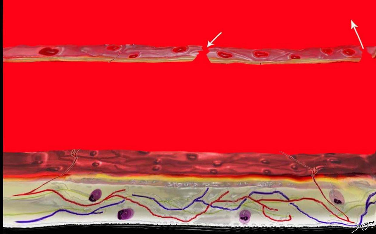
- Ulceration of an atheromatous plaque that
- extends deeply
- through the intima and
- into the aortic media
- may result in localized medial dissection
- may extend into the adventitia to form a pseudoaneurysm
- and is usually associated with a variable amount of hematoma
- distinguishing between
- aneurysm
- dissection
- ulceration is sometimes
- difficult,
- CT
- focal ulcer with adjacent
- subintimal hematoma and is often associated with
- aortic wall thickening or enhancement
-
42409c04La-b-c-scaled.jpg
Pathogenesis of Penetrating Diseases of the Aorta and Acute Aortic Syndrome
The lumen of the aorta with blood is demonstrated in red. The intima of the wall is the thin light pink layer just under the red luminal layer. The media or elastic layer is in teal, while the outer layer is a light yellow color
This diagram in a illustrates the normal aortic wall
Image b shows the development of a cholesterol plaque commonly associated with acute aortic syndrome.
Courtesy Ashley Davidoff MD
TheCommonVein.netInitially, atheromatous ulcers develop in patients with advanced atherosclerosis. At this stage, the lesions are usually asymptomatic and
- lesion confined to the intimal layer
- usually asymptomatic
-

Mural Hematoma
Ashley Davidoff
TheCommonVein.net
- extends deeply
- lesion progresses to the media
- usually symptomatic
-

Progressive Mural Hematoma
Ashley Davidoff
TheCommonVein.net
Penetrating Ulcer with Hematoma
Penetrating ulcer in the descending descending thoracic aorta (bright red ) with overlying hematoma (maroon) key words fx aortic ulcer fx atherosclerosis atheroma fx penetrating ulcer
acute aortic syndrome CTscan
48363
Courtesy Ashley Davidoff MD TheCommonVein.net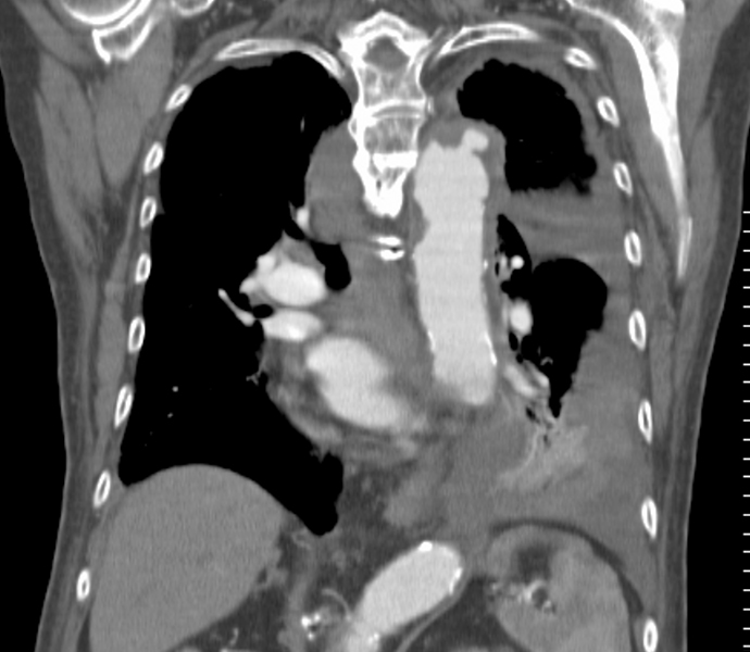
Penetrating ulcer in the descending descending thoracic aorta with overlying hematoma key words fx aortic ulcer fx atherosclerosis atheroma fx penetrating ulcer
acute aortic syndrome CTscan
Ashley Davidoff MD TheCommonVein.net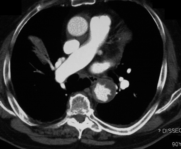
Penetrating Ulcer with Hematoma
Penetrating ulcer in the descending descending thoracic aorta with overlying hematoma in a patient with acute aortic syndrome
Courtesy Ashley Davidoff MD
TheCommonVein.net -
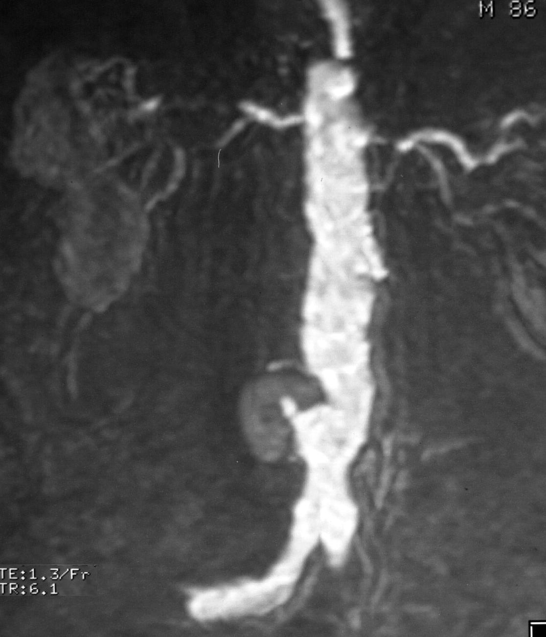
86 M with penetrating ulcer of the distal abdominal aorta associated with a hematoma
Ashley Davidoff
TheCommonVein.net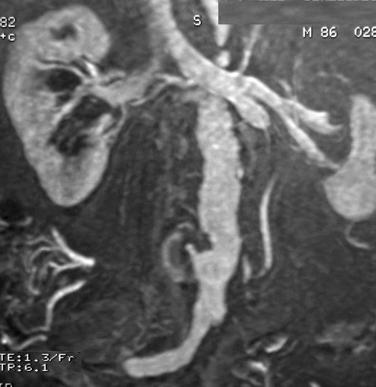
86 M with penetrating ulcer of the distal abdominal aorta associated with a hematoma
Ashley Davidoff
TheCommonVein.net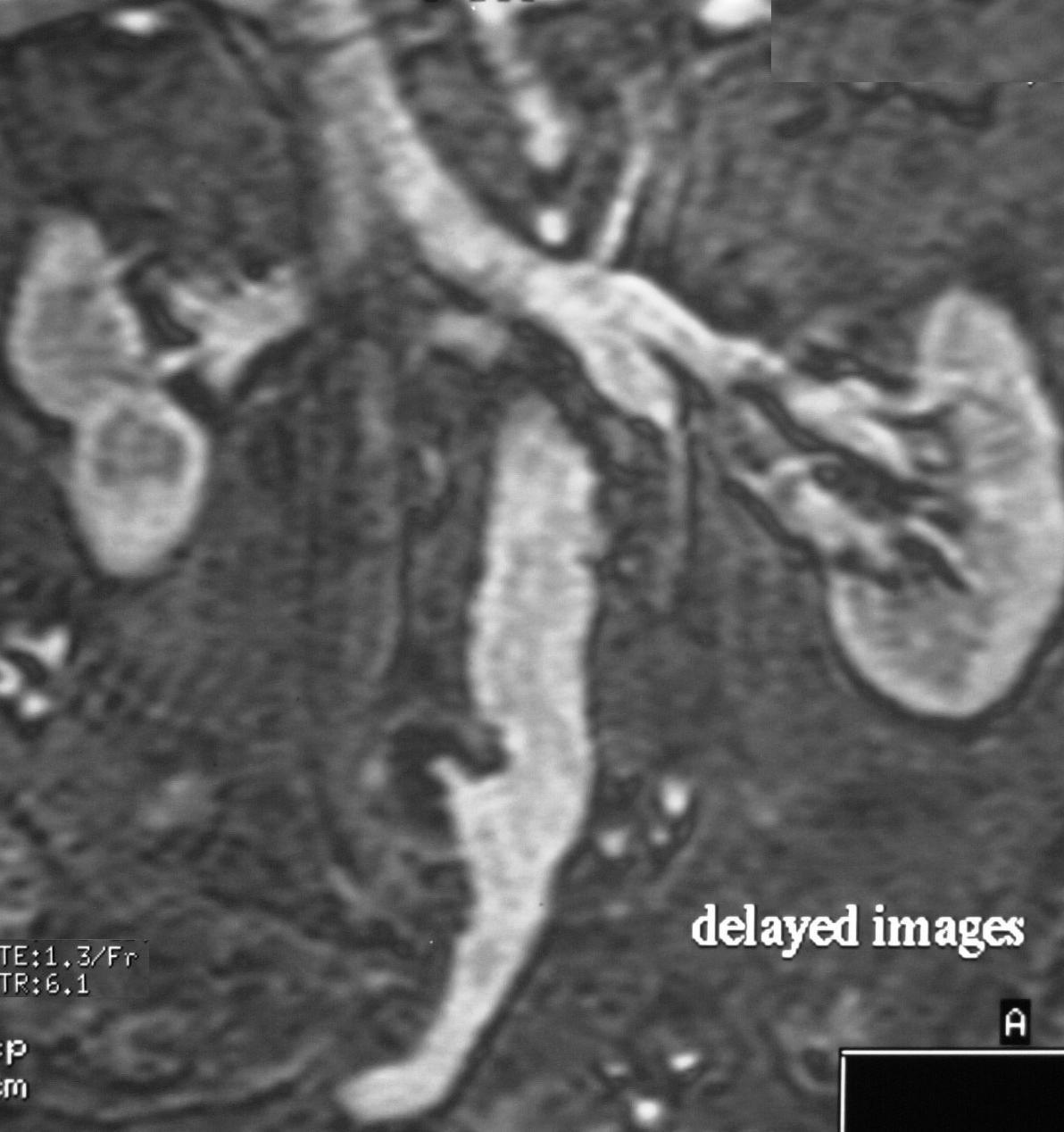
86 M with penetrating ulcer of the distal abdominal aorta associated with a hematoma
Ashley Davidoff
TheCommonVein.net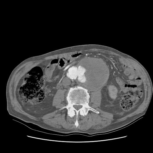
87 year old male with penetrating ulcer and expanding hematoma
in the descending descending thoracic aorta in a patient with acute aortic syndrome
Courtesy Ashley Davidoff MD TheCommonVein.net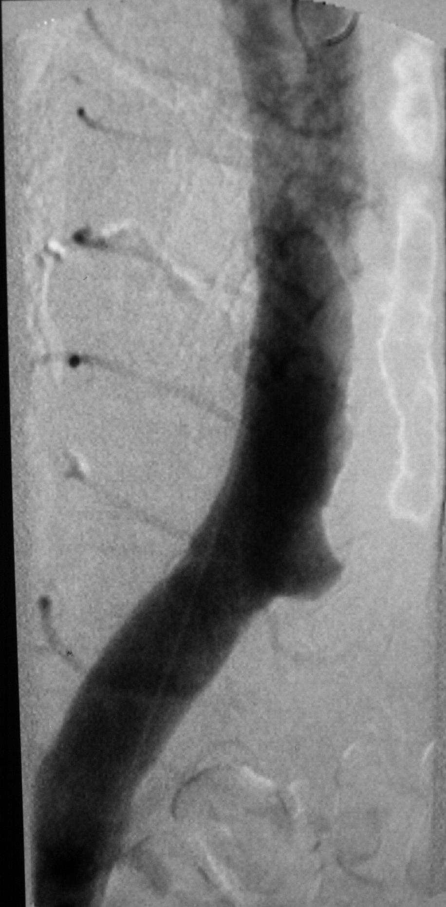
Angiogram of an atherosclerotic ulcer in the descending thoracic aorta on angiography
It is difficult to distinguish between an aneurysm of the aorta and ulcerated plaque
Ashley Davidoff
TheCommonVein.netMay focally dissect

Focal Dissection
A small focal dissection with flowing blood is seen, and this can either thrombose or progress to a full dissection
Ashley Davidoff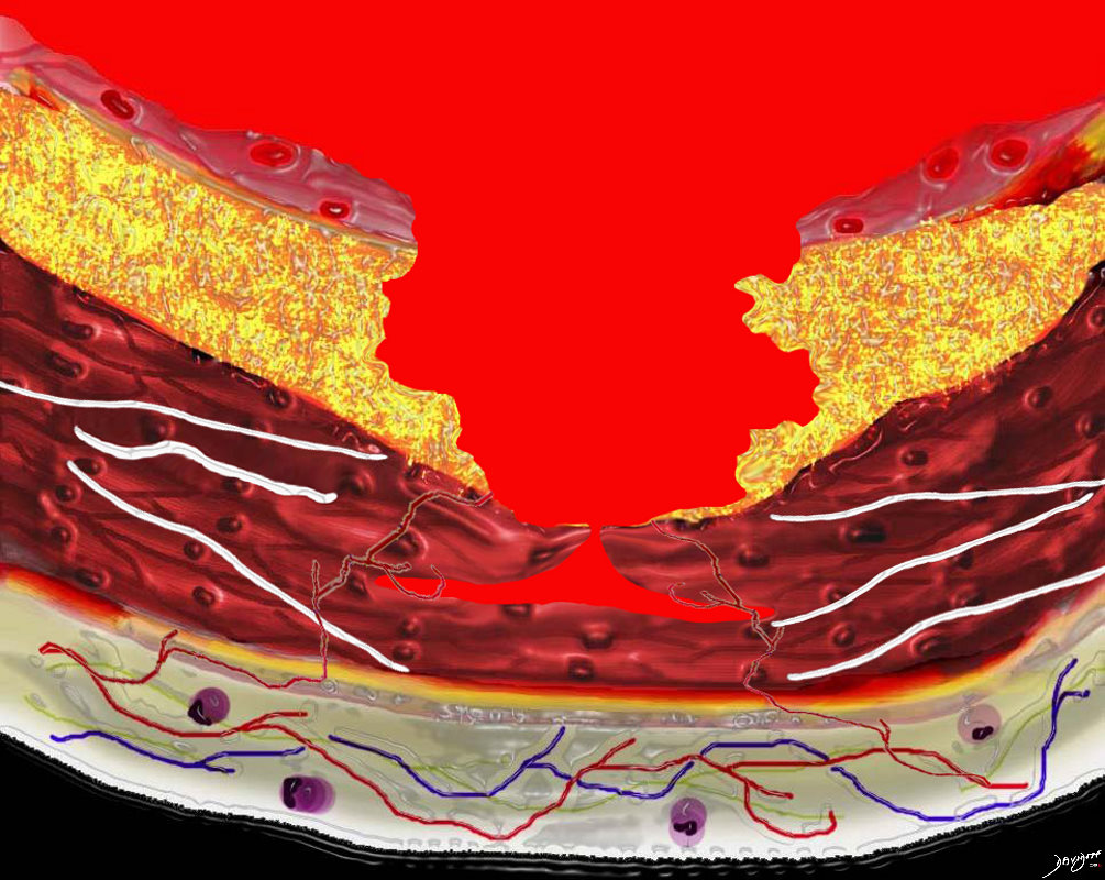
Focal Dissection
The penetrating ulcer can progress into the media and may have limited dissection due to the chronic inflammatory changes, including fibrosis in the media
Ashley Davidoff
TheCommonVein.net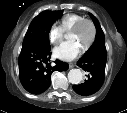
Limited Dissection .
Superiorly the aorta is mildly aneurysmal but no evidence of dissection
Ashley Davidoff MD
TheCommonVein.net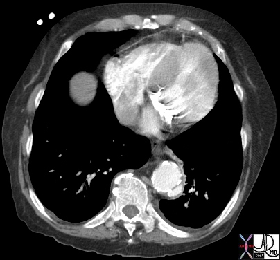
Limited Dissection .
Note the “flap ” of the dissection is thicker than the intimal flap of the classical dissection
Ashley Davidoff MD
TheCommonVein.net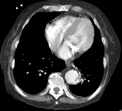
Limited Dissection .
Note the “flap ” of the dissection is thicker than the intimal flap of the classical dissection
Ashley Davidoff MD
TheCommonVein.net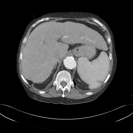
Inferiorly there is no evidence of dissection
Ashley Davidoff MD
TheCommonVein.netDissection could be progressive

Progressive Dissection
Progressive dissection with flowing blood is seen, and this can either thrombose or progress to a full dissection
Ashley Davidoff
TheCommonVein.net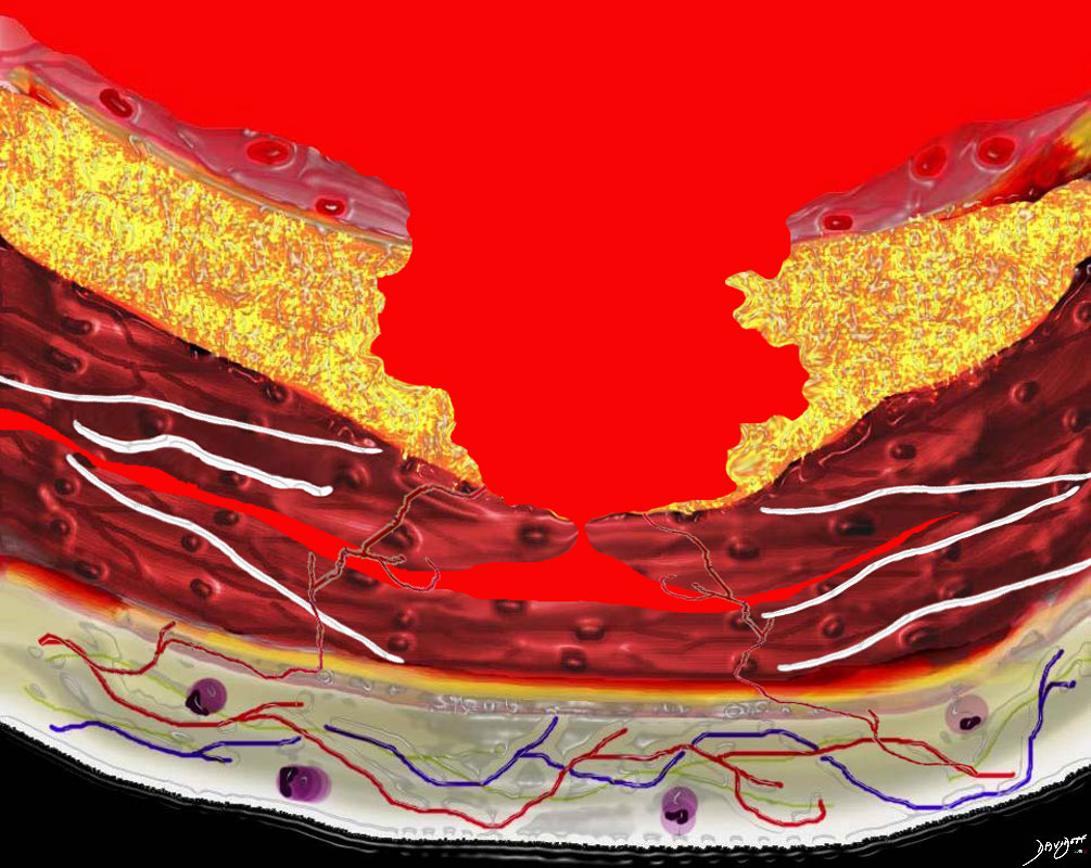
Acute Aotic Dissection in A Young Patient
In acute aortic dissecction, the event is usually in a younger patient without atherosclerotic disease and the associated inflammatory changes in the wall. Hence a small tear in the media can rapidly dissect antegrade or retrograde , with rentry points occurring
Ashley Davidoff
TheCommonVein.net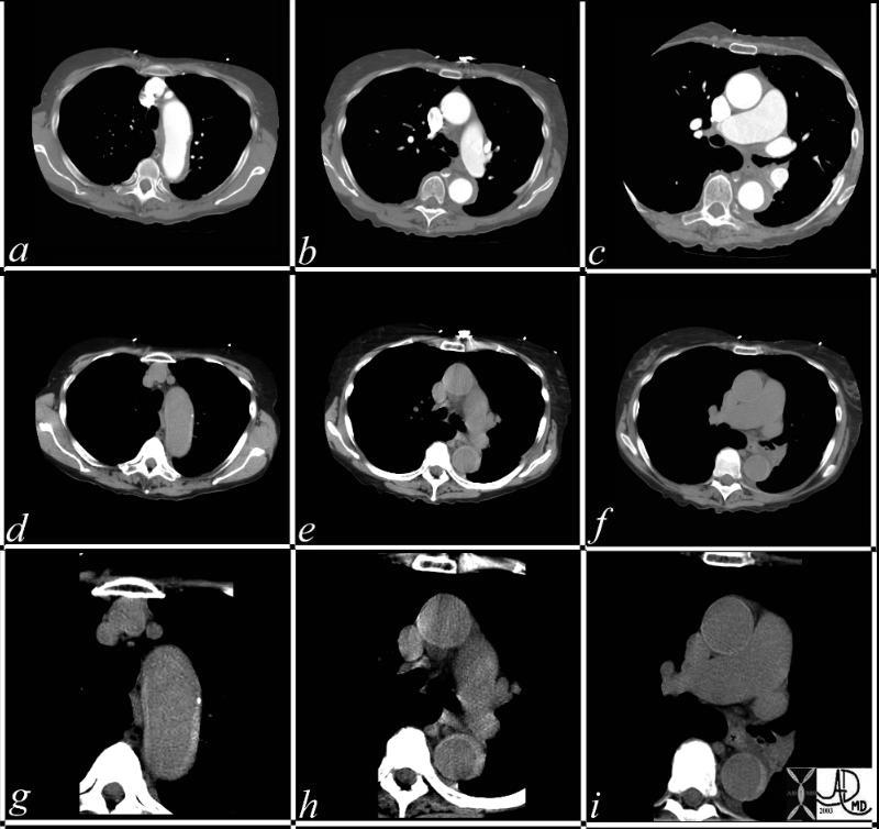
This series of CT scans were taken 1 day apart. The patient presented with chest pain and the soft tissue changes around contrast filled aorta (a,b,c) suggested a chronic dissection, or an acute thrombosed dissection. 1 day later the non-contrast CT clearly reveals an acute thrombosed dissection (d,e,f) that started in the arch and extended along the descending thoracic aorta. The last series (g,h,i) enhance the non contrats study. A non contrast CT followed by contrast injection, is an important tchnique to optimally characterise acute changes in this clincal setting. 36865c code CVS aorta thoracic arch descending dissection hyperdense cresecent sign acute thrombosed dissection 36865c new
Keywords:
CVS aorta thoracic arch descending dissection hyperdense cresecent sign acute thrombosed dissection 36865c new - Advance to the adventitia and form an aneurysm or pseudoaneurysm

Penetration to Adventitia
Ashley Davidoff
TheCommonVein.net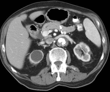
Expanding ulcer extending into the adventitia
Ashley Davidoff
TheCommonVein.net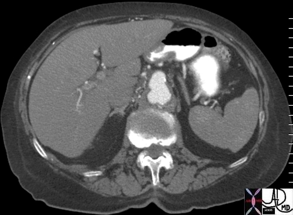
Expanding ulcer extending into the adventitia
Ashley Davidoff
TheCommonVein.net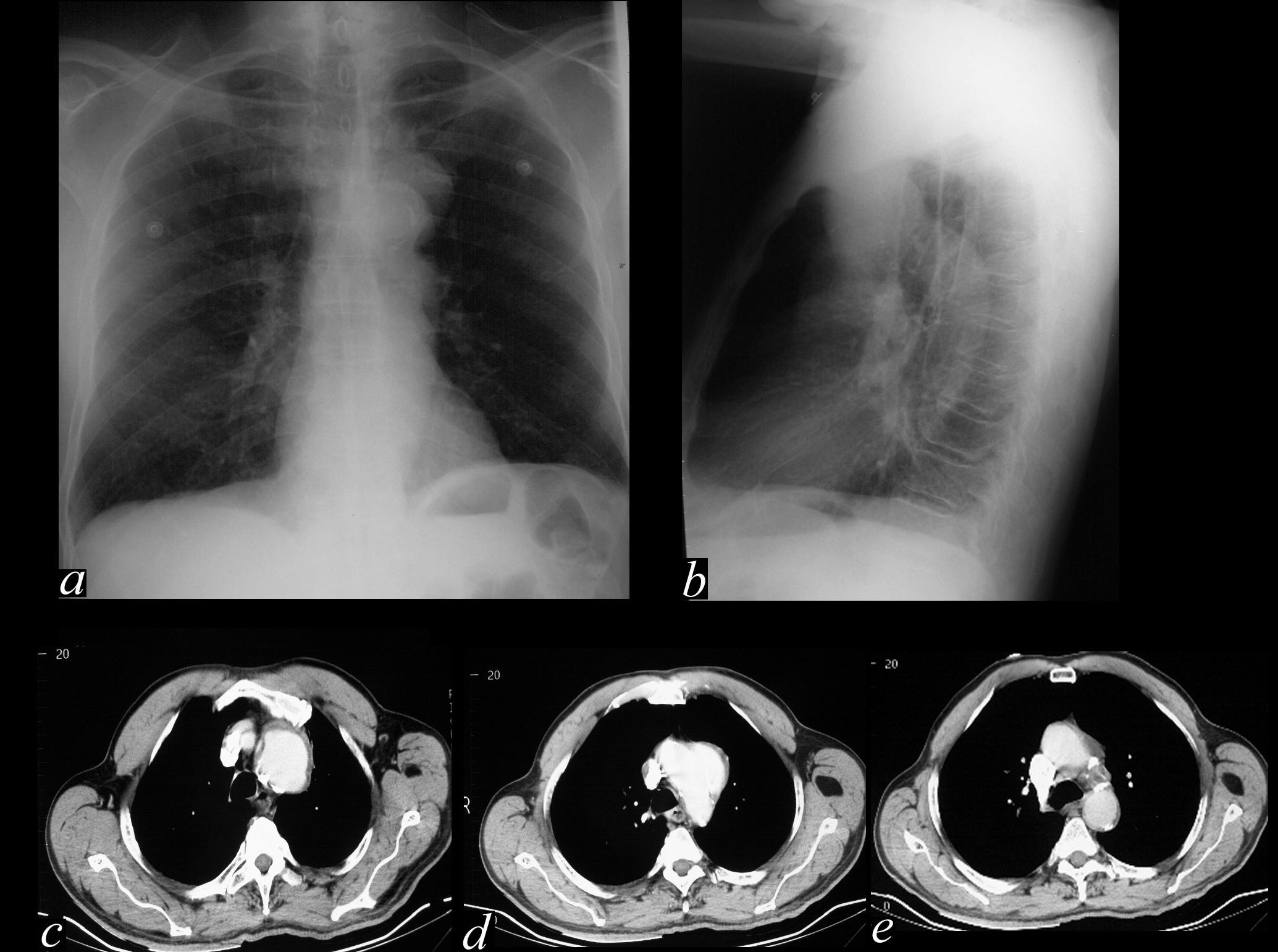
Expanding ulcer extending into the adventitia It is difficult to distinguish between an aneurysm of the aorta and expanding ulcerated plaque
Ashley Davidoff
TheCommonVein.net - Rupture
-

Rupture into the Mediastinum
Ashley Davidoff
TheCommonVein.net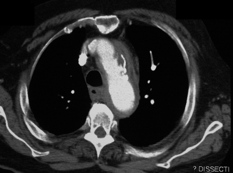
90 year old male with ulcerated plaque with rupture
Courtesy Ashley Davidoff MD TheCommonVein.net2nd Case
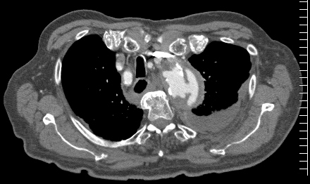
Ulcerated Plaque with Focal Dissection and Rupture into the Mediastinum
Courtesy Ashley Davidoff MD TheCommonVein.net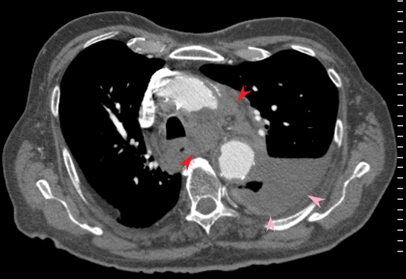
Rupture Rupture into the mediastinum (red arrow ) and complicated by a ipsilateral hemothorax (pink arrow)
Ashley Davidoff
TheCommonVein.net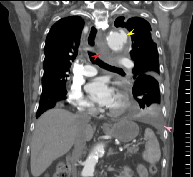
The focal disection (yellow arrow) into the wall and mediastinum (red arrow ) and complicated by a hemothorax (yellow arrow)
Ashley Davidoff TheCommonVein.net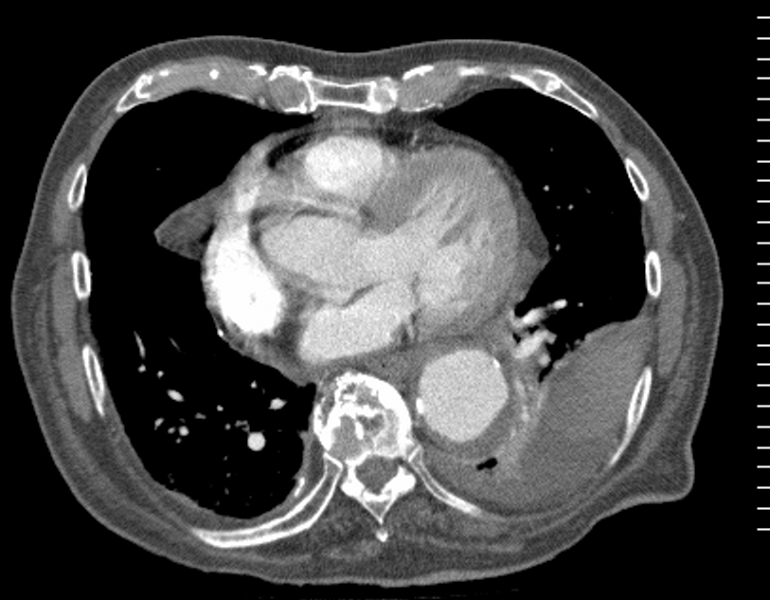
Hemothorax with clot in the left pleural space
Ashley Davidoff
TheCommonVein.net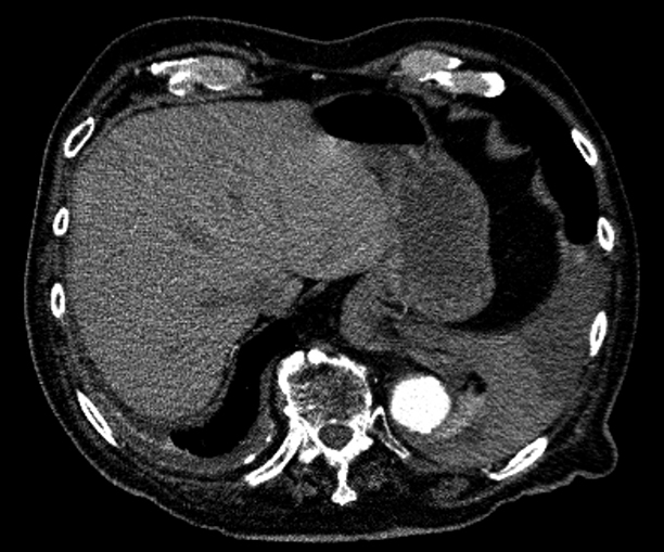
Hemothorax with clot in the left pleural space
Ashley Davidoff
TheCommonVein.net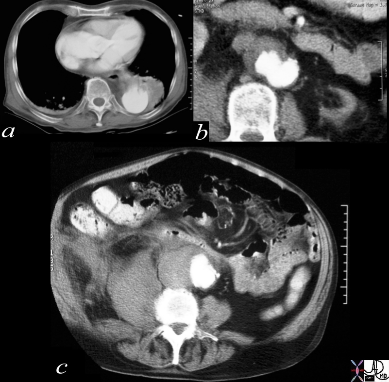
Penetrating Ulcer with Rupture
17529c01 artery descending thoracic aorta abdominal aorta dx rupture pseudoaneurysm ulcerating plaque mural hematoma ruptured through aortic wall hemorrhage hematoma retroperitoneum CTscan Courtesy Ashley Davidoff MD Ashley Davidoff MD
- Magnetic resonance imaging
- superior to conventional CT in
- differentiating
- acute intramural hematoma from
- atherosclerotic plaque and chronic intraluminal thrombus
Links and References
Cooke JP et al The Penetrating Ulcer
TCV

