DOMElement Object
(
    [schemaTypeInfo] =>
    [tagName] => table
    [firstElementChild] => (object value omitted)
    [lastElementChild] => (object value omitted)
    [childElementCount] => 1
    [previousElementSibling] => (object value omitted)
    [nextElementSibling] => (object value omitted)
    [nodeName] => table
    [nodeValue] =>


Ascending Aorta – Dilated and Calcified


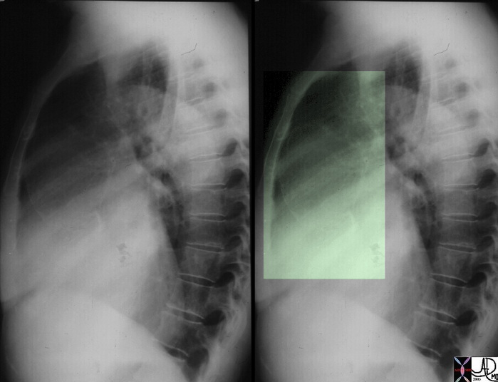
This lateral examination of the chest shows fine calcification in an ectatic ascending aorta associated with aortic annular calcification. Note the remarkable paucity of atherosclerotic change in the descending aorta. These findings are highly characteristic of tertiary syphilis of the aorta. Courtesy Ashley Davidoff MD. 00018c code CVS artery aorta ascending syphilis aneurysm calcification tortoise shell


    [nodeType] => 1
    [parentNode] => (object value omitted)
    [childNodes] => (object value omitted)
    [firstChild] => (object value omitted)
    [lastChild] => (object value omitted)
    [previousSibling] => (object value omitted)
    [nextSibling] => (object value omitted)
    [attributes] => (object value omitted)
    [ownerDocument] => (object value omitted)
    [namespaceURI] =>
    [prefix] =>
    [localName] => table
    [baseURI] =>
    [textContent] =>


Ascending Aorta – Dilated and Calcified


This lateral examination of the chest shows fine calcification in an ectatic ascending aorta associated with aortic annular calcification. Note the remarkable paucity of atherosclerotic change in the descending aorta. These findings are highly characteristic of tertiary syphilis of the aorta. Courtesy Ashley Davidoff MD. 00018c code CVS artery aorta ascending syphilis aneurysm calcification tortoise shell


)
DOMElement Object
(
    [schemaTypeInfo] =>
    [tagName] => td
    [firstElementChild] => (object value omitted)
    [lastElementChild] => (object value omitted)
    [childElementCount] => 1
    [previousElementSibling] =>
    [nextElementSibling] =>
    [nodeName] => td
    [nodeValue] => This lateral examination of the chest shows fine calcification in an ectatic ascending aorta associated with aortic annular calcification. Note the remarkable paucity of atherosclerotic change in the descending aorta. These findings are highly characteristic of tertiary syphilis of the aorta. Courtesy Ashley Davidoff MD. 00018c code CVS artery aorta ascending syphilis aneurysm calcification tortoise shell
    [nodeType] => 1
    [parentNode] => (object value omitted)
    [childNodes] => (object value omitted)
    [firstChild] => (object value omitted)
    [lastChild] => (object value omitted)
    [previousSibling] => (object value omitted)
    [nextSibling] => (object value omitted)
    [attributes] => (object value omitted)
    [ownerDocument] => (object value omitted)
    [namespaceURI] =>
    [prefix] =>
    [localName] => td
    [baseURI] =>
    [textContent] => This lateral examination of the chest shows fine calcification in an ectatic ascending aorta associated with aortic annular calcification. Note the remarkable paucity of atherosclerotic change in the descending aorta. These findings are highly characteristic of tertiary syphilis of the aorta. Courtesy Ashley Davidoff MD. 00018c code CVS artery aorta ascending syphilis aneurysm calcification tortoise shell
)
DOMElement Object
(
    [schemaTypeInfo] =>
    [tagName] => td
    [firstElementChild] => (object value omitted)
    [lastElementChild] => (object value omitted)
    [childElementCount] => 2
    [previousElementSibling] =>
    [nextElementSibling] =>
    [nodeName] => td
    [nodeValue] =>
Ascending Aorta – Dilated and Calcified

    [nodeType] => 1
    [parentNode] => (object value omitted)
    [childNodes] => (object value omitted)
    [firstChild] => (object value omitted)
    [lastChild] => (object value omitted)
    [previousSibling] => (object value omitted)
    [nextSibling] => (object value omitted)
    [attributes] => (object value omitted)
    [ownerDocument] => (object value omitted)
    [namespaceURI] =>
    [prefix] =>
    [localName] => td
    [baseURI] =>
    [textContent] =>
Ascending Aorta – Dilated and Calcified

)
DOMElement Object
(
    [schemaTypeInfo] =>
    [tagName] => table
    [firstElementChild] => (object value omitted)
    [lastElementChild] => (object value omitted)
    [childElementCount] => 1
    [previousElementSibling] => (object value omitted)
    [nextElementSibling] => (object value omitted)
    [nodeName] => table
    [nodeValue] =>


Mycotic Aneurysm


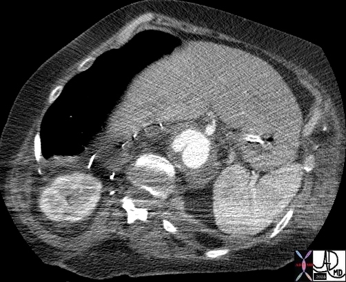
hx 68F with previous aortic surgery p/w back pain and fever aorta descending thoracic, thoracoabdominal fx periaortic fluid collection fx false aneurysm fx pseudoaneurysm fx paravertebral fluid collection bone vertebral body destruction dx mycotic aneurym with dx vertebral osteomyelitis CTscan
Courtesy Ashley Davidoff MD 44502


    [nodeType] => 1
    [parentNode] => (object value omitted)
    [childNodes] => (object value omitted)
    [firstChild] => (object value omitted)
    [lastChild] => (object value omitted)
    [previousSibling] => (object value omitted)
    [nextSibling] => (object value omitted)
    [attributes] => (object value omitted)
    [ownerDocument] => (object value omitted)
    [namespaceURI] =>
    [prefix] =>
    [localName] => table
    [baseURI] =>
    [textContent] =>


Mycotic Aneurysm


hx 68F with previous aortic surgery p/w back pain and fever aorta descending thoracic, thoracoabdominal fx periaortic fluid collection fx false aneurysm fx pseudoaneurysm fx paravertebral fluid collection bone vertebral body destruction dx mycotic aneurym with dx vertebral osteomyelitis CTscan
Courtesy Ashley Davidoff MD 44502


)
DOMElement Object
(
    [schemaTypeInfo] =>
    [tagName] => td
    [firstElementChild] => (object value omitted)
    [lastElementChild] => (object value omitted)
    [childElementCount] => 2
    [previousElementSibling] =>
    [nextElementSibling] =>
    [nodeName] => td
    [nodeValue] => hx 68F with previous aortic surgery p/w back pain and fever aorta descending thoracic, thoracoabdominal fx periaortic fluid collection fx false aneurysm fx pseudoaneurysm fx paravertebral fluid collection bone vertebral body destruction dx mycotic aneurym with dx vertebral osteomyelitis CTscan
Courtesy Ashley Davidoff MD 44502

    [nodeType] => 1
    [parentNode] => (object value omitted)
    [childNodes] => (object value omitted)
    [firstChild] => (object value omitted)
    [lastChild] => (object value omitted)
    [previousSibling] => (object value omitted)
    [nextSibling] => (object value omitted)
    [attributes] => (object value omitted)
    [ownerDocument] => (object value omitted)
    [namespaceURI] =>
    [prefix] =>
    [localName] => td
    [baseURI] =>
    [textContent] => hx 68F with previous aortic surgery p/w back pain and fever aorta descending thoracic, thoracoabdominal fx periaortic fluid collection fx false aneurysm fx pseudoaneurysm fx paravertebral fluid collection bone vertebral body destruction dx mycotic aneurym with dx vertebral osteomyelitis CTscan
Courtesy Ashley Davidoff MD 44502

)
DOMElement Object
(
    [schemaTypeInfo] =>
    [tagName] => td
    [firstElementChild] => (object value omitted)
    [lastElementChild] => (object value omitted)
    [childElementCount] => 2
    [previousElementSibling] =>
    [nextElementSibling] =>
    [nodeName] => td
    [nodeValue] =>
Mycotic Aneurysm

    [nodeType] => 1
    [parentNode] => (object value omitted)
    [childNodes] => (object value omitted)
    [firstChild] => (object value omitted)
    [lastChild] => (object value omitted)
    [previousSibling] => (object value omitted)
    [nextSibling] => (object value omitted)
    [attributes] => (object value omitted)
    [ownerDocument] => (object value omitted)
    [namespaceURI] =>
    [prefix] =>
    [localName] => td
    [baseURI] =>
    [textContent] =>
Mycotic Aneurysm

)
https://beta.thecommonvein.net/wp-content/uploads/2023/06/44502.jpg
http://thecommonvein.net/media/44502.JPG
DOMElement Object
(
    [schemaTypeInfo] =>
    [tagName] => table
    [firstElementChild] => (object value omitted)
    [lastElementChild] => (object value omitted)
    [childElementCount] => 1
    [previousElementSibling] => (object value omitted)
    [nextElementSibling] => (object value omitted)
    [nodeName] => table
    [nodeValue] =>


Mycotic Aneurysm with Osteomyelitis


This combination of images from a CTscan of the abdomen are of a middle aged man who presented with back pain and fever, with a remote history of AAA repair. The lateral scout film shows scalloping of vertebral bodies 2 and 3 (a) highighted in green overlay in b. The CTscan with soft tissue windows (c) and bone windows (d) show a complex fluid collection surrounding the aorta which proved to be a perigraft infection. Courtesy Ashley Davidoff MD. 22725c02 code CVS artery aorta abdomen abscess AA repair infection bone vertebra lumbar anterior scalloping


    [nodeType] => 1
    [parentNode] => (object value omitted)
    [childNodes] => (object value omitted)
    [firstChild] => (object value omitted)
    [lastChild] => (object value omitted)
    [previousSibling] => (object value omitted)
    [nextSibling] => (object value omitted)
    [attributes] => (object value omitted)
    [ownerDocument] => (object value omitted)
    [namespaceURI] =>
    [prefix] =>
    [localName] => table
    [baseURI] =>
    [textContent] =>


Mycotic Aneurysm with Osteomyelitis


This combination of images from a CTscan of the abdomen are of a middle aged man who presented with back pain and fever, with a remote history of AAA repair. The lateral scout film shows scalloping of vertebral bodies 2 and 3 (a) highighted in green overlay in b. The CTscan with soft tissue windows (c) and bone windows (d) show a complex fluid collection surrounding the aorta which proved to be a perigraft infection. Courtesy Ashley Davidoff MD. 22725c02 code CVS artery aorta abdomen abscess AA repair infection bone vertebra lumbar anterior scalloping


)
DOMElement Object
(
    [schemaTypeInfo] =>
    [tagName] => td
    [firstElementChild] => (object value omitted)
    [lastElementChild] => (object value omitted)
    [childElementCount] => 1
    [previousElementSibling] =>
    [nextElementSibling] =>
    [nodeName] => td
    [nodeValue] => This combination of images from a CTscan of the abdomen are of a middle aged man who presented with back pain and fever, with a remote history of AAA repair. The lateral scout film shows scalloping of vertebral bodies 2 and 3 (a) highighted in green overlay in b. The CTscan with soft tissue windows (c) and bone windows (d) show a complex fluid collection surrounding the aorta which proved to be a perigraft infection. Courtesy Ashley Davidoff MD. 22725c02 code CVS artery aorta abdomen abscess AA repair infection bone vertebra lumbar anterior scalloping
    [nodeType] => 1
    [parentNode] => (object value omitted)
    [childNodes] => (object value omitted)
    [firstChild] => (object value omitted)
    [lastChild] => (object value omitted)
    [previousSibling] => (object value omitted)
    [nextSibling] => (object value omitted)
    [attributes] => (object value omitted)
    [ownerDocument] => (object value omitted)
    [namespaceURI] =>
    [prefix] =>
    [localName] => td
    [baseURI] =>
    [textContent] => This combination of images from a CTscan of the abdomen are of a middle aged man who presented with back pain and fever, with a remote history of AAA repair. The lateral scout film shows scalloping of vertebral bodies 2 and 3 (a) highighted in green overlay in b. The CTscan with soft tissue windows (c) and bone windows (d) show a complex fluid collection surrounding the aorta which proved to be a perigraft infection. Courtesy Ashley Davidoff MD. 22725c02 code CVS artery aorta abdomen abscess AA repair infection bone vertebra lumbar anterior scalloping
)
DOMElement Object
(
    [schemaTypeInfo] =>
    [tagName] => td
    [firstElementChild] => (object value omitted)
    [lastElementChild] => (object value omitted)
    [childElementCount] => 2
    [previousElementSibling] =>
    [nextElementSibling] =>
    [nodeName] => td
    [nodeValue] =>
Mycotic Aneurysm with Osteomyelitis

    [nodeType] => 1
    [parentNode] => (object value omitted)
    [childNodes] => (object value omitted)
    [firstChild] => (object value omitted)
    [lastChild] => (object value omitted)
    [previousSibling] => (object value omitted)
    [nextSibling] => (object value omitted)
    [attributes] => (object value omitted)
    [ownerDocument] => (object value omitted)
    [namespaceURI] =>
    [prefix] =>
    [localName] => td
    [baseURI] =>
    [textContent] =>
Mycotic Aneurysm with Osteomyelitis

)
DOMElement Object
(
    [schemaTypeInfo] =>
    [tagName] => table
    [firstElementChild] => (object value omitted)
    [lastElementChild] => (object value omitted)
    [childElementCount] => 1
    [previousElementSibling] => (object value omitted)
    [nextElementSibling] => (object value omitted)
    [nodeName] => table
    [nodeValue] =>


Aortic Valve Vegetations


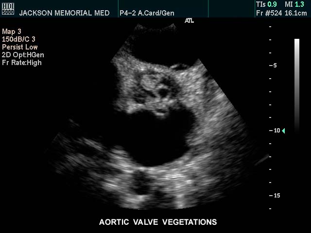
This gray scale echo of the heart showing a short-axis aorta left atrial view,, and demonstrating vegetations on the aortic valve. The patient has a diagnosis of bacterial endocarditis. Courtesy Philips Medical Systems 33126 code cardiac heart echo AO valve vegetations SBE bacterial endocarditis infection imaging cardiac echo


    [nodeType] => 1
    [parentNode] => (object value omitted)
    [childNodes] => (object value omitted)
    [firstChild] => (object value omitted)
    [lastChild] => (object value omitted)
    [previousSibling] => (object value omitted)
    [nextSibling] => (object value omitted)
    [attributes] => (object value omitted)
    [ownerDocument] => (object value omitted)
    [namespaceURI] =>
    [prefix] =>
    [localName] => table
    [baseURI] =>
    [textContent] =>


Aortic Valve Vegetations


This gray scale echo of the heart showing a short-axis aorta left atrial view,, and demonstrating vegetations on the aortic valve. The patient has a diagnosis of bacterial endocarditis. Courtesy Philips Medical Systems 33126 code cardiac heart echo AO valve vegetations SBE bacterial endocarditis infection imaging cardiac echo


)
DOMElement Object
(
    [schemaTypeInfo] =>
    [tagName] => td
    [firstElementChild] => (object value omitted)
    [lastElementChild] => (object value omitted)
    [childElementCount] => 1
    [previousElementSibling] =>
    [nextElementSibling] =>
    [nodeName] => td
    [nodeValue] => This gray scale echo of the heart showing a short-axis aorta left atrial view,, and demonstrating vegetations on the aortic valve. The patient has a diagnosis of bacterial endocarditis. Courtesy Philips Medical Systems 33126 code cardiac heart echo AO valve vegetations SBE bacterial endocarditis infection imaging cardiac echo
    [nodeType] => 1
    [parentNode] => (object value omitted)
    [childNodes] => (object value omitted)
    [firstChild] => (object value omitted)
    [lastChild] => (object value omitted)
    [previousSibling] => (object value omitted)
    [nextSibling] => (object value omitted)
    [attributes] => (object value omitted)
    [ownerDocument] => (object value omitted)
    [namespaceURI] =>
    [prefix] =>
    [localName] => td
    [baseURI] =>
    [textContent] => This gray scale echo of the heart showing a short-axis aorta left atrial view,, and demonstrating vegetations on the aortic valve. The patient has a diagnosis of bacterial endocarditis. Courtesy Philips Medical Systems 33126 code cardiac heart echo AO valve vegetations SBE bacterial endocarditis infection imaging cardiac echo
)
DOMElement Object
(
    [schemaTypeInfo] =>
    [tagName] => td
    [firstElementChild] => (object value omitted)
    [lastElementChild] => (object value omitted)
    [childElementCount] => 2
    [previousElementSibling] =>
    [nextElementSibling] =>
    [nodeName] => td
    [nodeValue] =>
Aortic Valve Vegetations

    [nodeType] => 1
    [parentNode] => (object value omitted)
    [childNodes] => (object value omitted)
    [firstChild] => (object value omitted)
    [lastChild] => (object value omitted)
    [previousSibling] => (object value omitted)
    [nextSibling] => (object value omitted)
    [attributes] => (object value omitted)
    [ownerDocument] => (object value omitted)
    [namespaceURI] =>
    [prefix] =>
    [localName] => td
    [baseURI] =>
    [textContent] =>
Aortic Valve Vegetations

)
DOMElement Object
(
    [schemaTypeInfo] =>
    [tagName] => table
    [firstElementChild] => (object value omitted)
    [lastElementChild] => (object value omitted)
    [childElementCount] => 1
    [previousElementSibling] =>
    [nextElementSibling] =>
    [nodeName] => table
    [nodeValue] =>


Aortic Infections
The Common Vein Copyright 2007
Ashley Davidoff MD
Cause
Aortic infection occurs through transmission of organisms via the vasa vasorum. Mycotic aneurysm, perigraft infection syphilis


Aortic Valve Vegetations


This gray scale echo of the heart showing a short-axis aorta left atrial view,, and demonstrating vegetations on the aortic valve. The patient has a diagnosis of bacterial endocarditis. Courtesy Philips Medical Systems 33126 code cardiac heart echo AO valve vegetations SBE bacterial endocarditis infection imaging cardiac echo


Mycotic Aneurysm with Osteomyelitis


This combination of images from a CTscan of the abdomen are of a middle aged man who presented with back pain and fever, with a remote history of AAA repair. The lateral scout film shows scalloping of vertebral bodies 2 and 3 (a) highighted in green overlay in b. The CTscan with soft tissue windows (c) and bone windows (d) show a complex fluid collection surrounding the aorta which proved to be a perigraft infection. Courtesy Ashley Davidoff MD. 22725c02 code CVS artery aorta abdomen abscess AA repair infection bone vertebra lumbar anterior scalloping


Mycotic Aneurysm


hx 68F with previous aortic surgery p/w back pain and fever aorta descending thoracic, thoracoabdominal fx periaortic fluid collection fx false aneurysm fx pseudoaneurysm fx paravertebral fluid collection bone vertebral body destruction dx mycotic aneurym with dx vertebral osteomyelitis CTscan
Courtesy Ashley Davidoff MD 44502


Screening?


Ascending Aorta – Dilated and Calcified


This lateral examination of the chest shows fine calcification in an ectatic ascending aorta associated with aortic annular calcification. Note the remarkable paucity of atherosclerotic change in the descending aorta. These findings are highly characteristic of tertiary syphilis of the aorta. Courtesy Ashley Davidoff MD. 00018c code CVS artery aorta ascending syphilis aneurysm calcification tortoise shell


Treatment


    [nodeType] => 1
    [parentNode] => (object value omitted)
    [childNodes] => (object value omitted)
    [firstChild] => (object value omitted)
    [lastChild] => (object value omitted)
    [previousSibling] =>
    [nextSibling] => (object value omitted)
    [attributes] => (object value omitted)
    [ownerDocument] => (object value omitted)
    [namespaceURI] =>
    [prefix] =>
    [localName] => table
    [baseURI] =>
    [textContent] =>


Aortic Infections
The Common Vein Copyright 2007
Ashley Davidoff MD
Cause
Aortic infection occurs through transmission of organisms via the vasa vasorum. Mycotic aneurysm, perigraft infection syphilis


Aortic Valve Vegetations


This gray scale echo of the heart showing a short-axis aorta left atrial view,, and demonstrating vegetations on the aortic valve. The patient has a diagnosis of bacterial endocarditis. Courtesy Philips Medical Systems 33126 code cardiac heart echo AO valve vegetations SBE bacterial endocarditis infection imaging cardiac echo


Mycotic Aneurysm with Osteomyelitis


This combination of images from a CTscan of the abdomen are of a middle aged man who presented with back pain and fever, with a remote history of AAA repair. The lateral scout film shows scalloping of vertebral bodies 2 and 3 (a) highighted in green overlay in b. The CTscan with soft tissue windows (c) and bone windows (d) show a complex fluid collection surrounding the aorta which proved to be a perigraft infection. Courtesy Ashley Davidoff MD. 22725c02 code CVS artery aorta abdomen abscess AA repair infection bone vertebra lumbar anterior scalloping


Mycotic Aneurysm


hx 68F with previous aortic surgery p/w back pain and fever aorta descending thoracic, thoracoabdominal fx periaortic fluid collection fx false aneurysm fx pseudoaneurysm fx paravertebral fluid collection bone vertebral body destruction dx mycotic aneurym with dx vertebral osteomyelitis CTscan
Courtesy Ashley Davidoff MD 44502


Screening?


Ascending Aorta – Dilated and Calcified


This lateral examination of the chest shows fine calcification in an ectatic ascending aorta associated with aortic annular calcification. Note the remarkable paucity of atherosclerotic change in the descending aorta. These findings are highly characteristic of tertiary syphilis of the aorta. Courtesy Ashley Davidoff MD. 00018c code CVS artery aorta ascending syphilis aneurysm calcification tortoise shell


Treatment


)
DOMElement Object
(
    [schemaTypeInfo] =>
    [tagName] => td
    [firstElementChild] => (object value omitted)
    [lastElementChild] => (object value omitted)
    [childElementCount] => 1
    [previousElementSibling] =>
    [nextElementSibling] =>
    [nodeName] => td
    [nodeValue] => This lateral examination of the chest shows fine calcification in an ectatic ascending aorta associated with aortic annular calcification. Note the remarkable paucity of atherosclerotic change in the descending aorta. These findings are highly characteristic of tertiary syphilis of the aorta. Courtesy Ashley Davidoff MD. 00018c code CVS artery aorta ascending syphilis aneurysm calcification tortoise shell
    [nodeType] => 1
    [parentNode] => (object value omitted)
    [childNodes] => (object value omitted)
    [firstChild] => (object value omitted)
    [lastChild] => (object value omitted)
    [previousSibling] => (object value omitted)
    [nextSibling] => (object value omitted)
    [attributes] => (object value omitted)
    [ownerDocument] => (object value omitted)
    [namespaceURI] =>
    [prefix] =>
    [localName] => td
    [baseURI] =>
    [textContent] => This lateral examination of the chest shows fine calcification in an ectatic ascending aorta associated with aortic annular calcification. Note the remarkable paucity of atherosclerotic change in the descending aorta. These findings are highly characteristic of tertiary syphilis of the aorta. Courtesy Ashley Davidoff MD. 00018c code CVS artery aorta ascending syphilis aneurysm calcification tortoise shell
)
DOMElement Object
(
    [schemaTypeInfo] =>
    [tagName] => td
    [firstElementChild] => (object value omitted)
    [lastElementChild] => (object value omitted)
    [childElementCount] => 2
    [previousElementSibling] =>
    [nextElementSibling] =>
    [nodeName] => td
    [nodeValue] =>
Ascending Aorta – Dilated and Calcified

    [nodeType] => 1
    [parentNode] => (object value omitted)
    [childNodes] => (object value omitted)
    [firstChild] => (object value omitted)
    [lastChild] => (object value omitted)
    [previousSibling] => (object value omitted)
    [nextSibling] => (object value omitted)
    [attributes] => (object value omitted)
    [ownerDocument] => (object value omitted)
    [namespaceURI] =>
    [prefix] =>
    [localName] => td
    [baseURI] =>
    [textContent] =>
Ascending Aorta – Dilated and Calcified

)
DOMElement Object
(
    [schemaTypeInfo] =>
    [tagName] => td
    [firstElementChild] => (object value omitted)
    [lastElementChild] => (object value omitted)
    [childElementCount] => 2
    [previousElementSibling] =>
    [nextElementSibling] =>
    [nodeName] => td
    [nodeValue] => hx 68F with previous aortic surgery p/w back pain and fever aorta descending thoracic, thoracoabdominal fx periaortic fluid collection fx false aneurysm fx pseudoaneurysm fx paravertebral fluid collection bone vertebral body destruction dx mycotic aneurym with dx vertebral osteomyelitis CTscan
Courtesy Ashley Davidoff MD 44502

    [nodeType] => 1
    [parentNode] => (object value omitted)
    [childNodes] => (object value omitted)
    [firstChild] => (object value omitted)
    [lastChild] => (object value omitted)
    [previousSibling] => (object value omitted)
    [nextSibling] => (object value omitted)
    [attributes] => (object value omitted)
    [ownerDocument] => (object value omitted)
    [namespaceURI] =>
    [prefix] =>
    [localName] => td
    [baseURI] =>
    [textContent] => hx 68F with previous aortic surgery p/w back pain and fever aorta descending thoracic, thoracoabdominal fx periaortic fluid collection fx false aneurysm fx pseudoaneurysm fx paravertebral fluid collection bone vertebral body destruction dx mycotic aneurym with dx vertebral osteomyelitis CTscan
Courtesy Ashley Davidoff MD 44502

)
https://beta.thecommonvein.net/wp-content/uploads/2023/04/00018c.jpg
DOMElement Object
(
    [schemaTypeInfo] =>
    [tagName] => td
    [firstElementChild] => (object value omitted)
    [lastElementChild] => (object value omitted)
    [childElementCount] => 3
    [previousElementSibling] =>
    [nextElementSibling] =>
    [nodeName] => td
    [nodeValue] =>
Mycotic Aneurysm

    [nodeType] => 1
    [parentNode] => (object value omitted)
    [childNodes] => (object value omitted)
    [firstChild] => (object value omitted)
    [lastChild] => (object value omitted)
    [previousSibling] => (object value omitted)
    [nextSibling] => (object value omitted)
    [attributes] => (object value omitted)
    [ownerDocument] => (object value omitted)
    [namespaceURI] =>
    [prefix] =>
    [localName] => td
    [baseURI] =>
    [textContent] =>
Mycotic Aneurysm

)
https://beta.thecommonvein.net/wp-content/uploads/2023/04/00018c.jpg
https://beta.thecommonvein.net/wp-content/uploads/2023/06/44502.jpg http://thecommonvein.net/media/44502.JPG
DOMElement Object
(
    [schemaTypeInfo] =>
    [tagName] => td
    [firstElementChild] => (object value omitted)
    [lastElementChild] => (object value omitted)
    [childElementCount] => 1
    [previousElementSibling] =>
    [nextElementSibling] =>
    [nodeName] => td
    [nodeValue] => This combination of images from a CTscan of the abdomen are of a middle aged man who presented with back pain and fever, with a remote history of AAA repair. The lateral scout film shows scalloping of vertebral bodies 2 and 3 (a) highighted in green overlay in b. The CTscan with soft tissue windows (c) and bone windows (d) show a complex fluid collection surrounding the aorta which proved to be a perigraft infection. Courtesy Ashley Davidoff MD. 22725c02 code CVS artery aorta abdomen abscess AA repair infection bone vertebra lumbar anterior scalloping
    [nodeType] => 1
    [parentNode] => (object value omitted)
    [childNodes] => (object value omitted)
    [firstChild] => (object value omitted)
    [lastChild] => (object value omitted)
    [previousSibling] => (object value omitted)
    [nextSibling] => (object value omitted)
    [attributes] => (object value omitted)
    [ownerDocument] => (object value omitted)
    [namespaceURI] =>
    [prefix] =>
    [localName] => td
    [baseURI] =>
    [textContent] => This combination of images from a CTscan of the abdomen are of a middle aged man who presented with back pain and fever, with a remote history of AAA repair. The lateral scout film shows scalloping of vertebral bodies 2 and 3 (a) highighted in green overlay in b. The CTscan with soft tissue windows (c) and bone windows (d) show a complex fluid collection surrounding the aorta which proved to be a perigraft infection. Courtesy Ashley Davidoff MD. 22725c02 code CVS artery aorta abdomen abscess AA repair infection bone vertebra lumbar anterior scalloping
)
https://beta.thecommonvein.net/wp-content/uploads/2023/04/00018c.jpg
DOMElement Object
(
    [schemaTypeInfo] =>
    [tagName] => td
    [firstElementChild] => (object value omitted)
    [lastElementChild] => (object value omitted)
    [childElementCount] => 2
    [previousElementSibling] =>
    [nextElementSibling] =>
    [nodeName] => td
    [nodeValue] =>
Mycotic Aneurysm with Osteomyelitis

    [nodeType] => 1
    [parentNode] => (object value omitted)
    [childNodes] => (object value omitted)
    [firstChild] => (object value omitted)
    [lastChild] => (object value omitted)
    [previousSibling] => (object value omitted)
    [nextSibling] => (object value omitted)
    [attributes] => (object value omitted)
    [ownerDocument] => (object value omitted)
    [namespaceURI] =>
    [prefix] =>
    [localName] => td
    [baseURI] =>
    [textContent] =>
Mycotic Aneurysm with Osteomyelitis

)
https://beta.thecommonvein.net/wp-content/uploads/2023/04/00018c.jpg
http://thecommonvein.net/media/22725c02.jpg
DOMElement Object
(
    [schemaTypeInfo] =>
    [tagName] => td
    [firstElementChild] => (object value omitted)
    [lastElementChild] => (object value omitted)
    [childElementCount] => 1
    [previousElementSibling] =>
    [nextElementSibling] =>
    [nodeName] => td
    [nodeValue] => This gray scale echo of the heart showing a short-axis aorta left atrial view,, and demonstrating vegetations on the aortic valve. The patient has a diagnosis of bacterial endocarditis. Courtesy Philips Medical Systems 33126 code cardiac heart echo AO valve vegetations SBE bacterial endocarditis infection imaging cardiac echo
    [nodeType] => 1
    [parentNode] => (object value omitted)
    [childNodes] => (object value omitted)
    [firstChild] => (object value omitted)
    [lastChild] => (object value omitted)
    [previousSibling] => (object value omitted)
    [nextSibling] => (object value omitted)
    [attributes] => (object value omitted)
    [ownerDocument] => (object value omitted)
    [namespaceURI] =>
    [prefix] =>
    [localName] => td
    [baseURI] =>
    [textContent] => This gray scale echo of the heart showing a short-axis aorta left atrial view,, and demonstrating vegetations on the aortic valve. The patient has a diagnosis of bacterial endocarditis. Courtesy Philips Medical Systems 33126 code cardiac heart echo AO valve vegetations SBE bacterial endocarditis infection imaging cardiac echo
)
https://beta.thecommonvein.net/wp-content/uploads/2023/04/00018c.jpg
DOMElement Object
(
    [schemaTypeInfo] =>
    [tagName] => td
    [firstElementChild] => (object value omitted)
    [lastElementChild] => (object value omitted)
    [childElementCount] => 2
    [previousElementSibling] =>
    [nextElementSibling] =>
    [nodeName] => td
    [nodeValue] =>
Aortic Valve Vegetations

    [nodeType] => 1
    [parentNode] => (object value omitted)
    [childNodes] => (object value omitted)
    [firstChild] => (object value omitted)
    [lastChild] => (object value omitted)
    [previousSibling] => (object value omitted)
    [nextSibling] => (object value omitted)
    [attributes] => (object value omitted)
    [ownerDocument] => (object value omitted)
    [namespaceURI] =>
    [prefix] =>
    [localName] => td
    [baseURI] =>
    [textContent] =>
Aortic Valve Vegetations

)
https://beta.thecommonvein.net/wp-content/uploads/2023/04/00018c.jpg
http://thecommonvein.net/media/33126.jpg
DOMElement Object
(
    [schemaTypeInfo] =>
    [tagName] => td
    [firstElementChild] => (object value omitted)
    [lastElementChild] => (object value omitted)
    [childElementCount] => 13
    [previousElementSibling] =>
    [nextElementSibling] =>
    [nodeName] => td
    [nodeValue] =>
Aortic Infections
The Common Vein Copyright 2007
Ashley Davidoff MD
Cause
Aortic infection occurs through transmission of organisms via the vasa vasorum. Mycotic aneurysm, perigraft infection syphilis


Aortic Valve Vegetations


This gray scale echo of the heart showing a short-axis aorta left atrial view,, and demonstrating vegetations on the aortic valve. The patient has a diagnosis of bacterial endocarditis. Courtesy Philips Medical Systems 33126 code cardiac heart echo AO valve vegetations SBE bacterial endocarditis infection imaging cardiac echo


Mycotic Aneurysm with Osteomyelitis


This combination of images from a CTscan of the abdomen are of a middle aged man who presented with back pain and fever, with a remote history of AAA repair. The lateral scout film shows scalloping of vertebral bodies 2 and 3 (a) highighted in green overlay in b. The CTscan with soft tissue windows (c) and bone windows (d) show a complex fluid collection surrounding the aorta which proved to be a perigraft infection. Courtesy Ashley Davidoff MD. 22725c02 code CVS artery aorta abdomen abscess AA repair infection bone vertebra lumbar anterior scalloping


Mycotic Aneurysm


hx 68F with previous aortic surgery p/w back pain and fever aorta descending thoracic, thoracoabdominal fx periaortic fluid collection fx false aneurysm fx pseudoaneurysm fx paravertebral fluid collection bone vertebral body destruction dx mycotic aneurym with dx vertebral osteomyelitis CTscan
Courtesy Ashley Davidoff MD 44502


Screening?


Ascending Aorta – Dilated and Calcified


This lateral examination of the chest shows fine calcification in an ectatic ascending aorta associated with aortic annular calcification. Note the remarkable paucity of atherosclerotic change in the descending aorta. These findings are highly characteristic of tertiary syphilis of the aorta. Courtesy Ashley Davidoff MD. 00018c code CVS artery aorta ascending syphilis aneurysm calcification tortoise shell


Treatment

    [nodeType] => 1
    [parentNode] => (object value omitted)
    [childNodes] => (object value omitted)
    [firstChild] => (object value omitted)
    [lastChild] => (object value omitted)
    [previousSibling] => (object value omitted)
    [nextSibling] => (object value omitted)
    [attributes] => (object value omitted)
    [ownerDocument] => (object value omitted)
    [namespaceURI] =>
    [prefix] =>
    [localName] => td
    [baseURI] =>
    [textContent] =>
Aortic Infections
The Common Vein Copyright 2007
Ashley Davidoff MD
Cause
Aortic infection occurs through transmission of organisms via the vasa vasorum. Mycotic aneurysm, perigraft infection syphilis


Aortic Valve Vegetations


This gray scale echo of the heart showing a short-axis aorta left atrial view,, and demonstrating vegetations on the aortic valve. The patient has a diagnosis of bacterial endocarditis. Courtesy Philips Medical Systems 33126 code cardiac heart echo AO valve vegetations SBE bacterial endocarditis infection imaging cardiac echo


Mycotic Aneurysm with Osteomyelitis


This combination of images from a CTscan of the abdomen are of a middle aged man who presented with back pain and fever, with a remote history of AAA repair. The lateral scout film shows scalloping of vertebral bodies 2 and 3 (a) highighted in green overlay in b. The CTscan with soft tissue windows (c) and bone windows (d) show a complex fluid collection surrounding the aorta which proved to be a perigraft infection. Courtesy Ashley Davidoff MD. 22725c02 code CVS artery aorta abdomen abscess AA repair infection bone vertebra lumbar anterior scalloping


Mycotic Aneurysm


hx 68F with previous aortic surgery p/w back pain and fever aorta descending thoracic, thoracoabdominal fx periaortic fluid collection fx false aneurysm fx pseudoaneurysm fx paravertebral fluid collection bone vertebral body destruction dx mycotic aneurym with dx vertebral osteomyelitis CTscan
Courtesy Ashley Davidoff MD 44502


Screening?


Ascending Aorta – Dilated and Calcified


This lateral examination of the chest shows fine calcification in an ectatic ascending aorta associated with aortic annular calcification. Note the remarkable paucity of atherosclerotic change in the descending aorta. These findings are highly characteristic of tertiary syphilis of the aorta. Courtesy Ashley Davidoff MD. 00018c code CVS artery aorta ascending syphilis aneurysm calcification tortoise shell


Treatment

)


