A Marriage Made In Heaven –
The story of Mr. Aortic and Miss Mitral
Ashley Davidoff MD Copyright 2007
The attraction of Mr. Aortic to Miss Mitral
goes back a long long way
As far back as fetal times ?
and in fact even beyond in evolution
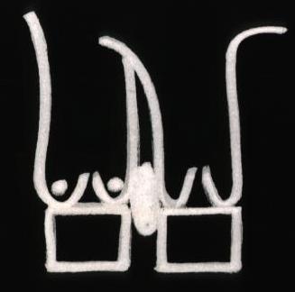
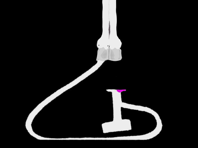
Aorta and Pulmonary Artery Side by Side |
| 01806 heart cardiac bilateral conus outflow tract infundibulum aorta pulmonary artery D-loop anatomy embryology Courtesy Ashley Davidoff drawing |
When the valve of Mr. Aortic eyed the developing Miss Mitral
He knew from that moment and on
That she was to be his bride
And that no other would come between them
He had to leave his fetal home
A throne shared with his twin Miss Pulmonary
And he knew that they would now move in different circles
Each to go their own way
His mission was unrelenting
As he continued down on his trip
Her anterior leaflet was his goal
As he flapped to the Lub of his master
He had a chance with her twin Tricuspid
She was pretty too
But Miss Mitral was the twinkle of his eye
Since her flap was in synch with his
And on a glorious day one summer
They bonded in fibrous union
In ecstasy they danced together
And never could be parted
From then on they lived their life
In a beautiful pulsatile glory
She gave him all that she received
and he in turn delivered ?
DOMElement Object
(
    [schemaTypeInfo] =>
    [tagName] => table
    [firstElementChild] => (object value omitted)
    [lastElementChild] => (object value omitted)
    [childElementCount] => 1
    [previousElementSibling] => (object value omitted)
    [nextElementSibling] => (object value omitted)
    [nodeName] => table
    [nodeValue] =>


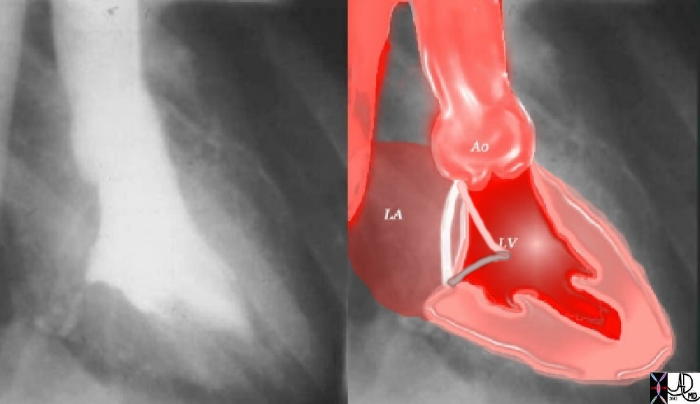
Prolapse of the Mitral Valve and Aortic Regurgitation  – Marfan’s Syndrome


This angiogram in RAO projection shows prolapse of both the anterior and posterior leaflets of the mitral valve with mitral regurgitation. Note also the bulbous appearance of the aortic sinuses. The back wall of the left atrium is flattened against the spine suggesting a narrow A-P diameter or pectus excavatum. These findings are characteristic of Maran’s syndrome Courtesy Ashley DAvidoff MD. 34810c code cardiac heart mitral valve MV anterior leaflet posterior leaflet prolapse mitral regurgitation MR incompetence aorta Marfan’s syndrome floppy mitral valve angiography LA shape MVP imaging radiology angiography disease overlay radiologists and detectives


    [nodeType] => 1
    [parentNode] => (object value omitted)
    [childNodes] => (object value omitted)
    [firstChild] => (object value omitted)
    [lastChild] => (object value omitted)
    [previousSibling] => (object value omitted)
    [nextSibling] => (object value omitted)
    [attributes] => (object value omitted)
    [ownerDocument] => (object value omitted)
    [namespaceURI] =>
    [prefix] =>
    [localName] => table
    [baseURI] =>
    [textContent] =>


Prolapse of the Mitral Valve and Aortic Regurgitation  – Marfan’s Syndrome


This angiogram in RAO projection shows prolapse of both the anterior and posterior leaflets of the mitral valve with mitral regurgitation. Note also the bulbous appearance of the aortic sinuses. The back wall of the left atrium is flattened against the spine suggesting a narrow A-P diameter or pectus excavatum. These findings are characteristic of Maran’s syndrome Courtesy Ashley DAvidoff MD. 34810c code cardiac heart mitral valve MV anterior leaflet posterior leaflet prolapse mitral regurgitation MR incompetence aorta Marfan’s syndrome floppy mitral valve angiography LA shape MVP imaging radiology angiography disease overlay radiologists and detectives


)
DOMElement Object
(
    [schemaTypeInfo] =>
    [tagName] => td
    [firstElementChild] =>
    [lastElementChild] =>
    [childElementCount] => 0
    [previousElementSibling] =>
    [nextElementSibling] =>
    [nodeName] => td
    [nodeValue] => This angiogram in RAO projection shows prolapse of both the anterior and posterior leaflets of the mitral valve with mitral regurgitation. Note also the bulbous appearance of the aortic sinuses. The back wall of the left atrium is flattened against the spine suggesting a narrow A-P diameter or pectus excavatum. These findings are characteristic of Maran’s syndrome Courtesy Ashley DAvidoff MD. 34810c code cardiac heart mitral valve MV anterior leaflet posterior leaflet prolapse mitral regurgitation MR incompetence aorta Marfan’s syndrome floppy mitral valve angiography LA shape MVP imaging radiology angiography disease overlay radiologists and detectives
    [nodeType] => 1
    [parentNode] => (object value omitted)
    [childNodes] => (object value omitted)
    [firstChild] => (object value omitted)
    [lastChild] => (object value omitted)
    [previousSibling] => (object value omitted)
    [nextSibling] => (object value omitted)
    [attributes] => (object value omitted)
    [ownerDocument] => (object value omitted)
    [namespaceURI] =>
    [prefix] =>
    [localName] => td
    [baseURI] =>
    [textContent] => This angiogram in RAO projection shows prolapse of both the anterior and posterior leaflets of the mitral valve with mitral regurgitation. Note also the bulbous appearance of the aortic sinuses. The back wall of the left atrium is flattened against the spine suggesting a narrow A-P diameter or pectus excavatum. These findings are characteristic of Maran’s syndrome Courtesy Ashley DAvidoff MD. 34810c code cardiac heart mitral valve MV anterior leaflet posterior leaflet prolapse mitral regurgitation MR incompetence aorta Marfan’s syndrome floppy mitral valve angiography LA shape MVP imaging radiology angiography disease overlay radiologists and detectives
)
DOMElement Object
(
    [schemaTypeInfo] =>
    [tagName] => td
    [firstElementChild] => (object value omitted)
    [lastElementChild] => (object value omitted)
    [childElementCount] => 1
    [previousElementSibling] =>
    [nextElementSibling] =>
    [nodeName] => td
    [nodeValue] => Prolapse of the Mitral Valve and Aortic Regurgitation  – Marfan’s Syndrome
    [nodeType] => 1
    [parentNode] => (object value omitted)
    [childNodes] => (object value omitted)
    [firstChild] => (object value omitted)
    [lastChild] => (object value omitted)
    [previousSibling] => (object value omitted)
    [nextSibling] => (object value omitted)
    [attributes] => (object value omitted)
    [ownerDocument] => (object value omitted)
    [namespaceURI] =>
    [prefix] =>
    [localName] => td
    [baseURI] =>
    [textContent] => Prolapse of the Mitral Valve and Aortic Regurgitation  – Marfan’s Syndrome
)
DOMElement Object
(
    [schemaTypeInfo] =>
    [tagName] => table
    [firstElementChild] => (object value omitted)
    [lastElementChild] => (object value omitted)
    [childElementCount] => 1
    [previousElementSibling] => (object value omitted)
    [nextElementSibling] => (object value omitted)
    [nodeName] => table
    [nodeValue] =>


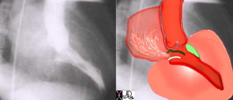
IHSS – Left Ventricular Muscular Obstruction with Mitral Regurgitation


This angiogram in RAO projection shows a hypercontractile left ventricle that has a ballet shoe appearance, with mitral regurgitation filling the left atrium. The drawing shows the significant LVH small cavity of the LV, the area of subaortic muscle bundle (green) and the mitral regurgitation caused by the systolic anterior motion of the mitral valve. Courtesy Ashley Davidoff 34805 cardiac heart MV interventriclar septum LVH IHSS SAM MR imaging radiology angiography disease overlay


    [nodeType] => 1
    [parentNode] => (object value omitted)
    [childNodes] => (object value omitted)
    [firstChild] => (object value omitted)
    [lastChild] => (object value omitted)
    [previousSibling] => (object value omitted)
    [nextSibling] => (object value omitted)
    [attributes] => (object value omitted)
    [ownerDocument] => (object value omitted)
    [namespaceURI] =>
    [prefix] =>
    [localName] => table
    [baseURI] =>
    [textContent] =>


IHSS – Left Ventricular Muscular Obstruction with Mitral Regurgitation


This angiogram in RAO projection shows a hypercontractile left ventricle that has a ballet shoe appearance, with mitral regurgitation filling the left atrium. The drawing shows the significant LVH small cavity of the LV, the area of subaortic muscle bundle (green) and the mitral regurgitation caused by the systolic anterior motion of the mitral valve. Courtesy Ashley Davidoff 34805 cardiac heart MV interventriclar septum LVH IHSS SAM MR imaging radiology angiography disease overlay


)
DOMElement Object
(
    [schemaTypeInfo] =>
    [tagName] => td
    [firstElementChild] =>
    [lastElementChild] =>
    [childElementCount] => 0
    [previousElementSibling] =>
    [nextElementSibling] =>
    [nodeName] => td
    [nodeValue] => This angiogram in RAO projection shows a hypercontractile left ventricle that has a ballet shoe appearance, with mitral regurgitation filling the left atrium. The drawing shows the significant LVH small cavity of the LV, the area of subaortic muscle bundle (green) and the mitral regurgitation caused by the systolic anterior motion of the mitral valve. Courtesy Ashley Davidoff 34805 cardiac heart MV interventriclar septum LVH IHSS SAM MR imaging radiology angiography disease overlay
    [nodeType] => 1
    [parentNode] => (object value omitted)
    [childNodes] => (object value omitted)
    [firstChild] => (object value omitted)
    [lastChild] => (object value omitted)
    [previousSibling] => (object value omitted)
    [nextSibling] => (object value omitted)
    [attributes] => (object value omitted)
    [ownerDocument] => (object value omitted)
    [namespaceURI] =>
    [prefix] =>
    [localName] => td
    [baseURI] =>
    [textContent] => This angiogram in RAO projection shows a hypercontractile left ventricle that has a ballet shoe appearance, with mitral regurgitation filling the left atrium. The drawing shows the significant LVH small cavity of the LV, the area of subaortic muscle bundle (green) and the mitral regurgitation caused by the systolic anterior motion of the mitral valve. Courtesy Ashley Davidoff 34805 cardiac heart MV interventriclar septum LVH IHSS SAM MR imaging radiology angiography disease overlay
)
DOMElement Object
(
    [schemaTypeInfo] =>
    [tagName] => td
    [firstElementChild] => (object value omitted)
    [lastElementChild] => (object value omitted)
    [childElementCount] => 1
    [previousElementSibling] =>
    [nextElementSibling] =>
    [nodeName] => td
    [nodeValue] => IHSS – Left Ventricular Muscular Obstruction with Mitral Regurgitation
    [nodeType] => 1
    [parentNode] => (object value omitted)
    [childNodes] => (object value omitted)
    [firstChild] => (object value omitted)
    [lastChild] => (object value omitted)
    [previousSibling] => (object value omitted)
    [nextSibling] => (object value omitted)
    [attributes] => (object value omitted)
    [ownerDocument] => (object value omitted)
    [namespaceURI] =>
    [prefix] =>
    [localName] => td
    [baseURI] =>
    [textContent] => IHSS – Left Ventricular Muscular Obstruction with Mitral Regurgitation
)
DOMElement Object
(
    [schemaTypeInfo] =>
    [tagName] => table
    [firstElementChild] => (object value omitted)
    [lastElementChild] => (object value omitted)
    [childElementCount] => 1
    [previousElementSibling] => (object value omitted)
    [nextElementSibling] => (object value omitted)
    [nodeName] => table
    [nodeValue] =>


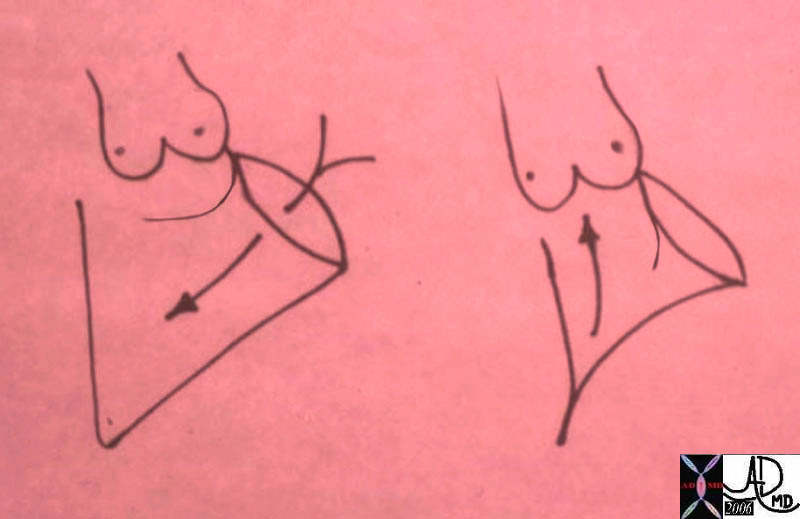
Working Together to Form an Inflow and an Outflow to teh Left Ventricle


The mitral valve participates in the inflow as well as the outflow of the LV. The LV does not have an infundibular chamber like the RV. It is the anterior leaflet of the MV that has this dual function. The first drawing represents LV diastole, showing the open anterior leaflet acting as the anterior, medial and rightward border of the inflow to the LV. The second drawing is the systolic phase where this same anterior leaflet acts as the leftward and lateral border of the outflow tract. Courtesy of Ashley Davidoff M.D. 32119b01


    [nodeType] => 1
    [parentNode] => (object value omitted)
    [childNodes] => (object value omitted)
    [firstChild] => (object value omitted)
    [lastChild] => (object value omitted)
    [previousSibling] => (object value omitted)
    [nextSibling] => (object value omitted)
    [attributes] => (object value omitted)
    [ownerDocument] => (object value omitted)
    [namespaceURI] =>
    [prefix] =>
    [localName] => table
    [baseURI] =>
    [textContent] =>


Working Together to Form an Inflow and an Outflow to teh Left Ventricle


The mitral valve participates in the inflow as well as the outflow of the LV. The LV does not have an infundibular chamber like the RV. It is the anterior leaflet of the MV that has this dual function. The first drawing represents LV diastole, showing the open anterior leaflet acting as the anterior, medial and rightward border of the inflow to the LV. The second drawing is the systolic phase where this same anterior leaflet acts as the leftward and lateral border of the outflow tract. Courtesy of Ashley Davidoff M.D. 32119b01


)
DOMElement Object
(
    [schemaTypeInfo] =>
    [tagName] => td
    [firstElementChild] =>
    [lastElementChild] =>
    [childElementCount] => 0
    [previousElementSibling] =>
    [nextElementSibling] =>
    [nodeName] => td
    [nodeValue] => The mitral valve participates in the inflow as well as the outflow of the LV. The LV does not have an infundibular chamber like the RV. It is the anterior leaflet of the MV that has this dual function. The first drawing represents LV diastole, showing the open anterior leaflet acting as the anterior, medial and rightward border of the inflow to the LV. The second drawing is the systolic phase where this same anterior leaflet acts as the leftward and lateral border of the outflow tract. Courtesy of Ashley Davidoff M.D. 32119b01
    [nodeType] => 1
    [parentNode] => (object value omitted)
    [childNodes] => (object value omitted)
    [firstChild] => (object value omitted)
    [lastChild] => (object value omitted)
    [previousSibling] => (object value omitted)
    [nextSibling] => (object value omitted)
    [attributes] => (object value omitted)
    [ownerDocument] => (object value omitted)
    [namespaceURI] =>
    [prefix] =>
    [localName] => td
    [baseURI] =>
    [textContent] => The mitral valve participates in the inflow as well as the outflow of the LV. The LV does not have an infundibular chamber like the RV. It is the anterior leaflet of the MV that has this dual function. The first drawing represents LV diastole, showing the open anterior leaflet acting as the anterior, medial and rightward border of the inflow to the LV. The second drawing is the systolic phase where this same anterior leaflet acts as the leftward and lateral border of the outflow tract. Courtesy of Ashley Davidoff M.D. 32119b01
)
DOMElement Object
(
    [schemaTypeInfo] =>
    [tagName] => td
    [firstElementChild] => (object value omitted)
    [lastElementChild] => (object value omitted)
    [childElementCount] => 1
    [previousElementSibling] =>
    [nextElementSibling] =>
    [nodeName] => td
    [nodeValue] => Working Together to Form an Inflow and an Outflow to teh Left Ventricle
    [nodeType] => 1
    [parentNode] => (object value omitted)
    [childNodes] => (object value omitted)
    [firstChild] => (object value omitted)
    [lastChild] => (object value omitted)
    [previousSibling] => (object value omitted)
    [nextSibling] => (object value omitted)
    [attributes] => (object value omitted)
    [ownerDocument] => (object value omitted)
    [namespaceURI] =>
    [prefix] =>
    [localName] => td
    [baseURI] =>
    [textContent] => Working Together to Form an Inflow and an Outflow to teh Left Ventricle
)
DOMElement Object
(
    [schemaTypeInfo] =>
    [tagName] => table
    [firstElementChild] => (object value omitted)
    [lastElementChild] => (object value omitted)
    [childElementCount] => 1
    [previousElementSibling] => (object value omitted)
    [nextElementSibling] => (object value omitted)
    [nodeName] => table
    [nodeValue] =>


Working together During an Angiogram


code cardiac heart MV anterior leaflet posterior leaflet normal anatomy physiology systole 34814 imaging radiology angiography normal overlay


    [nodeType] => 1
    [parentNode] => (object value omitted)
    [childNodes] => (object value omitted)
    [firstChild] => (object value omitted)
    [lastChild] => (object value omitted)
    [previousSibling] => (object value omitted)
    [nextSibling] => (object value omitted)
    [attributes] => (object value omitted)
    [ownerDocument] => (object value omitted)
    [namespaceURI] =>
    [prefix] =>
    [localName] => table
    [baseURI] =>
    [textContent] =>


Working together During an Angiogram


code cardiac heart MV anterior leaflet posterior leaflet normal anatomy physiology systole 34814 imaging radiology angiography normal overlay


)
DOMElement Object
(
    [schemaTypeInfo] =>
    [tagName] => td
    [firstElementChild] =>
    [lastElementChild] =>
    [childElementCount] => 0
    [previousElementSibling] =>
    [nextElementSibling] =>
    [nodeName] => td
    [nodeValue] => code cardiac heart MV anterior leaflet posterior leaflet normal anatomy physiology systole 34814 imaging radiology angiography normal overlay
    [nodeType] => 1
    [parentNode] => (object value omitted)
    [childNodes] => (object value omitted)
    [firstChild] => (object value omitted)
    [lastChild] => (object value omitted)
    [previousSibling] => (object value omitted)
    [nextSibling] => (object value omitted)
    [attributes] => (object value omitted)
    [ownerDocument] => (object value omitted)
    [namespaceURI] =>
    [prefix] =>
    [localName] => td
    [baseURI] =>
    [textContent] => code cardiac heart MV anterior leaflet posterior leaflet normal anatomy physiology systole 34814 imaging radiology angiography normal overlay
)
DOMElement Object
(
    [schemaTypeInfo] =>
    [tagName] => td
    [firstElementChild] => (object value omitted)
    [lastElementChild] => (object value omitted)
    [childElementCount] => 1
    [previousElementSibling] =>
    [nextElementSibling] =>
    [nodeName] => td
    [nodeValue] => Working together During an Angiogram
    [nodeType] => 1
    [parentNode] => (object value omitted)
    [childNodes] => (object value omitted)
    [firstChild] => (object value omitted)
    [lastChild] => (object value omitted)
    [previousSibling] => (object value omitted)
    [nextSibling] => (object value omitted)
    [attributes] => (object value omitted)
    [ownerDocument] => (object value omitted)
    [namespaceURI] =>
    [prefix] =>
    [localName] => td
    [baseURI] =>
    [textContent] => Working together During an Angiogram
)
DOMElement Object
(
    [schemaTypeInfo] =>
    [tagName] => table
    [firstElementChild] => (object value omitted)
    [lastElementChild] => (object value omitted)
    [childElementCount] => 1
    [previousElementSibling] => (object value omitted)
    [nextElementSibling] =>
    [nodeName] => table
    [nodeValue] =>


Working together During an Angiogram


code cardiac heart MV anterior leaflet posterior leaflet normal anatomy physiology systole 34814 imaging radiology angiography normal overlay


Working Together to Form an Inflow and an Outflow to teh Left Ventricle


The mitral valve participates in the inflow as well as the outflow of the LV. The LV does not have an infundibular chamber like the RV. It is the anterior leaflet of the MV that has this dual function. The first drawing represents LV diastole, showing the open anterior leaflet acting as the anterior, medial and rightward border of the inflow to the LV. The second drawing is the systolic phase where this same anterior leaflet acts as the leftward and lateral border of the outflow tract. Courtesy of Ashley Davidoff M.D. 32119b01


And through many years they worked hard in harmony
Toward a greater strength for the whole
Together they produced
One million beats and a volume of enormous score

Nothing could separate this bonded union
?cept the disease that wreaked its havoc
And in the end the disease of  him
affected her greatly too


IHSS – Left Ventricular Muscular Obstruction with Mitral Regurgitation


This angiogram in RAO projection shows a hypercontractile left ventricle that has a ballet shoe appearance, with mitral regurgitation filling the left atrium. The drawing shows the significant LVH small cavity of the LV, the area of subaortic muscle bundle (green) and the mitral regurgitation caused by the systolic anterior motion of the mitral valve. Courtesy Ashley Davidoff 34805 cardiac heart MV interventriclar septum LVH IHSS SAM MR imaging radiology angiography disease overlay


Prolapse of the Mitral Valve and Aortic Regurgitation  – Marfan’s Syndrome


This angiogram in RAO projection shows prolapse of both the anterior and posterior leaflets of the mitral valve with mitral regurgitation. Note also the bulbous appearance of the aortic sinuses. The back wall of the left atrium is flattened against the spine suggesting a narrow A-P diameter or pectus excavatum. These findings are characteristic of Maran’s syndrome Courtesy Ashley DAvidoff MD. 34810c code cardiac heart mitral valve MV anterior leaflet posterior leaflet prolapse mitral regurgitation MR incompetence aorta Marfan’s syndrome floppy mitral valve angiography LA shape MVP imaging radiology angiography disease overlay radiologists and detectives


Aged and happy they so declined
Still beating to the same drum
And they were of course happily assured
That their beat would repeat ? in the next of kin


    [nodeType] => 1
    [parentNode] => (object value omitted)
    [childNodes] => (object value omitted)
    [firstChild] => (object value omitted)
    [lastChild] => (object value omitted)
    [previousSibling] => (object value omitted)
    [nextSibling] => (object value omitted)
    [attributes] => (object value omitted)
    [ownerDocument] => (object value omitted)
    [namespaceURI] =>
    [prefix] =>
    [localName] => table
    [baseURI] =>
    [textContent] =>


Working together During an Angiogram


code cardiac heart MV anterior leaflet posterior leaflet normal anatomy physiology systole 34814 imaging radiology angiography normal overlay


Working Together to Form an Inflow and an Outflow to teh Left Ventricle


The mitral valve participates in the inflow as well as the outflow of the LV. The LV does not have an infundibular chamber like the RV. It is the anterior leaflet of the MV that has this dual function. The first drawing represents LV diastole, showing the open anterior leaflet acting as the anterior, medial and rightward border of the inflow to the LV. The second drawing is the systolic phase where this same anterior leaflet acts as the leftward and lateral border of the outflow tract. Courtesy of Ashley Davidoff M.D. 32119b01


And through many years they worked hard in harmony
Toward a greater strength for the whole
Together they produced
One million beats and a volume of enormous score

Nothing could separate this bonded union
?cept the disease that wreaked its havoc
And in the end the disease of  him
affected her greatly too


IHSS – Left Ventricular Muscular Obstruction with Mitral Regurgitation


This angiogram in RAO projection shows a hypercontractile left ventricle that has a ballet shoe appearance, with mitral regurgitation filling the left atrium. The drawing shows the significant LVH small cavity of the LV, the area of subaortic muscle bundle (green) and the mitral regurgitation caused by the systolic anterior motion of the mitral valve. Courtesy Ashley Davidoff 34805 cardiac heart MV interventriclar septum LVH IHSS SAM MR imaging radiology angiography disease overlay


Prolapse of the Mitral Valve and Aortic Regurgitation  – Marfan’s Syndrome


This angiogram in RAO projection shows prolapse of both the anterior and posterior leaflets of the mitral valve with mitral regurgitation. Note also the bulbous appearance of the aortic sinuses. The back wall of the left atrium is flattened against the spine suggesting a narrow A-P diameter or pectus excavatum. These findings are characteristic of Maran’s syndrome Courtesy Ashley DAvidoff MD. 34810c code cardiac heart mitral valve MV anterior leaflet posterior leaflet prolapse mitral regurgitation MR incompetence aorta Marfan’s syndrome floppy mitral valve angiography LA shape MVP imaging radiology angiography disease overlay radiologists and detectives


Aged and happy they so declined
Still beating to the same drum
And they were of course happily assured
That their beat would repeat ? in the next of kin


)
DOMElement Object
(
    [schemaTypeInfo] =>
    [tagName] => td
    [firstElementChild] =>
    [lastElementChild] =>
    [childElementCount] => 0
    [previousElementSibling] =>
    [nextElementSibling] =>
    [nodeName] => td
    [nodeValue] => This angiogram in RAO projection shows prolapse of both the anterior and posterior leaflets of the mitral valve with mitral regurgitation. Note also the bulbous appearance of the aortic sinuses. The back wall of the left atrium is flattened against the spine suggesting a narrow A-P diameter or pectus excavatum. These findings are characteristic of Maran’s syndrome Courtesy Ashley DAvidoff MD. 34810c code cardiac heart mitral valve MV anterior leaflet posterior leaflet prolapse mitral regurgitation MR incompetence aorta Marfan’s syndrome floppy mitral valve angiography LA shape MVP imaging radiology angiography disease overlay radiologists and detectives
    [nodeType] => 1
    [parentNode] => (object value omitted)
    [childNodes] => (object value omitted)
    [firstChild] => (object value omitted)
    [lastChild] => (object value omitted)
    [previousSibling] => (object value omitted)
    [nextSibling] => (object value omitted)
    [attributes] => (object value omitted)
    [ownerDocument] => (object value omitted)
    [namespaceURI] =>
    [prefix] =>
    [localName] => td
    [baseURI] =>
    [textContent] => This angiogram in RAO projection shows prolapse of both the anterior and posterior leaflets of the mitral valve with mitral regurgitation. Note also the bulbous appearance of the aortic sinuses. The back wall of the left atrium is flattened against the spine suggesting a narrow A-P diameter or pectus excavatum. These findings are characteristic of Maran’s syndrome Courtesy Ashley DAvidoff MD. 34810c code cardiac heart mitral valve MV anterior leaflet posterior leaflet prolapse mitral regurgitation MR incompetence aorta Marfan’s syndrome floppy mitral valve angiography LA shape MVP imaging radiology angiography disease overlay radiologists and detectives
)
DOMElement Object
(
    [schemaTypeInfo] =>
    [tagName] => td
    [firstElementChild] => (object value omitted)
    [lastElementChild] => (object value omitted)
    [childElementCount] => 1
    [previousElementSibling] =>
    [nextElementSibling] =>
    [nodeName] => td
    [nodeValue] => Prolapse of the Mitral Valve and Aortic Regurgitation  – Marfan’s Syndrome
    [nodeType] => 1
    [parentNode] => (object value omitted)
    [childNodes] => (object value omitted)
    [firstChild] => (object value omitted)
    [lastChild] => (object value omitted)
    [previousSibling] => (object value omitted)
    [nextSibling] => (object value omitted)
    [attributes] => (object value omitted)
    [ownerDocument] => (object value omitted)
    [namespaceURI] =>
    [prefix] =>
    [localName] => td
    [baseURI] =>
    [textContent] => Prolapse of the Mitral Valve and Aortic Regurgitation  – Marfan’s Syndrome
)
DOMElement Object
(
    [schemaTypeInfo] =>
    [tagName] => td
    [firstElementChild] =>
    [lastElementChild] =>
    [childElementCount] => 0
    [previousElementSibling] =>
    [nextElementSibling] =>
    [nodeName] => td
    [nodeValue] => This angiogram in RAO projection shows a hypercontractile left ventricle that has a ballet shoe appearance, with mitral regurgitation filling the left atrium. The drawing shows the significant LVH small cavity of the LV, the area of subaortic muscle bundle (green) and the mitral regurgitation caused by the systolic anterior motion of the mitral valve. Courtesy Ashley Davidoff 34805 cardiac heart MV interventriclar septum LVH IHSS SAM MR imaging radiology angiography disease overlay
    [nodeType] => 1
    [parentNode] => (object value omitted)
    [childNodes] => (object value omitted)
    [firstChild] => (object value omitted)
    [lastChild] => (object value omitted)
    [previousSibling] => (object value omitted)
    [nextSibling] => (object value omitted)
    [attributes] => (object value omitted)
    [ownerDocument] => (object value omitted)
    [namespaceURI] =>
    [prefix] =>
    [localName] => td
    [baseURI] =>
    [textContent] => This angiogram in RAO projection shows a hypercontractile left ventricle that has a ballet shoe appearance, with mitral regurgitation filling the left atrium. The drawing shows the significant LVH small cavity of the LV, the area of subaortic muscle bundle (green) and the mitral regurgitation caused by the systolic anterior motion of the mitral valve. Courtesy Ashley Davidoff 34805 cardiac heart MV interventriclar septum LVH IHSS SAM MR imaging radiology angiography disease overlay
)
DOMElement Object
(
    [schemaTypeInfo] =>
    [tagName] => td
    [firstElementChild] => (object value omitted)
    [lastElementChild] => (object value omitted)
    [childElementCount] => 1
    [previousElementSibling] =>
    [nextElementSibling] =>
    [nodeName] => td
    [nodeValue] => IHSS – Left Ventricular Muscular Obstruction with Mitral Regurgitation
    [nodeType] => 1
    [parentNode] => (object value omitted)
    [childNodes] => (object value omitted)
    [firstChild] => (object value omitted)
    [lastChild] => (object value omitted)
    [previousSibling] => (object value omitted)
    [nextSibling] => (object value omitted)
    [attributes] => (object value omitted)
    [ownerDocument] => (object value omitted)
    [namespaceURI] =>
    [prefix] =>
    [localName] => td
    [baseURI] =>
    [textContent] => IHSS – Left Ventricular Muscular Obstruction with Mitral Regurgitation
)
DOMElement Object
(
    [schemaTypeInfo] =>
    [tagName] => td
    [firstElementChild] =>
    [lastElementChild] =>
    [childElementCount] => 0
    [previousElementSibling] =>
    [nextElementSibling] =>
    [nodeName] => td
    [nodeValue] => The mitral valve participates in the inflow as well as the outflow of the LV. The LV does not have an infundibular chamber like the RV. It is the anterior leaflet of the MV that has this dual function. The first drawing represents LV diastole, showing the open anterior leaflet acting as the anterior, medial and rightward border of the inflow to the LV. The second drawing is the systolic phase where this same anterior leaflet acts as the leftward and lateral border of the outflow tract. Courtesy of Ashley Davidoff M.D. 32119b01
    [nodeType] => 1
    [parentNode] => (object value omitted)
    [childNodes] => (object value omitted)
    [firstChild] => (object value omitted)
    [lastChild] => (object value omitted)
    [previousSibling] => (object value omitted)
    [nextSibling] => (object value omitted)
    [attributes] => (object value omitted)
    [ownerDocument] => (object value omitted)
    [namespaceURI] =>
    [prefix] =>
    [localName] => td
    [baseURI] =>
    [textContent] => The mitral valve participates in the inflow as well as the outflow of the LV. The LV does not have an infundibular chamber like the RV. It is the anterior leaflet of the MV that has this dual function. The first drawing represents LV diastole, showing the open anterior leaflet acting as the anterior, medial and rightward border of the inflow to the LV. The second drawing is the systolic phase where this same anterior leaflet acts as the leftward and lateral border of the outflow tract. Courtesy of Ashley Davidoff M.D. 32119b01
)
DOMElement Object
(
    [schemaTypeInfo] =>
    [tagName] => td
    [firstElementChild] => (object value omitted)
    [lastElementChild] => (object value omitted)
    [childElementCount] => 1
    [previousElementSibling] =>
    [nextElementSibling] =>
    [nodeName] => td
    [nodeValue] => Working Together to Form an Inflow and an Outflow to teh Left Ventricle
    [nodeType] => 1
    [parentNode] => (object value omitted)
    [childNodes] => (object value omitted)
    [firstChild] => (object value omitted)
    [lastChild] => (object value omitted)
    [previousSibling] => (object value omitted)
    [nextSibling] => (object value omitted)
    [attributes] => (object value omitted)
    [ownerDocument] => (object value omitted)
    [namespaceURI] =>
    [prefix] =>
    [localName] => td
    [baseURI] =>
    [textContent] => Working Together to Form an Inflow and an Outflow to teh Left Ventricle
)
DOMElement Object
(
    [schemaTypeInfo] =>
    [tagName] => td
    [firstElementChild] =>
    [lastElementChild] =>
    [childElementCount] => 0
    [previousElementSibling] =>
    [nextElementSibling] =>
    [nodeName] => td
    [nodeValue] => code cardiac heart MV anterior leaflet posterior leaflet normal anatomy physiology systole 34814 imaging radiology angiography normal overlay
    [nodeType] => 1
    [parentNode] => (object value omitted)
    [childNodes] => (object value omitted)
    [firstChild] => (object value omitted)
    [lastChild] => (object value omitted)
    [previousSibling] => (object value omitted)
    [nextSibling] => (object value omitted)
    [attributes] => (object value omitted)
    [ownerDocument] => (object value omitted)
    [namespaceURI] =>
    [prefix] =>
    [localName] => td
    [baseURI] =>
    [textContent] => code cardiac heart MV anterior leaflet posterior leaflet normal anatomy physiology systole 34814 imaging radiology angiography normal overlay
)
DOMElement Object
(
    [schemaTypeInfo] =>
    [tagName] => td
    [firstElementChild] => (object value omitted)
    [lastElementChild] => (object value omitted)
    [childElementCount] => 2
    [previousElementSibling] =>
    [nextElementSibling] =>
    [nodeName] => td
    [nodeValue] => Working together During an Angiogram
    [nodeType] => 1
    [parentNode] => (object value omitted)
    [childNodes] => (object value omitted)
    [firstChild] => (object value omitted)
    [lastChild] => (object value omitted)
    [previousSibling] => (object value omitted)
    [nextSibling] => (object value omitted)
    [attributes] => (object value omitted)
    [ownerDocument] => (object value omitted)
    [namespaceURI] =>
    [prefix] =>
    [localName] => td
    [baseURI] =>
    [textContent] => Working together During an Angiogram
)
DOMElement Object
(
    [schemaTypeInfo] =>
    [tagName] => td
    [firstElementChild] => (object value omitted)
    [lastElementChild] => (object value omitted)
    [childElementCount] => 26
    [previousElementSibling] =>
    [nextElementSibling] =>
    [nodeName] => td
    [nodeValue] =>


Working together During an Angiogram


code cardiac heart MV anterior leaflet posterior leaflet normal anatomy physiology systole 34814 imaging radiology angiography normal overlay


Working Together to Form an Inflow and an Outflow to teh Left Ventricle


The mitral valve participates in the inflow as well as the outflow of the LV. The LV does not have an infundibular chamber like the RV. It is the anterior leaflet of the MV that has this dual function. The first drawing represents LV diastole, showing the open anterior leaflet acting as the anterior, medial and rightward border of the inflow to the LV. The second drawing is the systolic phase where this same anterior leaflet acts as the leftward and lateral border of the outflow tract. Courtesy of Ashley Davidoff M.D. 32119b01


And through many years they worked hard in harmony
Toward a greater strength for the whole
Together they produced
One million beats and a volume of enormous score

Nothing could separate this bonded union
?cept the disease that wreaked its havoc
And in the end the disease of  him
affected her greatly too


IHSS – Left Ventricular Muscular Obstruction with Mitral Regurgitation


This angiogram in RAO projection shows a hypercontractile left ventricle that has a ballet shoe appearance, with mitral regurgitation filling the left atrium. The drawing shows the significant LVH small cavity of the LV, the area of subaortic muscle bundle (green) and the mitral regurgitation caused by the systolic anterior motion of the mitral valve. Courtesy Ashley Davidoff 34805 cardiac heart MV interventriclar septum LVH IHSS SAM MR imaging radiology angiography disease overlay


Prolapse of the Mitral Valve and Aortic Regurgitation  – Marfan’s Syndrome


This angiogram in RAO projection shows prolapse of both the anterior and posterior leaflets of the mitral valve with mitral regurgitation. Note also the bulbous appearance of the aortic sinuses. The back wall of the left atrium is flattened against the spine suggesting a narrow A-P diameter or pectus excavatum. These findings are characteristic of Maran’s syndrome Courtesy Ashley DAvidoff MD. 34810c code cardiac heart mitral valve MV anterior leaflet posterior leaflet prolapse mitral regurgitation MR incompetence aorta Marfan’s syndrome floppy mitral valve angiography LA shape MVP imaging radiology angiography disease overlay radiologists and detectives


Aged and happy they so declined
Still beating to the same drum
And they were of course happily assured
That their beat would repeat ? in the next of kin

    [nodeType] => 1
    [parentNode] => (object value omitted)
    [childNodes] => (object value omitted)
    [firstChild] => (object value omitted)
    [lastChild] => (object value omitted)
    [previousSibling] => (object value omitted)
    [nextSibling] => (object value omitted)
    [attributes] => (object value omitted)
    [ownerDocument] => (object value omitted)
    [namespaceURI] =>
    [prefix] =>
    [localName] => td
    [baseURI] =>
    [textContent] =>


Working together During an Angiogram


code cardiac heart MV anterior leaflet posterior leaflet normal anatomy physiology systole 34814 imaging radiology angiography normal overlay


Working Together to Form an Inflow and an Outflow to teh Left Ventricle


The mitral valve participates in the inflow as well as the outflow of the LV. The LV does not have an infundibular chamber like the RV. It is the anterior leaflet of the MV that has this dual function. The first drawing represents LV diastole, showing the open anterior leaflet acting as the anterior, medial and rightward border of the inflow to the LV. The second drawing is the systolic phase where this same anterior leaflet acts as the leftward and lateral border of the outflow tract. Courtesy of Ashley Davidoff M.D. 32119b01


And through many years they worked hard in harmony
Toward a greater strength for the whole
Together they produced
One million beats and a volume of enormous score

Nothing could separate this bonded union
?cept the disease that wreaked its havoc
And in the end the disease of  him
affected her greatly too


IHSS – Left Ventricular Muscular Obstruction with Mitral Regurgitation


This angiogram in RAO projection shows a hypercontractile left ventricle that has a ballet shoe appearance, with mitral regurgitation filling the left atrium. The drawing shows the significant LVH small cavity of the LV, the area of subaortic muscle bundle (green) and the mitral regurgitation caused by the systolic anterior motion of the mitral valve. Courtesy Ashley Davidoff 34805 cardiac heart MV interventriclar septum LVH IHSS SAM MR imaging radiology angiography disease overlay


Prolapse of the Mitral Valve and Aortic Regurgitation  – Marfan’s Syndrome


This angiogram in RAO projection shows prolapse of both the anterior and posterior leaflets of the mitral valve with mitral regurgitation. Note also the bulbous appearance of the aortic sinuses. The back wall of the left atrium is flattened against the spine suggesting a narrow A-P diameter or pectus excavatum. These findings are characteristic of Maran’s syndrome Courtesy Ashley DAvidoff MD. 34810c code cardiac heart mitral valve MV anterior leaflet posterior leaflet prolapse mitral regurgitation MR incompetence aorta Marfan’s syndrome floppy mitral valve angiography LA shape MVP imaging radiology angiography disease overlay radiologists and detectives


Aged and happy they so declined
Still beating to the same drum
And they were of course happily assured
That their beat would repeat ? in the next of kin

)
DOMElement Object
(
    [schemaTypeInfo] =>
    [tagName] => table
    [firstElementChild] => (object value omitted)
    [lastElementChild] => (object value omitted)
    [childElementCount] => 1
    [previousElementSibling] => (object value omitted)
    [nextElementSibling] => (object value omitted)
    [nodeName] => table
    [nodeValue] =>


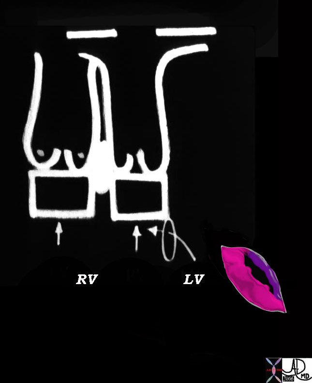
Bonding in Fibrous Union


07427b02 heart cardiac bilateral conus outflow tract infundibulum aorta pulmonary artery D-loop RV LV conoventricular defect anatomy embryology subaortic conus subpulmonary conus fibrous continuity anterior leaflet posterior leaflet of the mitral valve MV Courtesy Ashley Davidoff Davidoff drawing


    [nodeType] => 1
    [parentNode] => (object value omitted)
    [childNodes] => (object value omitted)
    [firstChild] => (object value omitted)
    [lastChild] => (object value omitted)
    [previousSibling] => (object value omitted)
    [nextSibling] => (object value omitted)
    [attributes] => (object value omitted)
    [ownerDocument] => (object value omitted)
    [namespaceURI] =>
    [prefix] =>
    [localName] => table
    [baseURI] =>
    [textContent] =>


Bonding in Fibrous Union


07427b02 heart cardiac bilateral conus outflow tract infundibulum aorta pulmonary artery D-loop RV LV conoventricular defect anatomy embryology subaortic conus subpulmonary conus fibrous continuity anterior leaflet posterior leaflet of the mitral valve MV Courtesy Ashley Davidoff Davidoff drawing


)
DOMElement Object
(
    [schemaTypeInfo] =>
    [tagName] => td
    [firstElementChild] =>
    [lastElementChild] =>
    [childElementCount] => 0
    [previousElementSibling] =>
    [nextElementSibling] =>
    [nodeName] => td
    [nodeValue] => 07427b02 heart cardiac bilateral conus outflow tract infundibulum aorta pulmonary artery D-loop RV LV conoventricular defect anatomy embryology subaortic conus subpulmonary conus fibrous continuity anterior leaflet posterior leaflet of the mitral valve MV Courtesy Ashley Davidoff Davidoff drawing
    [nodeType] => 1
    [parentNode] => (object value omitted)
    [childNodes] => (object value omitted)
    [firstChild] => (object value omitted)
    [lastChild] => (object value omitted)
    [previousSibling] => (object value omitted)
    [nextSibling] => (object value omitted)
    [attributes] => (object value omitted)
    [ownerDocument] => (object value omitted)
    [namespaceURI] =>
    [prefix] =>
    [localName] => td
    [baseURI] =>
    [textContent] => 07427b02 heart cardiac bilateral conus outflow tract infundibulum aorta pulmonary artery D-loop RV LV conoventricular defect anatomy embryology subaortic conus subpulmonary conus fibrous continuity anterior leaflet posterior leaflet of the mitral valve MV Courtesy Ashley Davidoff Davidoff drawing
)
DOMElement Object
(
    [schemaTypeInfo] =>
    [tagName] => td
    [firstElementChild] => (object value omitted)
    [lastElementChild] => (object value omitted)
    [childElementCount] => 1
    [previousElementSibling] =>
    [nextElementSibling] =>
    [nodeName] => td
    [nodeValue] => Bonding in Fibrous Union
    [nodeType] => 1
    [parentNode] => (object value omitted)
    [childNodes] => (object value omitted)
    [firstChild] => (object value omitted)
    [lastChild] => (object value omitted)
    [previousSibling] => (object value omitted)
    [nextSibling] => (object value omitted)
    [attributes] => (object value omitted)
    [ownerDocument] => (object value omitted)
    [namespaceURI] =>
    [prefix] =>
    [localName] => td
    [baseURI] =>
    [textContent] => Bonding in Fibrous Union
)
DOMElement Object
(
    [schemaTypeInfo] =>
    [tagName] => table
    [firstElementChild] => (object value omitted)
    [lastElementChild] => (object value omitted)
    [childElementCount] => 1
    [previousElementSibling] => (object value omitted)
    [nextElementSibling] => (object value omitted)
    [nodeName] => table
    [nodeValue] =>


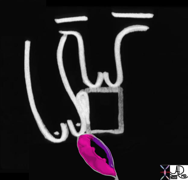
Resorbtion of the Subaortic Conus Allows Mr Aorta to get Close to Ms Mitral


06370b03 heart cardiac bilateral conus outflow tract infundibulum aorta pulmonary artery D-loop RV LV conoventricular defect bilateral conus atrioventricular endocardial cushion mitral valve anatomy embryology subaortic conus subpulmonary conus fibrous continuity mitral valve MV Courtesy Ashley Davidoff Davidoff drawing


    [nodeType] => 1
    [parentNode] => (object value omitted)
    [childNodes] => (object value omitted)
    [firstChild] => (object value omitted)
    [lastChild] => (object value omitted)
    [previousSibling] => (object value omitted)
    [nextSibling] => (object value omitted)
    [attributes] => (object value omitted)
    [ownerDocument] => (object value omitted)
    [namespaceURI] =>
    [prefix] =>
    [localName] => table
    [baseURI] =>
    [textContent] =>


Resorbtion of the Subaortic Conus Allows Mr Aorta to get Close to Ms Mitral


06370b03 heart cardiac bilateral conus outflow tract infundibulum aorta pulmonary artery D-loop RV LV conoventricular defect bilateral conus atrioventricular endocardial cushion mitral valve anatomy embryology subaortic conus subpulmonary conus fibrous continuity mitral valve MV Courtesy Ashley Davidoff Davidoff drawing


)
DOMElement Object
(
    [schemaTypeInfo] =>
    [tagName] => td
    [firstElementChild] =>
    [lastElementChild] =>
    [childElementCount] => 0
    [previousElementSibling] =>
    [nextElementSibling] =>
    [nodeName] => td
    [nodeValue] => 06370b03 heart cardiac bilateral conus outflow tract infundibulum aorta pulmonary artery D-loop RV LV conoventricular defect bilateral conus atrioventricular endocardial cushion mitral valve anatomy embryology subaortic conus subpulmonary conus fibrous continuity mitral valve MV Courtesy Ashley Davidoff Davidoff drawing
    [nodeType] => 1
    [parentNode] => (object value omitted)
    [childNodes] => (object value omitted)
    [firstChild] => (object value omitted)
    [lastChild] => (object value omitted)
    [previousSibling] => (object value omitted)
    [nextSibling] => (object value omitted)
    [attributes] => (object value omitted)
    [ownerDocument] => (object value omitted)
    [namespaceURI] =>
    [prefix] =>
    [localName] => td
    [baseURI] =>
    [textContent] => 06370b03 heart cardiac bilateral conus outflow tract infundibulum aorta pulmonary artery D-loop RV LV conoventricular defect bilateral conus atrioventricular endocardial cushion mitral valve anatomy embryology subaortic conus subpulmonary conus fibrous continuity mitral valve MV Courtesy Ashley Davidoff Davidoff drawing
)
DOMElement Object
(
    [schemaTypeInfo] =>
    [tagName] => td
    [firstElementChild] => (object value omitted)
    [lastElementChild] => (object value omitted)
    [childElementCount] => 1
    [previousElementSibling] =>
    [nextElementSibling] =>
    [nodeName] => td
    [nodeValue] => Resorbtion of the Subaortic Conus Allows Mr Aorta to get Close to Ms Mitral
    [nodeType] => 1
    [parentNode] => (object value omitted)
    [childNodes] => (object value omitted)
    [firstChild] => (object value omitted)
    [lastChild] => (object value omitted)
    [previousSibling] => (object value omitted)
    [nextSibling] => (object value omitted)
    [attributes] => (object value omitted)
    [ownerDocument] => (object value omitted)
    [namespaceURI] =>
    [prefix] =>
    [localName] => td
    [baseURI] =>
    [textContent] => Resorbtion of the Subaortic Conus Allows Mr Aorta to get Close to Ms Mitral
)
DOMElement Object
(
    [schemaTypeInfo] =>
    [tagName] => table
    [firstElementChild] => (object value omitted)
    [lastElementChild] => (object value omitted)
    [childElementCount] => 1
    [previousElementSibling] => (object value omitted)
    [nextElementSibling] => (object value omitted)
    [nodeName] => table
    [nodeValue] =>


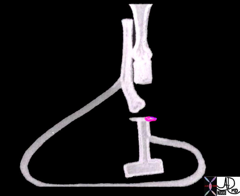
Ms Mitral Looking More Attractive
How is Mr Aorta Going to Reach Her


07427b10 heart cardiac bilateral conus outflow tract infundibulum aorta pulmonary artery D-loop RV LV conoventricular defect anatomy embryology subaortic conus subpulmonary conus fibrous continuity anterior leaflet posterior leaflet of the mitral valve MV Courtesy Ashley Davidoff Davidoff drawing


    [nodeType] => 1
    [parentNode] => (object value omitted)
    [childNodes] => (object value omitted)
    [firstChild] => (object value omitted)
    [lastChild] => (object value omitted)
    [previousSibling] => (object value omitted)
    [nextSibling] => (object value omitted)
    [attributes] => (object value omitted)
    [ownerDocument] => (object value omitted)
    [namespaceURI] =>
    [prefix] =>
    [localName] => table
    [baseURI] =>
    [textContent] =>


Ms Mitral Looking More Attractive
How is Mr Aorta Going to Reach Her


07427b10 heart cardiac bilateral conus outflow tract infundibulum aorta pulmonary artery D-loop RV LV conoventricular defect anatomy embryology subaortic conus subpulmonary conus fibrous continuity anterior leaflet posterior leaflet of the mitral valve MV Courtesy Ashley Davidoff Davidoff drawing


)
DOMElement Object
(
    [schemaTypeInfo] =>
    [tagName] => td
    [firstElementChild] =>
    [lastElementChild] =>
    [childElementCount] => 0
    [previousElementSibling] =>
    [nextElementSibling] =>
    [nodeName] => td
    [nodeValue] => 07427b10 heart cardiac bilateral conus outflow tract infundibulum aorta pulmonary artery D-loop RV LV conoventricular defect anatomy embryology subaortic conus subpulmonary conus fibrous continuity anterior leaflet posterior leaflet of the mitral valve MV Courtesy Ashley Davidoff Davidoff drawing
    [nodeType] => 1
    [parentNode] => (object value omitted)
    [childNodes] => (object value omitted)
    [firstChild] => (object value omitted)
    [lastChild] => (object value omitted)
    [previousSibling] => (object value omitted)
    [nextSibling] => (object value omitted)
    [attributes] => (object value omitted)
    [ownerDocument] => (object value omitted)
    [namespaceURI] =>
    [prefix] =>
    [localName] => td
    [baseURI] =>
    [textContent] => 07427b10 heart cardiac bilateral conus outflow tract infundibulum aorta pulmonary artery D-loop RV LV conoventricular defect anatomy embryology subaortic conus subpulmonary conus fibrous continuity anterior leaflet posterior leaflet of the mitral valve MV Courtesy Ashley Davidoff Davidoff drawing
)
DOMElement Object
(
    [schemaTypeInfo] =>
    [tagName] => td
    [firstElementChild] => (object value omitted)
    [lastElementChild] => (object value omitted)
    [childElementCount] => 2
    [previousElementSibling] =>
    [nextElementSibling] =>
    [nodeName] => td
    [nodeValue] => Ms Mitral Looking More Attractive
How is Mr Aorta Going to Reach Her
    [nodeType] => 1
    [parentNode] => (object value omitted)
    [childNodes] => (object value omitted)
    [firstChild] => (object value omitted)
    [lastChild] => (object value omitted)
    [previousSibling] => (object value omitted)
    [nextSibling] => (object value omitted)
    [attributes] => (object value omitted)
    [ownerDocument] => (object value omitted)
    [namespaceURI] =>
    [prefix] =>
    [localName] => td
    [baseURI] =>
    [textContent] => Ms Mitral Looking More Attractive
How is Mr Aorta Going to Reach Her
)
DOMElement Object
(
    [schemaTypeInfo] =>
    [tagName] => table
    [firstElementChild] => (object value omitted)
    [lastElementChild] => (object value omitted)
    [childElementCount] => 1
    [previousElementSibling] => (object value omitted)
    [nextElementSibling] => (object value omitted)
    [nodeName] => table
    [nodeValue] =>


Ms Mitral in Pink


06370a02 heart cardiac bilateral conus outflow tract infundibulum aorta pulmonary artery D-loop RV LV conoventricular defect bilateral conus atrioventricular endocardial cushion mitral valve anatomy embryology subaortic conus subpulmonary conus fibrous continuity mitral valve MV Courtesy Ashley Davidoff Davidoff drawing


    [nodeType] => 1
    [parentNode] => (object value omitted)
    [childNodes] => (object value omitted)
    [firstChild] => (object value omitted)
    [lastChild] => (object value omitted)
    [previousSibling] => (object value omitted)
    [nextSibling] => (object value omitted)
    [attributes] => (object value omitted)
    [ownerDocument] => (object value omitted)
    [namespaceURI] =>
    [prefix] =>
    [localName] => table
    [baseURI] =>
    [textContent] =>


Ms Mitral in Pink


06370a02 heart cardiac bilateral conus outflow tract infundibulum aorta pulmonary artery D-loop RV LV conoventricular defect bilateral conus atrioventricular endocardial cushion mitral valve anatomy embryology subaortic conus subpulmonary conus fibrous continuity mitral valve MV Courtesy Ashley Davidoff Davidoff drawing


)
DOMElement Object
(
    [schemaTypeInfo] =>
    [tagName] => td
    [firstElementChild] =>
    [lastElementChild] =>
    [childElementCount] => 0
    [previousElementSibling] =>
    [nextElementSibling] =>
    [nodeName] => td
    [nodeValue] => 06370a02 heart cardiac bilateral conus outflow tract infundibulum aorta pulmonary artery D-loop RV LV conoventricular defect bilateral conus atrioventricular endocardial cushion mitral valve anatomy embryology subaortic conus subpulmonary conus fibrous continuity mitral valve MV Courtesy Ashley Davidoff Davidoff drawing
    [nodeType] => 1
    [parentNode] => (object value omitted)
    [childNodes] => (object value omitted)
    [firstChild] => (object value omitted)
    [lastChild] => (object value omitted)
    [previousSibling] => (object value omitted)
    [nextSibling] => (object value omitted)
    [attributes] => (object value omitted)
    [ownerDocument] => (object value omitted)
    [namespaceURI] =>
    [prefix] =>
    [localName] => td
    [baseURI] =>
    [textContent] => 06370a02 heart cardiac bilateral conus outflow tract infundibulum aorta pulmonary artery D-loop RV LV conoventricular defect bilateral conus atrioventricular endocardial cushion mitral valve anatomy embryology subaortic conus subpulmonary conus fibrous continuity mitral valve MV Courtesy Ashley Davidoff Davidoff drawing
)
DOMElement Object
(
    [schemaTypeInfo] =>
    [tagName] => td
    [firstElementChild] => (object value omitted)
    [lastElementChild] => (object value omitted)
    [childElementCount] => 1
    [previousElementSibling] =>
    [nextElementSibling] =>
    [nodeName] => td
    [nodeValue] => Ms Mitral in Pink
    [nodeType] => 1
    [parentNode] => (object value omitted)
    [childNodes] => (object value omitted)
    [firstChild] => (object value omitted)
    [lastChild] => (object value omitted)
    [previousSibling] => (object value omitted)
    [nextSibling] => (object value omitted)
    [attributes] => (object value omitted)
    [ownerDocument] => (object value omitted)
    [namespaceURI] =>
    [prefix] =>
    [localName] => td
    [baseURI] =>
    [textContent] => Ms Mitral in Pink
)
DOMElement Object
(
    [schemaTypeInfo] =>
    [tagName] => table
    [firstElementChild] => (object value omitted)
    [lastElementChild] => (object value omitted)
    [childElementCount] => 1
    [previousElementSibling] => (object value omitted)
    [nextElementSibling] => (object value omitted)
    [nodeName] => table
    [nodeValue] =>


Aorta and Pulmonary Artery Side by Side


01806 heart cardiac bilateral conus outflow tract infundibulum aorta pulmonary artery D-loop anatomy embryology Courtesy Ashley Davidoff drawing


    [nodeType] => 1
    [parentNode] => (object value omitted)
    [childNodes] => (object value omitted)
    [firstChild] => (object value omitted)
    [lastChild] => (object value omitted)
    [previousSibling] => (object value omitted)
    [nextSibling] => (object value omitted)
    [attributes] => (object value omitted)
    [ownerDocument] => (object value omitted)
    [namespaceURI] =>
    [prefix] =>
    [localName] => table
    [baseURI] =>
    [textContent] =>


Aorta and Pulmonary Artery Side by Side


01806 heart cardiac bilateral conus outflow tract infundibulum aorta pulmonary artery D-loop anatomy embryology Courtesy Ashley Davidoff drawing


)
DOMElement Object
(
    [schemaTypeInfo] =>
    [tagName] => td
    [firstElementChild] =>
    [lastElementChild] =>
    [childElementCount] => 0
    [previousElementSibling] =>
    [nextElementSibling] =>
    [nodeName] => td
    [nodeValue] => 01806 heart cardiac bilateral conus outflow tract infundibulum aorta pulmonary artery D-loop anatomy embryology Courtesy Ashley Davidoff drawing
    [nodeType] => 1
    [parentNode] => (object value omitted)
    [childNodes] => (object value omitted)
    [firstChild] => (object value omitted)
    [lastChild] => (object value omitted)
    [previousSibling] => (object value omitted)
    [nextSibling] => (object value omitted)
    [attributes] => (object value omitted)
    [ownerDocument] => (object value omitted)
    [namespaceURI] =>
    [prefix] =>
    [localName] => td
    [baseURI] =>
    [textContent] => 01806 heart cardiac bilateral conus outflow tract infundibulum aorta pulmonary artery D-loop anatomy embryology Courtesy Ashley Davidoff drawing
)
DOMElement Object
(
    [schemaTypeInfo] =>
    [tagName] => td
    [firstElementChild] => (object value omitted)
    [lastElementChild] => (object value omitted)
    [childElementCount] => 2
    [previousElementSibling] =>
    [nextElementSibling] =>
    [nodeName] => td
    [nodeValue] =>
Aorta and Pulmonary Artery Side by Side
    [nodeType] => 1
    [parentNode] => (object value omitted)
    [childNodes] => (object value omitted)
    [firstChild] => (object value omitted)
    [lastChild] => (object value omitted)
    [previousSibling] => (object value omitted)
    [nextSibling] => (object value omitted)
    [attributes] => (object value omitted)
    [ownerDocument] => (object value omitted)
    [namespaceURI] =>
    [prefix] =>
    [localName] => td
    [baseURI] =>
    [textContent] =>
Aorta and Pulmonary Artery Side by Side
)

