Two Boys Fishing
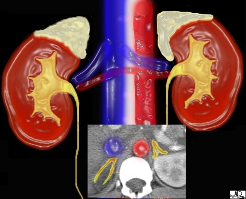
Imagine two young boys with straw hats fishing by the river.
The boy in red is napping and has his hat on his forehead ? the left adrenal.
The other boy in blue is wide awake and has his hat atop his head ? the right adrenal.
This is how the adrenals are positioned relative to the superior poles of the kidneys.
Ashley Davidoff MD 2018
39535
adrenals-0015
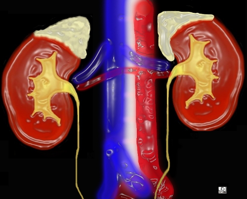
Now take a look at the straw hats as they are positioned on the kidneys. The left adrenal lies more medial and is therefore more compressed by surrounding structures including the medially positioned spine. The right adrenal is more able to take on its naturally triangular shape.Courtesy of: Ashley Davidoff, M.D.

The adrenal glands are usually difficult to find. Since the left kidney is slightly more superior than the right, the left adrenal is usually more superior. The left adrenal can be found when the splenic vein crosses behind the pancreas from the spleen to the portal vein, and the right adrenal can be found when the IVC frees itself from its intrahepatic portion inferiorly. The gland must be identified superiorly and inferiorly until no part of the gland remains, since exophytic tumors off the gland are not uncommon.The adrenals or adrenal like tissue can be found in places other than its normal position. This is termed ‘ectopic’ adrenal tissue.

In these images, the right adrenal is best seen as the IVC (blue overlay) emerges from the liver. Parts of the left adrenal are visualized as the splenic vein (blue overlay) courses behind the pancreas to the portal vein.Courtesy of: Ashley Davidoff, M.D.

Relations

The anatomic location of the adrenals, sandwiches them between several organs, and on the border between two major cavities. The adrenals are positioned differently in relation to the kidneys and to other asymmetric organs such as the liver, pancreas, and spleen.
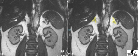
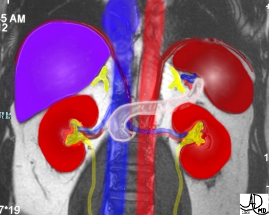
These coronal MRI images show the adrenals surrounded by retroperitoneal fat, padded by the liver on the right and the spleen on the left, and displaced from the superior aspect of the kidneys by a “mop” of fat.Courtesy of: Ashley Davidoff, M.D.


This is a larger and more colorful version of the previous set. Note the relationship of the tail of the pancreas (in light pink) to the left adrenal and the medial relationship of the great vessels (“finger on the pulse”) and crura.(slithers of maroon extending from the hemidiaphragms).Courtesy of: Ashley Davidoff, M.D.
This embedded cross section reflects the true relationship of the great vessels to the adrenals. The IVC really is positioned anterior to the right adrenal while the aorta lies medial to the left adrenal gland .Courtesy of: Ashley Davidoff, M.D..

Anatomy and Physiology of the Adrenal Glands: Relations – Right adrenal

The right adrenal is framed by the following:·         anteriorly – the inferior vena cava and the caudate lobe of the liver

·         posteriorly – the diaphragm

·         superiorly – the bare area of the right lobe of the liver, diaphragm and chest cavity

·         inferiorly – the superior pole of the right kidney

·         medially – the crus of the diaphragm and the spine

·         laterally – the bare area of the right lobe of the liver
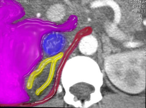
This cross sectional image shows the crus of the diaphragm (maroon), IVC (blue), and the liver (purple), surrounding the right adrenal (yellow).Courtesy of: Ashley Davidoff, M.D.

Anatomy and Physiology of the Adrenal Glands: Relations – Left adrenal

The left adrenal is framed by the following:·         anteriorly – the end of stomach above and the pancreas below and branches of the splenic artery and vein

·         posteriorly – the left kidney, spleen, and the diaphragm

·         superiorly – the diaphragm and chest cavity

·         inferiorly – left kidney

·         medially – left crus of the diaphragm, aorta, and spine

·         laterally – the spleen and left kidney

This cross sectional image of the superior aspect of the left upper abdomen shows the spleen (dark pink), splenic artery and vein in the splenic hilum, the pancreas (light pink), crus of the diaphragm (maroon), aorta (red), and the spine surrounding the left adrenal (yellow).Courtesy of: Ashley Davidoff, M.D.
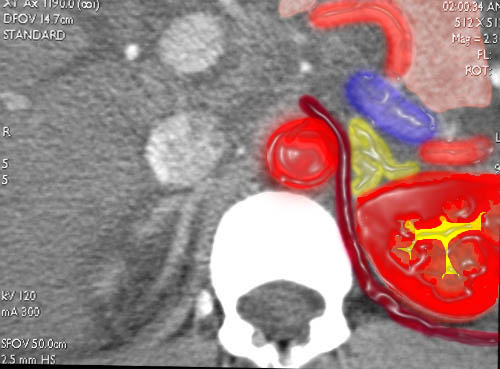
This coned down cross-sectional image, more inferiorly, shows the left kidney (bright red), the tubular wisps of the splenic artery and vein anterolaterally, the pancreas (pink), crus of the diaphragm (maroon), aorta (red), and the spine surrounding left adrenal (yellow).Courtesy of: Ashley Davidoff, M.D.