The adrenal glands are small and mighty. They are tucked away in the back of the abdomen, just below the chest cavity, and literally have their finger on the pulse as they sit alongside the highway systems of the aorta, vena cava, and autonomic nerve chains. They sit on top of the waterworks in close contact with the kidneys, and within the functional loop of the renin – angiotensin – aldosterone hormonal system that controls blood pressure, volume and salt metabolism. Some of the most dramatic responses of the body originate in these small and sometimes hidden glands.The adrenals have a reputation in the imaging world of being hard and difficult. Neither of these statements is true. First, they are so soft that they get pushed around by all the other organs that surround them. Secondly, like so many things that appear difficult initially, with appropriate attention, the structural complexity and eccentricities unfold.
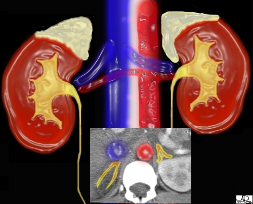
This image combines the coronal view with the axial view and reflects the intimate relationships that the adrenals have with the kidneys as well as the great vessels of the abdomen. They literally have their fingers on the pulse of the aorta (red overlay)and the inferior vena cava (IVC) (blue overlay).

Images courtesy of: Ashley Davidoff, M.D.
Anatomy and Physiology of the Adrenal Glands: Normal Frontal View
The adrenal glands are located retroperitoneally in the abdomen. They sit on the upper poles of the kidneys on the posterior abdominal wall and extend roughly from the level of the 11th thoracic rib to the first lumbar vertebrae.The right adrenal is slightly more lateral than the left. Since the adrenals sit on top of kidneys, and the right kidney is usually lower than the left, the right adrenal is usually more inferior.

Anatomy and Physiology of the Adrenal Glands: Normal Transverse View
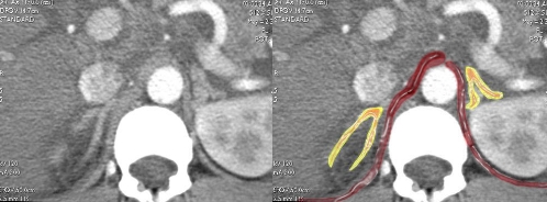
The adrenals can be found just lateral to the crus of the diaphragm. They have a ‘wishbone’ shape, or upside down ‘y’ shape and are surrounded by fat. In general the right adrenal is the long and thin one of family, and the left the short and chubby one.Often the lateral limb of the right adrenal gland is pressed against the liver and cannot be clearly seen. Since the limbs are soft, compressible, and malleable, they often are distorted by normal surrounding structures producing “bends” in them. This sometimes results in difficulty distinguishing cases of abnormal nodules and hyperplasia (increased size) from normal distortion.


