Character on CT Scan
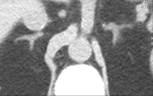
The adrenal gland is of soft tissue density on non contrast scan, measuring between 10 and 30 HU on the non contrast study. Because of its rich blood supply, a contrast study will enhance the gland significantly, particularly in stressful situations where blood flow to the gland is increased.

CT Imaging of the Adrenal Glands: Abnormal Adrenal on CT scan
CT is a highly sensitive modality, although slightly less specific than MRI. The CT may be able to see a disorder, but often cannot exactly define the disorder. However, the CT can easily identify fat, which is present in conditions such as incidentaloma and myelolipoma. If an incidentaloma is suspected, non-contrast study is indicated. CT is more sensitive to the presence of calcification than MRI.It is very important for the radiologist to make a distinction between unilateral disease and bilateral disease. Cushing?s disease, for example, affects both adrenal glands and treatment may relate to surgical excision of a tumor of the pituitary gland. A unilateral nodule in Conn’s syndrome would require excision only of the affected adrenal gland.
In adrenal hemorrhage, serial CT examination will reveal evolution and usually resorption, and hence a progressive decrease in the size of the gland. The issue of adrenal mass or hemorrhage arises in the neonatal setting as well as following trauma in the adult.
MR Imaging of the Adrenal Glands: Overview
MRI is slightly more specific than CT but less sensitive to morphologic disorder. It is well suited to characterize fat, hemorrhage, and cystic changes, and MRI is the study of choice in patients with suspected pheochromocytoma or adrenal cysts. The advantage of being able to acquire images in many planes enables the radiologist and referring physicians to have a different perspective that sometimes provides relevant clinical information.MRI of the pituitary is superior to CT imaging, which has relevance in adrenal disease that is secondary to pituitary disease. Hence, in planning trans-sphenoidal surgery of the pituitary in patients with pituitary adenoma and resultant Cushing’s disease, it would be important to visualise the optic chiasm, the optic nerves, and the cavernous sinuses. This evaluation is best performed by MRI.
If a study of the adrenals is required during pregnancy, MRI is the diagnostic option of choice.
Visualization of calcification and bony structures is limited on MRI. On the other hand, MRI is very useful for defining vascular structures and the perfusional nature of adrenal lesions.
MR Imaging of the Adrenal Glands: Normal Character on MRI
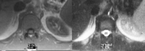
Because the normal adrenal gland is surrounded by retroperitoneal fat, the relatively low intensity of the gland is seen in sharp contrast to the fat, particularly on T1 weighted images. MRI is best used in the characterization of adrenal gland masses.
The T1-weighted sequence is performed to optimize the morphology of the gland and the “in-phase” and “out-of-phase” techniques are also useful in defining the incidentaloma. The T2-weighted sequence enables the characterization of lesions with high water content as well as pheochromocytomas.


MR Imaging of the Adrenal Glands: Abnormal Adrenal on MRI
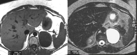
MRI is very useful in the relatively frequently asked indication to “rule out pheochromocytoma”. It is said that the adrenal gland is like a “lightbulb” on T2-weighted imaging in patients with pheochromocytoma – meaning of course that it is very bright. This is not always the case in pheochromocytoma, but if you really want to see a lightbulb, take a look at the adrenal cyst. It will hurt your eyes.
“In-phase” and “out-of-phase” T1 sequences are sensitive to the presence of steroidal fat present in the incidentaloma, and MRI competes well with CT for the diagnosis of this condition. Characterization of another fatty lesion called the myelolipoma is also easily solved by fat sensitive MRI sequences.

US Imaging of the Adrenal Glands: Overview
Ultrasound is quick, and a relatively inexpensive technology, but it has significant limitations in adrenal scanning. The adrenal glands are difficult to examine by ultrasound because of their small size, their location high in the abdomen under the rib cage, and the presence of retroperitoneal fat and bowel gas. In thin patients, the normal adrenal may be identified.An anterior or lateral approach may be necessary, particularly for the right adrenal gland, which occasionally can be seen through the liver. Bowel gas often impairs visualization of the left adrenal gland. A variety of patient positions and scanning windows are often required to adequately examine the glands.
Since CT and MRI have so many diagnostic advantages, ultrasound is not used as the primary modality for the adrenal glands. Ultrasound can help in the characterization of large adrenal masses, as well as to define whether a suprarenal mass originates from the kidney or the adrenal.

This image represents a solid mass that is half the size of the kidney below the mass. A mass of this size typically indicates a primary carcinoma, however, this case represents a large pheochromocytoma.Courtesy of: Ashley Davidoff, M.D.

