Blood Supply of the Adrenal Glands
Ashley Davidoff MD
The Common Vein Copyright 2012
Introduction
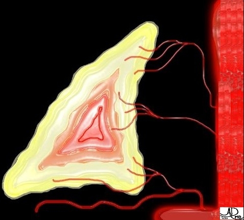
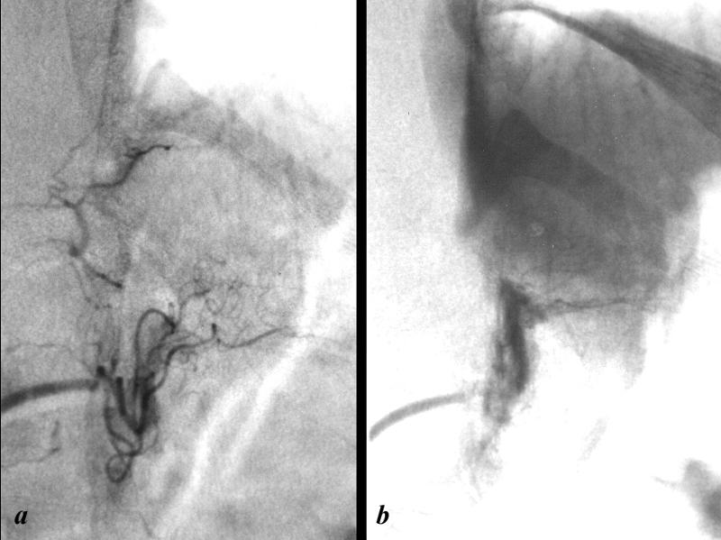
The arterial supply of the adrenal gland consists of three major arteries;
superior adrenal artery
middle adrenal artery
inferior adrenal artery
Each vessel enters the gland at various points around their periphery.
The superior adrenal artery arises from the inferior phrenic artery, the middle adrenal artery arises from the abdominal aorta, while the inferior adrenal artery arises from the renal artery.
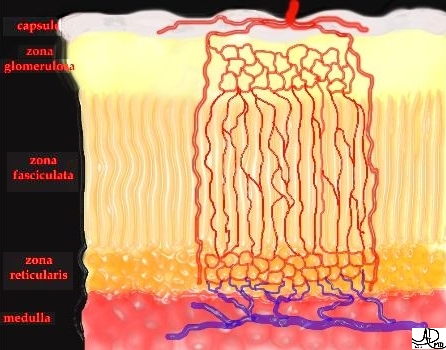
The branches of these arteries form a subcapsular plexus from which arise 3 groups vessels;
arteries of the capsule
arteries of the cortex
arteries of the medulla
The arteries of the capsule and the arteries of the cortex, branch repeatedly to form the capillary bed between parenchymal cells and then drain into medullary capillaries;
The arteries of the medulla, pass through the cortex before breaking up to form part of medulla’s extensive capillary network. This dual vascular supply provides the medulla with both arterial and venous blood. The endothelium of these capillaries is extremely attenuated and interrupted by small fenestrae that are closed by thin diaphragms.
DOMElement Object
(
    [schemaTypeInfo] =>
    [tagName] => table
    [firstElementChild] => (object value omitted)
    [lastElementChild] => (object value omitted)
    [childElementCount] => 1
    [previousElementSibling] => (object value omitted)
    [nextElementSibling] =>
    [nodeName] => table
    [nodeValue] =>


The Arteriole Circulation of the Adrenal Gland


This image shows a schematic of the histologic distribution of the adrenal arterial supply and venous drainage. The dominant area of arterial supply is the cortex, and the dominant.
Copyright 2012 Courtesy Ashley Davidoff  39518


    [nodeType] => 1
    [parentNode] => (object value omitted)
    [childNodes] => (object value omitted)
    [firstChild] => (object value omitted)
    [lastChild] => (object value omitted)
    [previousSibling] => (object value omitted)
    [nextSibling] => (object value omitted)
    [attributes] => (object value omitted)
    [ownerDocument] => (object value omitted)
    [namespaceURI] =>
    [prefix] =>
    [localName] => table
    [baseURI] =>
    [textContent] =>


The Arteriole Circulation of the Adrenal Gland


This image shows a schematic of the histologic distribution of the adrenal arterial supply and venous drainage. The dominant area of arterial supply is the cortex, and the dominant.
Copyright 2012 Courtesy Ashley Davidoff  39518


)
DOMElement Object
(
    [schemaTypeInfo] =>
    [tagName] => td
    [firstElementChild] => (object value omitted)
    [lastElementChild] => (object value omitted)
    [childElementCount] => 2
    [previousElementSibling] =>
    [nextElementSibling] =>
    [nodeName] => td
    [nodeValue] =>
This image shows a schematic of the histologic distribution of the adrenal arterial supply and venous drainage. The dominant area of arterial supply is the cortex, and the dominant.
Copyright 2012 Courtesy Ashley Davidoff  39518

    [nodeType] => 1
    [parentNode] => (object value omitted)
    [childNodes] => (object value omitted)
    [firstChild] => (object value omitted)
    [lastChild] => (object value omitted)
    [previousSibling] => (object value omitted)
    [nextSibling] => (object value omitted)
    [attributes] => (object value omitted)
    [ownerDocument] => (object value omitted)
    [namespaceURI] =>
    [prefix] =>
    [localName] => td
    [baseURI] =>
    [textContent] =>
This image shows a schematic of the histologic distribution of the adrenal arterial supply and venous drainage. The dominant area of arterial supply is the cortex, and the dominant.
Copyright 2012 Courtesy Ashley Davidoff  39518

)
DOMElement Object
(
    [schemaTypeInfo] =>
    [tagName] => td
    [firstElementChild] => (object value omitted)
    [lastElementChild] => (object value omitted)
    [childElementCount] => 2
    [previousElementSibling] =>
    [nextElementSibling] =>
    [nodeName] => td
    [nodeValue] =>
The Arteriole Circulation of the Adrenal Gland

    [nodeType] => 1
    [parentNode] => (object value omitted)
    [childNodes] => (object value omitted)
    [firstChild] => (object value omitted)
    [lastChild] => (object value omitted)
    [previousSibling] => (object value omitted)
    [nextSibling] => (object value omitted)
    [attributes] => (object value omitted)
    [ownerDocument] => (object value omitted)
    [namespaceURI] =>
    [prefix] =>
    [localName] => td
    [baseURI] =>
    [textContent] =>
The Arteriole Circulation of the Adrenal Gland

)
DOMElement Object
(
    [schemaTypeInfo] =>
    [tagName] => table
    [firstElementChild] => (object value omitted)
    [lastElementChild] => (object value omitted)
    [childElementCount] => 1
    [previousElementSibling] => (object value omitted)
    [nextElementSibling] => (object value omitted)
    [nodeName] => table
    [nodeValue] =>


Wedge Arteriogram
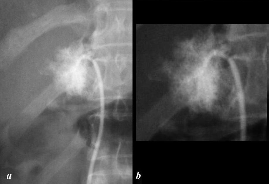
Parenchymogram – Arteriolar and Capillary Phase Combined


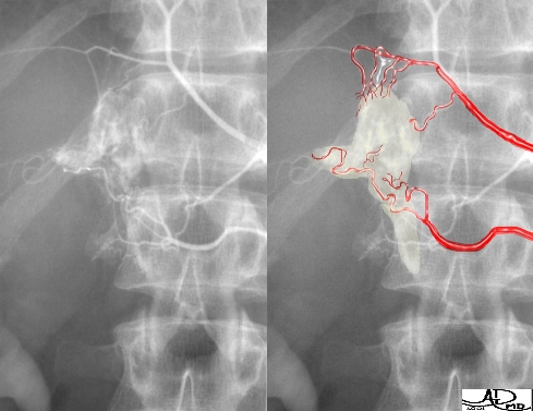
The angiogram of the adrenal artery with the catheter wedged into the artery (a) shows a parenchymal pattern magnified and highlighted in (b), and reflects opacification of a combination of the arteriole and capillary phases.
Copyright 2012 Courtesy Ashley Davidoff MD 26822cL.9b


    [nodeType] => 1
    [parentNode] => (object value omitted)
    [childNodes] => (object value omitted)
    [firstChild] => (object value omitted)
    [lastChild] => (object value omitted)
    [previousSibling] => (object value omitted)
    [nextSibling] => (object value omitted)
    [attributes] => (object value omitted)
    [ownerDocument] => (object value omitted)
    [namespaceURI] =>
    [prefix] =>
    [localName] => table
    [baseURI] =>
    [textContent] =>


Wedge Arteriogram
Parenchymogram – Arteriolar and Capillary Phase Combined


The angiogram of the adrenal artery with the catheter wedged into the artery (a) shows a parenchymal pattern magnified and highlighted in (b), and reflects opacification of a combination of the arteriole and capillary phases.
Copyright 2012 Courtesy Ashley Davidoff MD 26822cL.9b


)
DOMElement Object
(
    [schemaTypeInfo] =>
    [tagName] => td
    [firstElementChild] => (object value omitted)
    [lastElementChild] => (object value omitted)
    [childElementCount] => 1
    [previousElementSibling] =>
    [nextElementSibling] =>
    [nodeName] => td
    [nodeValue] => The angiogram of the adrenal artery with the catheter wedged into the artery (a) shows a parenchymal pattern magnified and highlighted in (b), and reflects opacification of a combination of the arteriole and capillary phases.
Copyright 2012 Courtesy Ashley Davidoff MD 26822cL.9b

    [nodeType] => 1
    [parentNode] => (object value omitted)
    [childNodes] => (object value omitted)
    [firstChild] => (object value omitted)
    [lastChild] => (object value omitted)
    [previousSibling] => (object value omitted)
    [nextSibling] => (object value omitted)
    [attributes] => (object value omitted)
    [ownerDocument] => (object value omitted)
    [namespaceURI] =>
    [prefix] =>
    [localName] => td
    [baseURI] =>
    [textContent] => The angiogram of the adrenal artery with the catheter wedged into the artery (a) shows a parenchymal pattern magnified and highlighted in (b), and reflects opacification of a combination of the arteriole and capillary phases.
Copyright 2012 Courtesy Ashley Davidoff MD 26822cL.9b

)
DOMElement Object
(
    [schemaTypeInfo] =>
    [tagName] => td
    [firstElementChild] => (object value omitted)
    [lastElementChild] => (object value omitted)
    [childElementCount] => 3
    [previousElementSibling] =>
    [nextElementSibling] =>
    [nodeName] => td
    [nodeValue] =>
Wedge Arteriogram
Parenchymogram – Arteriolar and Capillary Phase Combined

    [nodeType] => 1
    [parentNode] => (object value omitted)
    [childNodes] => (object value omitted)
    [firstChild] => (object value omitted)
    [lastChild] => (object value omitted)
    [previousSibling] => (object value omitted)
    [nextSibling] => (object value omitted)
    [attributes] => (object value omitted)
    [ownerDocument] => (object value omitted)
    [namespaceURI] =>
    [prefix] =>
    [localName] => td
    [baseURI] =>
    [textContent] =>
Wedge Arteriogram
Parenchymogram – Arteriolar and Capillary Phase Combined

)
DOMElement Object
(
    [schemaTypeInfo] =>
    [tagName] => table
    [firstElementChild] => (object value omitted)
    [lastElementChild] => (object value omitted)
    [childElementCount] => 1
    [previousElementSibling] => (object value omitted)
    [nextElementSibling] => (object value omitted)
    [nodeName] => table
    [nodeValue] =>


Arterial and Capillary Phase of the Adrenal Circulation


The angiogram of the left inferior adrenal artery shows a normal arterial phase (a) and a normal capillary phase revealing the triangular shape of the adrenal gland.
Copyright 2012 Courtesy Ashley Davidoff MD 26552bcL.8


    [nodeType] => 1
    [parentNode] => (object value omitted)
    [childNodes] => (object value omitted)
    [firstChild] => (object value omitted)
    [lastChild] => (object value omitted)
    [previousSibling] => (object value omitted)
    [nextSibling] => (object value omitted)
    [attributes] => (object value omitted)
    [ownerDocument] => (object value omitted)
    [namespaceURI] =>
    [prefix] =>
    [localName] => table
    [baseURI] =>
    [textContent] =>


Arterial and Capillary Phase of the Adrenal Circulation


The angiogram of the left inferior adrenal artery shows a normal arterial phase (a) and a normal capillary phase revealing the triangular shape of the adrenal gland.
Copyright 2012 Courtesy Ashley Davidoff MD 26552bcL.8


)
DOMElement Object
(
    [schemaTypeInfo] =>
    [tagName] => td
    [firstElementChild] => (object value omitted)
    [lastElementChild] => (object value omitted)
    [childElementCount] => 2
    [previousElementSibling] =>
    [nextElementSibling] =>
    [nodeName] => td
    [nodeValue] =>
The angiogram of the left inferior adrenal artery shows a normal arterial phase (a) and a normal capillary phase revealing the triangular shape of the adrenal gland.
Copyright 2012 Courtesy Ashley Davidoff MD 26552bcL.8

    [nodeType] => 1
    [parentNode] => (object value omitted)
    [childNodes] => (object value omitted)
    [firstChild] => (object value omitted)
    [lastChild] => (object value omitted)
    [previousSibling] => (object value omitted)
    [nextSibling] => (object value omitted)
    [attributes] => (object value omitted)
    [ownerDocument] => (object value omitted)
    [namespaceURI] =>
    [prefix] =>
    [localName] => td
    [baseURI] =>
    [textContent] =>
The angiogram of the left inferior adrenal artery shows a normal arterial phase (a) and a normal capillary phase revealing the triangular shape of the adrenal gland.
Copyright 2012 Courtesy Ashley Davidoff MD 26552bcL.8

)
DOMElement Object
(
    [schemaTypeInfo] =>
    [tagName] => td
    [firstElementChild] => (object value omitted)
    [lastElementChild] => (object value omitted)
    [childElementCount] => 2
    [previousElementSibling] =>
    [nextElementSibling] =>
    [nodeName] => td
    [nodeValue] =>
Arterial and Capillary Phase of the Adrenal Circulation
    [nodeType] => 1
    [parentNode] => (object value omitted)
    [childNodes] => (object value omitted)
    [firstChild] => (object value omitted)
    [lastChild] => (object value omitted)
    [previousSibling] => (object value omitted)
    [nextSibling] => (object value omitted)
    [attributes] => (object value omitted)
    [ownerDocument] => (object value omitted)
    [namespaceURI] =>
    [prefix] =>
    [localName] => td
    [baseURI] =>
    [textContent] =>
Arterial and Capillary Phase of the Adrenal Circulation
)
DOMElement Object
(
    [schemaTypeInfo] =>
    [tagName] => table
    [firstElementChild] => (object value omitted)
    [lastElementChild] => (object value omitted)
    [childElementCount] => 1
    [previousElementSibling] => (object value omitted)
    [nextElementSibling] => (object value omitted)
    [nodeName] => table
    [nodeValue] =>


39539


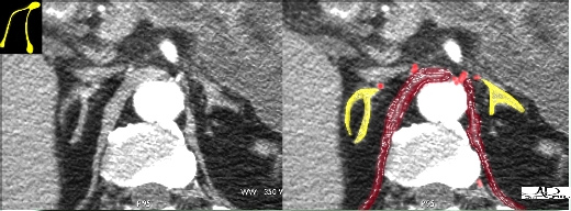
The CT scan shows the inverted “V” shaped adrenal glands reminiscent of the chicken breast overlaid in yellow in the second image.  The CT scan is in the arterial phase adrenal blood supply arteries artery CTscan imaging radiology
Copyright 2012 Courtesy Ashley DAvidoff MD 39539


    [nodeType] => 1
    [parentNode] => (object value omitted)
    [childNodes] => (object value omitted)
    [firstChild] => (object value omitted)
    [lastChild] => (object value omitted)
    [previousSibling] => (object value omitted)
    [nextSibling] => (object value omitted)
    [attributes] => (object value omitted)
    [ownerDocument] => (object value omitted)
    [namespaceURI] =>
    [prefix] =>
    [localName] => table
    [baseURI] =>
    [textContent] =>


39539


The CT scan shows the inverted “V” shaped adrenal glands reminiscent of the chicken breast overlaid in yellow in the second image.  The CT scan is in the arterial phase adrenal blood supply arteries artery CTscan imaging radiology
Copyright 2012 Courtesy Ashley DAvidoff MD 39539


)
DOMElement Object
(
    [schemaTypeInfo] =>
    [tagName] => td
    [firstElementChild] => (object value omitted)
    [lastElementChild] => (object value omitted)
    [childElementCount] => 2
    [previousElementSibling] =>
    [nextElementSibling] =>
    [nodeName] => td
    [nodeValue] =>
The CT scan shows the inverted “V” shaped adrenal glands reminiscent of the chicken breast overlaid in yellow in the second image.  The CT scan is in the arterial phase adrenal blood supply arteries artery CTscan imaging radiology
Copyright 2012 Courtesy Ashley DAvidoff MD 39539

    [nodeType] => 1
    [parentNode] => (object value omitted)
    [childNodes] => (object value omitted)
    [firstChild] => (object value omitted)
    [lastChild] => (object value omitted)
    [previousSibling] => (object value omitted)
    [nextSibling] => (object value omitted)
    [attributes] => (object value omitted)
    [ownerDocument] => (object value omitted)
    [namespaceURI] =>
    [prefix] =>
    [localName] => td
    [baseURI] =>
    [textContent] =>
The CT scan shows the inverted “V” shaped adrenal glands reminiscent of the chicken breast overlaid in yellow in the second image.  The CT scan is in the arterial phase adrenal blood supply arteries artery CTscan imaging radiology
Copyright 2012 Courtesy Ashley DAvidoff MD 39539

)
DOMElement Object
(
    [schemaTypeInfo] =>
    [tagName] => td
    [firstElementChild] => (object value omitted)
    [lastElementChild] => (object value omitted)
    [childElementCount] => 2
    [previousElementSibling] =>
    [nextElementSibling] =>
    [nodeName] => td
    [nodeValue] =>
39539

    [nodeType] => 1
    [parentNode] => (object value omitted)
    [childNodes] => (object value omitted)
    [firstChild] => (object value omitted)
    [lastChild] => (object value omitted)
    [previousSibling] => (object value omitted)
    [nextSibling] => (object value omitted)
    [attributes] => (object value omitted)
    [ownerDocument] => (object value omitted)
    [namespaceURI] =>
    [prefix] =>
    [localName] => td
    [baseURI] =>
    [textContent] =>
39539

)
DOMElement Object
(
    [schemaTypeInfo] =>
    [tagName] => table
    [firstElementChild] => (object value omitted)
    [lastElementChild] => (object value omitted)
    [childElementCount] => 1
    [previousElementSibling] => (object value omitted)
    [nextElementSibling] => (object value omitted)
    [nodeName] => table
    [nodeValue] =>


The Middle Adrenal Artery Arising from the Inferior Phrenic Artery


The angiogram of the right middle adrenal artery shows a normal arterial phase (a), magnified in b, and a normal capillary phase revealing the triangular shape of the adrenal gland (c yellow overlay).
Copyright 2012 Courtesy Ashley Davidoff MD 26566cL01.8


    [nodeType] => 1
    [parentNode] => (object value omitted)
    [childNodes] => (object value omitted)
    [firstChild] => (object value omitted)
    [lastChild] => (object value omitted)
    [previousSibling] => (object value omitted)
    [nextSibling] => (object value omitted)
    [attributes] => (object value omitted)
    [ownerDocument] => (object value omitted)
    [namespaceURI] =>
    [prefix] =>
    [localName] => table
    [baseURI] =>
    [textContent] =>


The Middle Adrenal Artery Arising from the Inferior Phrenic Artery


The angiogram of the right middle adrenal artery shows a normal arterial phase (a), magnified in b, and a normal capillary phase revealing the triangular shape of the adrenal gland (c yellow overlay).
Copyright 2012 Courtesy Ashley Davidoff MD 26566cL01.8


)
DOMElement Object
(
    [schemaTypeInfo] =>
    [tagName] => td
    [firstElementChild] => (object value omitted)
    [lastElementChild] => (object value omitted)
    [childElementCount] => 2
    [previousElementSibling] =>
    [nextElementSibling] =>
    [nodeName] => td
    [nodeValue] =>
The angiogram of the right middle adrenal artery shows a normal arterial phase (a), magnified in b, and a normal capillary phase revealing the triangular shape of the adrenal gland (c yellow overlay).
Copyright 2012 Courtesy Ashley Davidoff MD 26566cL01.8

    [nodeType] => 1
    [parentNode] => (object value omitted)
    [childNodes] => (object value omitted)
    [firstChild] => (object value omitted)
    [lastChild] => (object value omitted)
    [previousSibling] => (object value omitted)
    [nextSibling] => (object value omitted)
    [attributes] => (object value omitted)
    [ownerDocument] => (object value omitted)
    [namespaceURI] =>
    [prefix] =>
    [localName] => td
    [baseURI] =>
    [textContent] =>
The angiogram of the right middle adrenal artery shows a normal arterial phase (a), magnified in b, and a normal capillary phase revealing the triangular shape of the adrenal gland (c yellow overlay).
Copyright 2012 Courtesy Ashley Davidoff MD 26566cL01.8

)
DOMElement Object
(
    [schemaTypeInfo] =>
    [tagName] => td
    [firstElementChild] => (object value omitted)
    [lastElementChild] => (object value omitted)
    [childElementCount] => 2
    [previousElementSibling] =>
    [nextElementSibling] =>
    [nodeName] => td
    [nodeValue] =>
The Middle Adrenal Artery Arising from the Inferior Phrenic Artery

    [nodeType] => 1
    [parentNode] => (object value omitted)
    [childNodes] => (object value omitted)
    [firstChild] => (object value omitted)
    [lastChild] => (object value omitted)
    [previousSibling] => (object value omitted)
    [nextSibling] => (object value omitted)
    [attributes] => (object value omitted)
    [ownerDocument] => (object value omitted)
    [namespaceURI] =>
    [prefix] =>
    [localName] => td
    [baseURI] =>
    [textContent] =>
The Middle Adrenal Artery Arising from the Inferior Phrenic Artery

)
DOMElement Object
(
    [schemaTypeInfo] =>
    [tagName] => table
    [firstElementChild] => (object value omitted)
    [lastElementChild] => (object value omitted)
    [childElementCount] => 1
    [previousElementSibling] => (object value omitted)
    [nextElementSibling] => (object value omitted)
    [nodeName] => table
    [nodeValue] =>


The Inferior Adrenal Artery


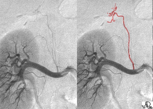
This image reflects the right renal artery with the capsular branch arising from within 2 cm. from the aortic origin. The inferior adrenal artery is overlayed in red and can be seen terminating in a tuft of vessels at the adrenal gland.
Copyright 2012 Image courtesy of Ashley Davidoff M.D. 39522


    [nodeType] => 1
    [parentNode] => (object value omitted)
    [childNodes] => (object value omitted)
    [firstChild] => (object value omitted)
    [lastChild] => (object value omitted)
    [previousSibling] => (object value omitted)
    [nextSibling] => (object value omitted)
    [attributes] => (object value omitted)
    [ownerDocument] => (object value omitted)
    [namespaceURI] =>
    [prefix] =>
    [localName] => table
    [baseURI] =>
    [textContent] =>


The Inferior Adrenal Artery


This image reflects the right renal artery with the capsular branch arising from within 2 cm. from the aortic origin. The inferior adrenal artery is overlayed in red and can be seen terminating in a tuft of vessels at the adrenal gland.
Copyright 2012 Image courtesy of Ashley Davidoff M.D. 39522


)
DOMElement Object
(
    [schemaTypeInfo] =>
    [tagName] => td
    [firstElementChild] => (object value omitted)
    [lastElementChild] => (object value omitted)
    [childElementCount] => 2
    [previousElementSibling] =>
    [nextElementSibling] =>
    [nodeName] => td
    [nodeValue] =>
This image reflects the right renal artery with the capsular branch arising from within 2 cm. from the aortic origin. The inferior adrenal artery is overlayed in red and can be seen terminating in a tuft of vessels at the adrenal gland.
Copyright 2012 Image courtesy of Ashley Davidoff M.D. 39522

    [nodeType] => 1
    [parentNode] => (object value omitted)
    [childNodes] => (object value omitted)
    [firstChild] => (object value omitted)
    [lastChild] => (object value omitted)
    [previousSibling] => (object value omitted)
    [nextSibling] => (object value omitted)
    [attributes] => (object value omitted)
    [ownerDocument] => (object value omitted)
    [namespaceURI] =>
    [prefix] =>
    [localName] => td
    [baseURI] =>
    [textContent] =>
This image reflects the right renal artery with the capsular branch arising from within 2 cm. from the aortic origin. The inferior adrenal artery is overlayed in red and can be seen terminating in a tuft of vessels at the adrenal gland.
Copyright 2012 Image courtesy of Ashley Davidoff M.D. 39522

)
DOMElement Object
(
    [schemaTypeInfo] =>
    [tagName] => td
    [firstElementChild] => (object value omitted)
    [lastElementChild] => (object value omitted)
    [childElementCount] => 2
    [previousElementSibling] =>
    [nextElementSibling] =>
    [nodeName] => td
    [nodeValue] =>
The Inferior Adrenal Artery

    [nodeType] => 1
    [parentNode] => (object value omitted)
    [childNodes] => (object value omitted)
    [firstChild] => (object value omitted)
    [lastChild] => (object value omitted)
    [previousSibling] => (object value omitted)
    [nextSibling] => (object value omitted)
    [attributes] => (object value omitted)
    [ownerDocument] => (object value omitted)
    [namespaceURI] =>
    [prefix] =>
    [localName] => td
    [baseURI] =>
    [textContent] =>
The Inferior Adrenal Artery

)
DOMElement Object
(
    [schemaTypeInfo] =>
    [tagName] => table
    [firstElementChild] => (object value omitted)
    [lastElementChild] => (object value omitted)
    [childElementCount] => 1
    [previousElementSibling] => (object value omitted)
    [nextElementSibling] => (object value omitted)
    [nodeName] => table
    [nodeValue] =>


Superior and Middle Adrenal Arteries


The arteriogram shows the inferior phrenic artery (upper branch) and the middle phrenic artery (lower branch).   The inferior phrenic artery gives rise to the superior adrenal artery.  The middle adrenal artery is also shown as a red overlay, arises directly off the aorta (not shown) and supplies the mid portion of the the right adrenal gland (yellow overlay).
Copyright 2012 Image courtesy of Ashley Davidoff M.D. 39525


    [nodeType] => 1
    [parentNode] => (object value omitted)
    [childNodes] => (object value omitted)
    [firstChild] => (object value omitted)
    [lastChild] => (object value omitted)
    [previousSibling] => (object value omitted)
    [nextSibling] => (object value omitted)
    [attributes] => (object value omitted)
    [ownerDocument] => (object value omitted)
    [namespaceURI] =>
    [prefix] =>
    [localName] => table
    [baseURI] =>
    [textContent] =>


Superior and Middle Adrenal Arteries


The arteriogram shows the inferior phrenic artery (upper branch) and the middle phrenic artery (lower branch).   The inferior phrenic artery gives rise to the superior adrenal artery.  The middle adrenal artery is also shown as a red overlay, arises directly off the aorta (not shown) and supplies the mid portion of the the right adrenal gland (yellow overlay).
Copyright 2012 Image courtesy of Ashley Davidoff M.D. 39525


)
DOMElement Object
(
    [schemaTypeInfo] =>
    [tagName] => td
    [firstElementChild] => (object value omitted)
    [lastElementChild] => (object value omitted)
    [childElementCount] => 2
    [previousElementSibling] =>
    [nextElementSibling] =>
    [nodeName] => td
    [nodeValue] =>
The arteriogram shows the inferior phrenic artery (upper branch) and the middle phrenic artery (lower branch).   The inferior phrenic artery gives rise to the superior adrenal artery.  The middle adrenal artery is also shown as a red overlay, arises directly off the aorta (not shown) and supplies the mid portion of the the right adrenal gland (yellow overlay).
Copyright 2012 Image courtesy of Ashley Davidoff M.D. 39525

    [nodeType] => 1
    [parentNode] => (object value omitted)
    [childNodes] => (object value omitted)
    [firstChild] => (object value omitted)
    [lastChild] => (object value omitted)
    [previousSibling] => (object value omitted)
    [nextSibling] => (object value omitted)
    [attributes] => (object value omitted)
    [ownerDocument] => (object value omitted)
    [namespaceURI] =>
    [prefix] =>
    [localName] => td
    [baseURI] =>
    [textContent] =>
The arteriogram shows the inferior phrenic artery (upper branch) and the middle phrenic artery (lower branch).   The inferior phrenic artery gives rise to the superior adrenal artery.  The middle adrenal artery is also shown as a red overlay, arises directly off the aorta (not shown) and supplies the mid portion of the the right adrenal gland (yellow overlay).
Copyright 2012 Image courtesy of Ashley Davidoff M.D. 39525

)
DOMElement Object
(
    [schemaTypeInfo] =>
    [tagName] => td
    [firstElementChild] => (object value omitted)
    [lastElementChild] => (object value omitted)
    [childElementCount] => 2
    [previousElementSibling] =>
    [nextElementSibling] =>
    [nodeName] => td
    [nodeValue] =>
Superior and Middle Adrenal Arteries

    [nodeType] => 1
    [parentNode] => (object value omitted)
    [childNodes] => (object value omitted)
    [firstChild] => (object value omitted)
    [lastChild] => (object value omitted)
    [previousSibling] => (object value omitted)
    [nextSibling] => (object value omitted)
    [attributes] => (object value omitted)
    [ownerDocument] => (object value omitted)
    [namespaceURI] =>
    [prefix] =>
    [localName] => td
    [baseURI] =>
    [textContent] =>
Superior and Middle Adrenal Arteries

)
DOMElement Object
(
    [schemaTypeInfo] =>
    [tagName] => table
    [firstElementChild] => (object value omitted)
    [lastElementChild] => (object value omitted)
    [childElementCount] => 1
    [previousElementSibling] => (object value omitted)
    [nextElementSibling] => (object value omitted)
    [nodeName] => table
    [nodeValue] =>


Blood Supply of the Adrenal Gland


There are three adrenal arteries – the superior adrenal artery arises from the inferior phrenic artery; the middle adrenal artery that arises directly off the aorta; and the inferior adrenal artery that arises from the renal artery usually as a branch of the capsular artery. This schematic only shows 3 branches per vessel, but in reality there is extensive branching before each artery actually enters the gland.
Copyright 2012 Image courtesy of Ashley Davidoff M.D. 39512


    [nodeType] => 1
    [parentNode] => (object value omitted)
    [childNodes] => (object value omitted)
    [firstChild] => (object value omitted)
    [lastChild] => (object value omitted)
    [previousSibling] => (object value omitted)
    [nextSibling] => (object value omitted)
    [attributes] => (object value omitted)
    [ownerDocument] => (object value omitted)
    [namespaceURI] =>
    [prefix] =>
    [localName] => table
    [baseURI] =>
    [textContent] =>


Blood Supply of the Adrenal Gland


There are three adrenal arteries – the superior adrenal artery arises from the inferior phrenic artery; the middle adrenal artery that arises directly off the aorta; and the inferior adrenal artery that arises from the renal artery usually as a branch of the capsular artery. This schematic only shows 3 branches per vessel, but in reality there is extensive branching before each artery actually enters the gland.
Copyright 2012 Image courtesy of Ashley Davidoff M.D. 39512


)
DOMElement Object
(
    [schemaTypeInfo] =>
    [tagName] => td
    [firstElementChild] => (object value omitted)
    [lastElementChild] => (object value omitted)
    [childElementCount] => 2
    [previousElementSibling] =>
    [nextElementSibling] =>
    [nodeName] => td
    [nodeValue] =>
There are three adrenal arteries – the superior adrenal artery arises from the inferior phrenic artery; the middle adrenal artery that arises directly off the aorta; and the inferior adrenal artery that arises from the renal artery usually as a branch of the capsular artery. This schematic only shows 3 branches per vessel, but in reality there is extensive branching before each artery actually enters the gland.
Copyright 2012 Image courtesy of Ashley Davidoff M.D. 39512

    [nodeType] => 1
    [parentNode] => (object value omitted)
    [childNodes] => (object value omitted)
    [firstChild] => (object value omitted)
    [lastChild] => (object value omitted)
    [previousSibling] => (object value omitted)
    [nextSibling] => (object value omitted)
    [attributes] => (object value omitted)
    [ownerDocument] => (object value omitted)
    [namespaceURI] =>
    [prefix] =>
    [localName] => td
    [baseURI] =>
    [textContent] =>
There are three adrenal arteries – the superior adrenal artery arises from the inferior phrenic artery; the middle adrenal artery that arises directly off the aorta; and the inferior adrenal artery that arises from the renal artery usually as a branch of the capsular artery. This schematic only shows 3 branches per vessel, but in reality there is extensive branching before each artery actually enters the gland.
Copyright 2012 Image courtesy of Ashley Davidoff M.D. 39512

)
DOMElement Object
(
    [schemaTypeInfo] =>
    [tagName] => td
    [firstElementChild] => (object value omitted)
    [lastElementChild] => (object value omitted)
    [childElementCount] => 2
    [previousElementSibling] =>
    [nextElementSibling] =>
    [nodeName] => td
    [nodeValue] =>
Blood Supply of the Adrenal Gland

    [nodeType] => 1
    [parentNode] => (object value omitted)
    [childNodes] => (object value omitted)
    [firstChild] => (object value omitted)
    [lastChild] => (object value omitted)
    [previousSibling] => (object value omitted)
    [nextSibling] => (object value omitted)
    [attributes] => (object value omitted)
    [ownerDocument] => (object value omitted)
    [namespaceURI] =>
    [prefix] =>
    [localName] => td
    [baseURI] =>
    [textContent] =>
Blood Supply of the Adrenal Gland

)

