Histology
The Common Vein
Sumit Karia MD Ashley Davidoff MD
Copyright 2010
Introduction
The brain is divided into two main macsroscopic and microscopic regions
gray matter and white matter
Gray Matter
|

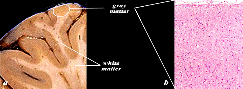
Histology 10X of the Gray Matter |
|
The image shows a gross specimen photographed to show the gyral pattern and the distinction between the rim of gray matter (light brown) and the white matter (cream colored) (a). The microsopic section (b), shows the histological equivalent of the gray matter as might be seen in the white circle consisting of a superficial layer of meninges and the deep layer of gray matter . A variety of cell types and sizes can be distinguished evn at this microscopic level.
Image Courtesy of Thomas W.Smith, MD; Department of Pathology, University of Massachusetts Medical School. 97630bc.8
|
White Matter
|

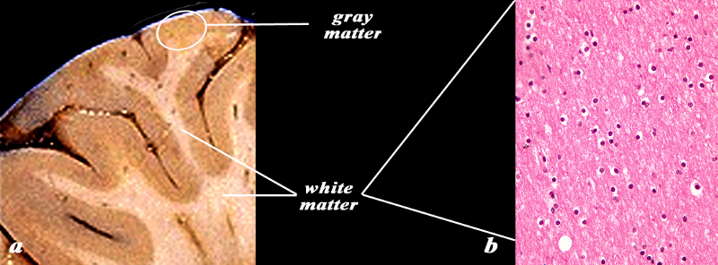
Histology of the White Matter (40X) |
|
The image shows a gross specimen (a) photographed to show the gyral pattern and the distinction between the rim of gray matter (light brown) and the white matter (cream colored). The microscopic section (b) is 40X magnified, and shows the histological equivalent of the white matter consisting mainly of oligodendrocytes characterized by cells with a white halo. No neurons are present
Image Courtesy of Thomas W.Smith, MD; Department of Pathology, University of Massachusetts Medical School. 97630bc02.8
|
The nervous system is composed of two major cellular types:
Neurons, highly differentiated cells, equipped with varied functional activities
Glial cells, which are the most numerous cells of the nervous system and ensure the physical and nutritional support of neurons
The function of the central nervous system is, in short, to collect stimuli (internal and external), to integrate it, analyze it, and provide a suitable response via the effector organs.
All of this rests on a fundamental property of neurons: “excitability” and their organization in a cellular network interconnected.

The Cerebral Cortex
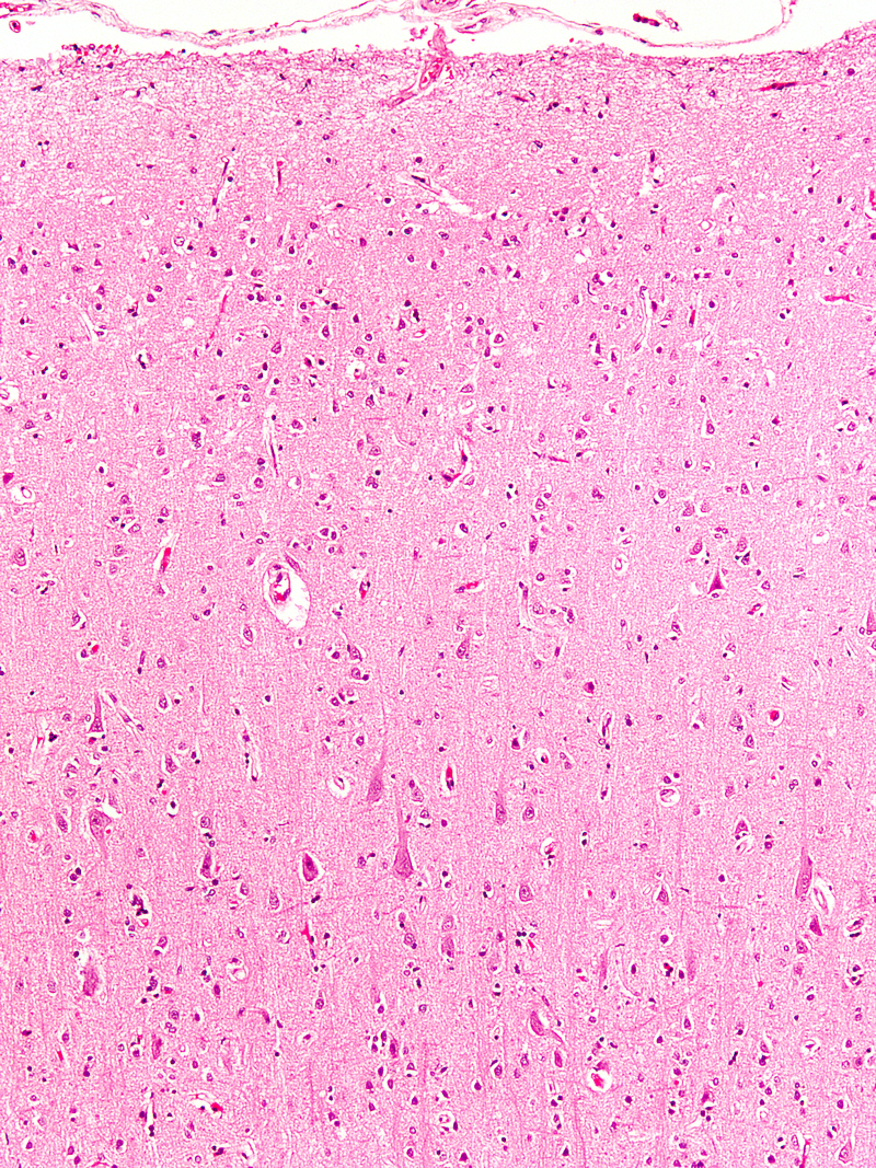
10X Magnification
|
| This image of the cerebral cortex suggests an organizational pattern of the 6 layers of the cortex but the exact depiction of these layers requires th use of special stains. The larger cells in the deper layers represent large neurons. In this section the stain used is hemotoxylin and eosin (H&E).
Courtesy of Thomas W.Smith, MD; Department of Pathology, University of Massachusetts Medical School. 97377
|

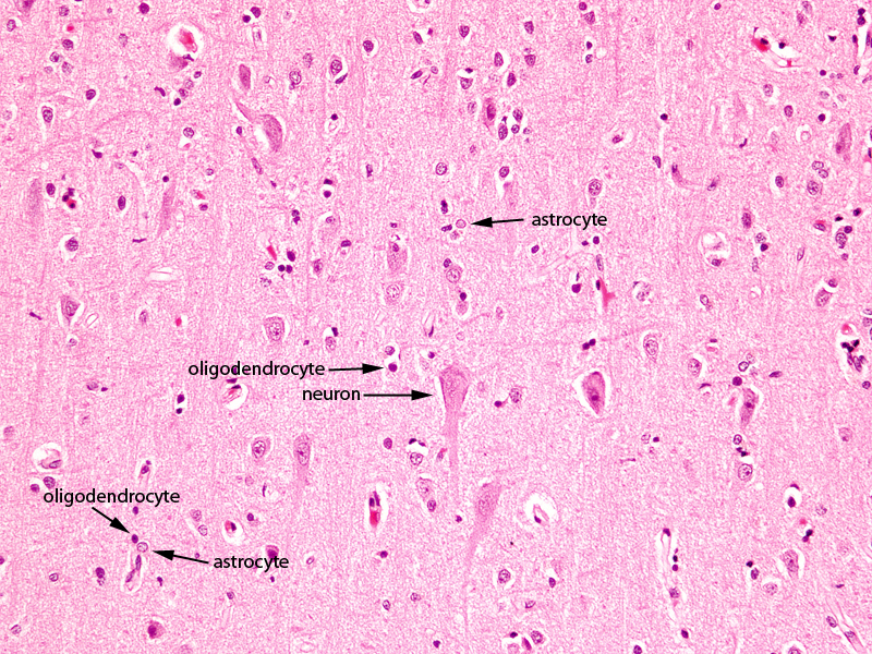
Higher Power Cerebral Cortex |
|
The image shows a histological section of the brain at 20X magnification. The section shows the 3 of the common basic types of brain cells; The largest is the neuronal cell which is only found in gray matter. They are 10 times larger but less common than the second type of cell called the glial cell. The astrocytes is the second largest cell and often lies in close association with the blood vessels. As third type of cell, also a glial cell is the oligodendrocyte. It is smaller than the other type of glial cell the astrocytre and it is responsible to myelinate the white matter. It is the predominant cell of white matter
Courtesy of Thomas W.Smith, MD; Department of Pathology, University of Massachusetts Medical School. 97799
|
Neurons
Although there are different types of neurons and variations from one region of the CNS to another, neurons have common features.
They are cells with a large cell body which contains a nucleus with a prominent nucleolus and dispersed chromatin visible.
The peri-nuclear cytoplasm is characterized by the presence of a large endoplasmic reticulum in clusters, making the Nissl bodies. The cytoplasm also contains numerous mitochondria and a cytoskeleton, composed of intermediate filaments and microfilaments.
The unique feature of neurons is represented by their cell processes, initially described in the early twentieth century by two pioneers in neurohistology (Golgi and Cajal). Neurons have two types of processes: dendrites and axons. Dendrites are usually short, multiple and highly branched, while the axon is a single extension, often with multiple endings. The axon is surrounded by a sheath ? myelin (composed of lipids) – created by a type of glial cells, the oligodendrocytes.
Neurons are separated by highly specialized intercellular junctions: the synapses, which allow communications between neurons or effector cells.
Glial Cells
The function of neurons is conditioned by glial cells: astrocytes are essential for neuron function since they collect the substrates from the blood required for metabolism of neurons and they regulate the composition of the extracellular environment of the nervous system.
Oligodendrocytes are responsible for myelination of axons.
Another type of glial cell, called microglia, corresponds to mononuclear cells (monocyte-macrophage). They are visible only in case of damage to the nervous system to ensure its defense.
Astrocytes
Astrocytes are the most numerous cells of the central nervous system. They are so called because they have branched extensions that occupy the space between neurons. They also have cell processes which are involve vessels and contribute to the formation of the blood-brain barrier.
The macroscopic appearance reveals two aspects: gray matter and white matter. The gray matter corresponds to the cell bodies of neurons, while white matter corresponds to axons (nerve fibers). The white color is due to the lipid content of the myelin sheath. The distribution and proportions of white matter / gray matter will change in different regions of the nervous system.
90% of the cerebral cortex has 6 layers (motor, sensory and associative areas). There is a 3-layer cortex (olfactory cortex and limbic system), which represents 10% of the cerebral cortex.
From an architectural point of view, the neocortex contains five different types of neurons that will fit together in 6 layers. These layers have no clear demarcation between themselves, but there is a difference on the appearance, size, density of neurons and variations among cortical regions, depending on the thickness of the cortex and its function.
Types of Neurons in the Cerbral Cortex
The pyramidal cells have a cell body with a pyramidal shape, with an apex oriented towards the surface of the cortex. The axon arises from the base and goes across the white matter. Pyramidal cells have multiple dendrites: a thick dendrite which ramifies on the surface and several small dendrites branched laterally. They vary in size; the smaller are more present in the surface while larger pyramidal cells in the motor cortex (Betz cells) are deeply located.
The pyramidal cells have glutamate as a mediator, which stimulates their influx.
Granular cells are small stellate cells, equipped with multiple dendrites, and a short vertical axon. These cells mediate GABA that inhibits excitability.
Martinotti cells are small polygonal neurons with few dendrites, a thin axon that goes to the surface where it travels horizontally;
Spindle cells are perpendicular to the cortical surface, the dendrites are branched laterally and the axon heads to the surface;
Cells of Cajal are horizontal, fusiform and oriented parallel to the cortical surface and can be found in the surface layer of the cortex, where their axon synapses with pyramidal cells.
The Different Layers in the Cerbral cortex
The five types of neurons will fit together in 6 cell layers.
From outside to inside, there are:
Layer I or “plexiform layer” that is scarce in cells; it contains few Cajal cells, but mostly dendrites and axons of cortical neurons;
Layer II: “External Granular Layer” contains a dense population of cells involving granular neurons and also small pyramidal cells
Layer III: ?External Pyramidal Layer? is populated by medium-sized pyramidal cells;
Layer IV: ?Internal Granular Layer? is very dense, made of granular neurons
Layer V: “Internal Pyramidal Layer” or ?Ganglionic Layer? contains large pyramidal cells, and also small stellate cells and Martinotti cells;
Layer VI: “Multiforme”, contains a mixture of small cells (stellate, pyramidal, Martinotti).
neuroglial glue cells hold brain together turn over astrocytes do not conduct
DOMElement Object
(
    [schemaTypeInfo] =>
    [tagName] => table
    [firstElementChild] => (object value omitted)
    [lastElementChild] => (object value omitted)
    [childElementCount] => 1
    [previousElementSibling] => (object value omitted)
    [nextElementSibling] => (object value omitted)
    [nodeName] => table
    [nodeValue] =>


Higher Power Cerebral Cortex


The image shows a histological section of the brain at 20X magnification. The section shows the 3 of the common basic types of brain cells; The largest is the neuronal cell which is only found in gray matter. They are 10 times larger but less common than the second type of cell called the glial cell. The astrocytes is the second largest cell and often lies in close association with the blood vessels. As third type of cell, also a glial cell is the oligodendrocyte. It is smaller than the other type of glial cell the astrocytre and it is responsible to myelinate the white matter. It is the predominant cell of white matter
Courtesy of Thomas W.Smith, MD; Department of Pathology, University of Massachusetts Medical School. 97799


    [nodeType] => 1
    [parentNode] => (object value omitted)
    [childNodes] => (object value omitted)
    [firstChild] => (object value omitted)
    [lastChild] => (object value omitted)
    [previousSibling] => (object value omitted)
    [nextSibling] => (object value omitted)
    [attributes] => (object value omitted)
    [ownerDocument] => (object value omitted)
    [namespaceURI] =>
    [prefix] =>
    [localName] => table
    [baseURI] =>
    [textContent] =>


Higher Power Cerebral Cortex


The image shows a histological section of the brain at 20X magnification. The section shows the 3 of the common basic types of brain cells; The largest is the neuronal cell which is only found in gray matter. They are 10 times larger but less common than the second type of cell called the glial cell. The astrocytes is the second largest cell and often lies in close association with the blood vessels. As third type of cell, also a glial cell is the oligodendrocyte. It is smaller than the other type of glial cell the astrocytre and it is responsible to myelinate the white matter. It is the predominant cell of white matter
Courtesy of Thomas W.Smith, MD; Department of Pathology, University of Massachusetts Medical School. 97799


)
DOMElement Object
(
    [schemaTypeInfo] =>
    [tagName] => td
    [firstElementChild] => (object value omitted)
    [lastElementChild] => (object value omitted)
    [childElementCount] => 2
    [previousElementSibling] =>
    [nextElementSibling] =>
    [nodeName] => td
    [nodeValue] =>
The image shows a histological section of the brain at 20X magnification. The section shows the 3 of the common basic types of brain cells; The largest is the neuronal cell which is only found in gray matter. They are 10 times larger but less common than the second type of cell called the glial cell. The astrocytes is the second largest cell and often lies in close association with the blood vessels. As third type of cell, also a glial cell is the oligodendrocyte. It is smaller than the other type of glial cell the astrocytre and it is responsible to myelinate the white matter. It is the predominant cell of white matter
Courtesy of Thomas W.Smith, MD; Department of Pathology, University of Massachusetts Medical School. 97799

    [nodeType] => 1
    [parentNode] => (object value omitted)
    [childNodes] => (object value omitted)
    [firstChild] => (object value omitted)
    [lastChild] => (object value omitted)
    [previousSibling] => (object value omitted)
    [nextSibling] => (object value omitted)
    [attributes] => (object value omitted)
    [ownerDocument] => (object value omitted)
    [namespaceURI] =>
    [prefix] =>
    [localName] => td
    [baseURI] =>
    [textContent] =>
The image shows a histological section of the brain at 20X magnification. The section shows the 3 of the common basic types of brain cells; The largest is the neuronal cell which is only found in gray matter. They are 10 times larger but less common than the second type of cell called the glial cell. The astrocytes is the second largest cell and often lies in close association with the blood vessels. As third type of cell, also a glial cell is the oligodendrocyte. It is smaller than the other type of glial cell the astrocytre and it is responsible to myelinate the white matter. It is the predominant cell of white matter
Courtesy of Thomas W.Smith, MD; Department of Pathology, University of Massachusetts Medical School. 97799

)
DOMElement Object
(
    [schemaTypeInfo] =>
    [tagName] => td
    [firstElementChild] => (object value omitted)
    [lastElementChild] => (object value omitted)
    [childElementCount] => 2
    [previousElementSibling] =>
    [nextElementSibling] =>
    [nodeName] => td
    [nodeValue] =>
Higher Power Cerebral Cortex
    [nodeType] => 1
    [parentNode] => (object value omitted)
    [childNodes] => (object value omitted)
    [firstChild] => (object value omitted)
    [lastChild] => (object value omitted)
    [previousSibling] => (object value omitted)
    [nextSibling] => (object value omitted)
    [attributes] => (object value omitted)
    [ownerDocument] => (object value omitted)
    [namespaceURI] =>
    [prefix] =>
    [localName] => td
    [baseURI] =>
    [textContent] =>
Higher Power Cerebral Cortex
)
DOMElement Object
(
    [schemaTypeInfo] =>
    [tagName] => td
    [firstElementChild] => (object value omitted)
    [lastElementChild] => (object value omitted)
    [childElementCount] => 3
    [previousElementSibling] =>
    [nextElementSibling] =>
    [nodeName] => td
    [nodeValue] =>
The Cerebral Cortex
10X Magnification

    [nodeType] => 1
    [parentNode] => (object value omitted)
    [childNodes] => (object value omitted)
    [firstChild] => (object value omitted)
    [lastChild] => (object value omitted)
    [previousSibling] => (object value omitted)
    [nextSibling] => (object value omitted)
    [attributes] => (object value omitted)
    [ownerDocument] => (object value omitted)
    [namespaceURI] =>
    [prefix] =>
    [localName] => td
    [baseURI] =>
    [textContent] =>
The Cerebral Cortex
10X Magnification

)
DOMElement Object
(
    [schemaTypeInfo] =>
    [tagName] => table
    [firstElementChild] => (object value omitted)
    [lastElementChild] => (object value omitted)
    [childElementCount] => 1
    [previousElementSibling] => (object value omitted)
    [nextElementSibling] => (object value omitted)
    [nodeName] => table
    [nodeValue] =>


Histology of the White Matter (40X)


The image shows a gross specimen (a) photographed to show the gyral pattern and the distinction between the rim of gray matter (light brown) and the white matter (cream colored). The microscopic section (b) is 40X magnified, and shows the histological equivalent of the white matter consisting mainly of oligodendrocytes characterized by cells with a white halo. No neurons are present
Image Courtesy of Thomas W.Smith, MD; Department of Pathology, University of Massachusetts Medical School. 97630bc02.8


    [nodeType] => 1
    [parentNode] => (object value omitted)
    [childNodes] => (object value omitted)
    [firstChild] => (object value omitted)
    [lastChild] => (object value omitted)
    [previousSibling] => (object value omitted)
    [nextSibling] => (object value omitted)
    [attributes] => (object value omitted)
    [ownerDocument] => (object value omitted)
    [namespaceURI] =>
    [prefix] =>
    [localName] => table
    [baseURI] =>
    [textContent] =>


Histology of the White Matter (40X)


The image shows a gross specimen (a) photographed to show the gyral pattern and the distinction between the rim of gray matter (light brown) and the white matter (cream colored). The microscopic section (b) is 40X magnified, and shows the histological equivalent of the white matter consisting mainly of oligodendrocytes characterized by cells with a white halo. No neurons are present
Image Courtesy of Thomas W.Smith, MD; Department of Pathology, University of Massachusetts Medical School. 97630bc02.8


)
DOMElement Object
(
    [schemaTypeInfo] =>
    [tagName] => td
    [firstElementChild] => (object value omitted)
    [lastElementChild] => (object value omitted)
    [childElementCount] => 2
    [previousElementSibling] =>
    [nextElementSibling] =>
    [nodeName] => td
    [nodeValue] =>
The image shows a gross specimen (a) photographed to show the gyral pattern and the distinction between the rim of gray matter (light brown) and the white matter (cream colored). The microscopic section (b) is 40X magnified, and shows the histological equivalent of the white matter consisting mainly of oligodendrocytes characterized by cells with a white halo. No neurons are present
Image Courtesy of Thomas W.Smith, MD; Department of Pathology, University of Massachusetts Medical School. 97630bc02.8

    [nodeType] => 1
    [parentNode] => (object value omitted)
    [childNodes] => (object value omitted)
    [firstChild] => (object value omitted)
    [lastChild] => (object value omitted)
    [previousSibling] => (object value omitted)
    [nextSibling] => (object value omitted)
    [attributes] => (object value omitted)
    [ownerDocument] => (object value omitted)
    [namespaceURI] =>
    [prefix] =>
    [localName] => td
    [baseURI] =>
    [textContent] =>
The image shows a gross specimen (a) photographed to show the gyral pattern and the distinction between the rim of gray matter (light brown) and the white matter (cream colored). The microscopic section (b) is 40X magnified, and shows the histological equivalent of the white matter consisting mainly of oligodendrocytes characterized by cells with a white halo. No neurons are present
Image Courtesy of Thomas W.Smith, MD; Department of Pathology, University of Massachusetts Medical School. 97630bc02.8

)
DOMElement Object
(
    [schemaTypeInfo] =>
    [tagName] => td
    [firstElementChild] => (object value omitted)
    [lastElementChild] => (object value omitted)
    [childElementCount] => 2
    [previousElementSibling] =>
    [nextElementSibling] =>
    [nodeName] => td
    [nodeValue] =>

Histology of the White Matter (40X)
    [nodeType] => 1
    [parentNode] => (object value omitted)
    [childNodes] => (object value omitted)
    [firstChild] => (object value omitted)
    [lastChild] => (object value omitted)
    [previousSibling] => (object value omitted)
    [nextSibling] => (object value omitted)
    [attributes] => (object value omitted)
    [ownerDocument] => (object value omitted)
    [namespaceURI] =>
    [prefix] =>
    [localName] => td
    [baseURI] =>
    [textContent] =>

Histology of the White Matter (40X)
)
DOMElement Object
(
    [schemaTypeInfo] =>
    [tagName] => table
    [firstElementChild] => (object value omitted)
    [lastElementChild] => (object value omitted)
    [childElementCount] => 1
    [previousElementSibling] => (object value omitted)
    [nextElementSibling] => (object value omitted)
    [nodeName] => table
    [nodeValue] =>


Histology 10X of the Gray Matter


The image shows a gross specimen photographed to show the gyral pattern and the distinction between the rim of gray matter (light brown) and the white matter (cream colored) (a). The microsopic section (b), shows the histological equivalent of the gray matter as might be seen in the white circle consisting of a superficial layer of meninges and the deep layer of gray matter .  A variety of cell types and sizes can be distinguished evn at this microscopic level.
Image Courtesy of Thomas W.Smith, MD; Department of Pathology, University of Massachusetts Medical School. 97630bc.8


    [nodeType] => 1
    [parentNode] => (object value omitted)
    [childNodes] => (object value omitted)
    [firstChild] => (object value omitted)
    [lastChild] => (object value omitted)
    [previousSibling] => (object value omitted)
    [nextSibling] => (object value omitted)
    [attributes] => (object value omitted)
    [ownerDocument] => (object value omitted)
    [namespaceURI] =>
    [prefix] =>
    [localName] => table
    [baseURI] =>
    [textContent] =>


Histology 10X of the Gray Matter


The image shows a gross specimen photographed to show the gyral pattern and the distinction between the rim of gray matter (light brown) and the white matter (cream colored) (a). The microsopic section (b), shows the histological equivalent of the gray matter as might be seen in the white circle consisting of a superficial layer of meninges and the deep layer of gray matter .  A variety of cell types and sizes can be distinguished evn at this microscopic level.
Image Courtesy of Thomas W.Smith, MD; Department of Pathology, University of Massachusetts Medical School. 97630bc.8


)
DOMElement Object
(
    [schemaTypeInfo] =>
    [tagName] => td
    [firstElementChild] => (object value omitted)
    [lastElementChild] => (object value omitted)
    [childElementCount] => 2
    [previousElementSibling] =>
    [nextElementSibling] =>
    [nodeName] => td
    [nodeValue] =>
The image shows a gross specimen photographed to show the gyral pattern and the distinction between the rim of gray matter (light brown) and the white matter (cream colored) (a). The microsopic section (b), shows the histological equivalent of the gray matter as might be seen in the white circle consisting of a superficial layer of meninges and the deep layer of gray matter .  A variety of cell types and sizes can be distinguished evn at this microscopic level.
Image Courtesy of Thomas W.Smith, MD; Department of Pathology, University of Massachusetts Medical School. 97630bc.8

    [nodeType] => 1
    [parentNode] => (object value omitted)
    [childNodes] => (object value omitted)
    [firstChild] => (object value omitted)
    [lastChild] => (object value omitted)
    [previousSibling] => (object value omitted)
    [nextSibling] => (object value omitted)
    [attributes] => (object value omitted)
    [ownerDocument] => (object value omitted)
    [namespaceURI] =>
    [prefix] =>
    [localName] => td
    [baseURI] =>
    [textContent] =>
The image shows a gross specimen photographed to show the gyral pattern and the distinction between the rim of gray matter (light brown) and the white matter (cream colored) (a). The microsopic section (b), shows the histological equivalent of the gray matter as might be seen in the white circle consisting of a superficial layer of meninges and the deep layer of gray matter .  A variety of cell types and sizes can be distinguished evn at this microscopic level.
Image Courtesy of Thomas W.Smith, MD; Department of Pathology, University of Massachusetts Medical School. 97630bc.8

)
DOMElement Object
(
    [schemaTypeInfo] =>
    [tagName] => td
    [firstElementChild] => (object value omitted)
    [lastElementChild] => (object value omitted)
    [childElementCount] => 2
    [previousElementSibling] =>
    [nextElementSibling] =>
    [nodeName] => td
    [nodeValue] =>

Histology 10X of the Gray Matter
    [nodeType] => 1
    [parentNode] => (object value omitted)
    [childNodes] => (object value omitted)
    [firstChild] => (object value omitted)
    [lastChild] => (object value omitted)
    [previousSibling] => (object value omitted)
    [nextSibling] => (object value omitted)
    [attributes] => (object value omitted)
    [ownerDocument] => (object value omitted)
    [namespaceURI] =>
    [prefix] =>
    [localName] => td
    [baseURI] =>
    [textContent] =>

Histology 10X of the Gray Matter
)


