Gyri
Ashley Davidoff MD
The Common Vein Copyright 2010
Definition
Gyri are the ridges of brain tissue seen on the surface of the brain that are separated by fissures and sulci consisting of mound of gray and white matted that have been thrown into folds enabling to increase the surface area of the brain in order to optimise space and hence optimise function.
The folds structurally have characteristic morphological features that allow them to be recognized, named and attributed certain functions.

Overall View of the Gyri |
|
The MRI represents a sagittal image of the brain in an elderly patient where the associated atrophy accentuates the gyri. This general view reveals the gyri of the frontal lobe (light green, parietal lobe (mauve), and occipital lobe (darker purlple) with the inner cingulate gyrus seen in darker green) Which exemplirifies the gyra The specimen shows the approximate position of the 3 major frontal gyri in the anterior location including the superior, middle and inferior gyrus. The cingulate gyrus is not really part of the forebrain but part of the limbic system. It holds a medial deep and very central position.
Courtesy Ashley Davidoff MD Copyright 2010 92170b06b01.8sgL.8
|
The precentral gyrus that lies in front of the central sulcus is the motor cortex for example; and the postcentral gyrus that lies behind the central sulcus is the sensory cortex. These gyri run in an almost vertical orientation, and run parallel to each other and parallel to the central sulcus (fissure of Rolando), precentral sulcus and postcentral sulcus.
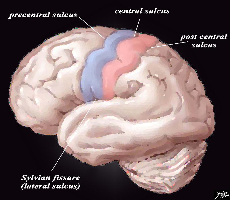
The Precentral Gyrus (blue) and Postcentral Gyrus (pink) |
|
The lateral view of the external brain shows the most easily recognized gyri – the precentral gyrus motor cortex (blue) and the post central sensory gyrus (pink) surrounded by the sulci. There are 4 important central sulci (aka fissures), 3 of which are vertically oriented and 1 of which is horizontal. The vertical sulci include the central sulcus, precentral sulcus and post central sulcus. The central sulcus is important because it separates the frontal lobe from the parietal lobe. The post central sulcus is important because together with the central sulcus it forms the borders of the post central gyrus (pink) which is the sensory cortex. The precentral sulcus together with the central sulcus forms the borders post central gyrus (blue) which is the motor cortex. The Sylvian fissure or lateral sulcus runs horizontally and separates the temporal lobe below from the frontal lobe and parietal lobe above.
Courtesy Ashley Davidoff copyright 2010 all rights reserved 83029b01b01b02g01L.8s
|
The gyri are best described as they are seen from the lateral examination of the brain and from a sagittal view of the brain
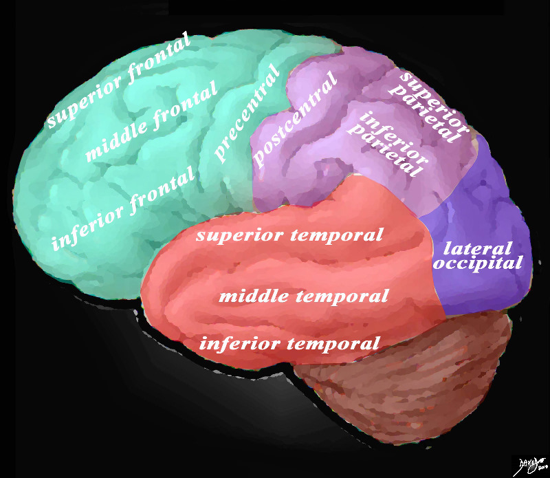
Overview of the Gyri from the Lateral External View |
|
The lateral view of the brain shows the frontal lobe (green) parietal lobe (light mauve), the occipital lobe (purple) and the temporal lobe (red) In this view the frontal lobe gyri that are visible are; superior frontal, middle frontal, inferior frontal and precentral gyri. The parietal gyri include the superior parietal and inferior parietal. The occipital gyrus that is visible is the lateral occipital. The temporal lobe gyri include the superior, middle and inferior temporal gyri.
Courtesy Ashley Davidoff MD Copyright 2010 83029d05g01.8s
|
|
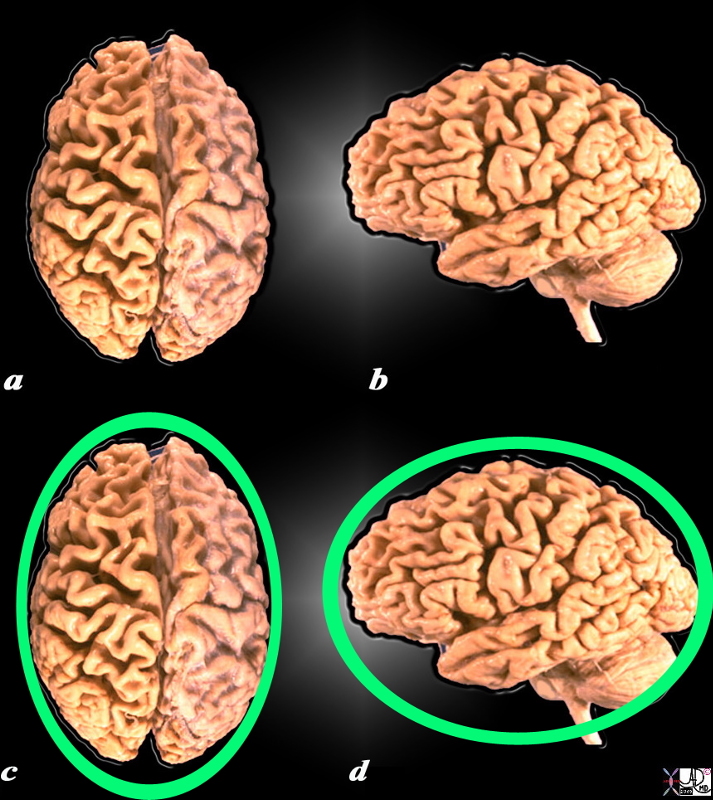
General Shape of the Brain
External Surface |
|
The ovoid shape is easily appreciated when the external surface of the brain is viewed from above, and although it in general it has an ovoid shape when viewed from the side, more complex shapes start to appear, as the temporal and cerebellum start to appear.
source unknown modified by Davidoff 52981b05.8sd01
|
|
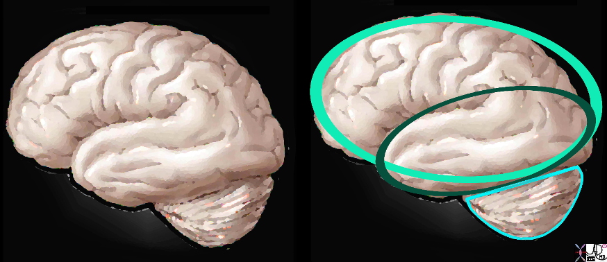
Basic Shapes
Lateral Projection |
|
When the external surface of the brain is viewed from its lateral aspect, , the ovoid shape of the conglomerate frontal parietal, occipital and part of the temporal lobe is still maintained. The part of the temporal lobe that projects into the middle cranial fossa adds a second oval directed slightly inferiorly. The medulla adds a new dimension having the shape somewhere between a half a sphere and the bottom of a top.
Davidoff art All Rights reserved copyright 2010 83029d03c01.9s
|
As one explores the surface of the brain the external shape becomes more complex by virtue of the ridges in the brain surface. The ridges and clefts – gyri and sulci have characteristic shape and position and each named according to structure and function.
There are not many other structures that look like the brain in nature but there are two that have similar convolutional appearance including the walnut as described above and brain coral.
|
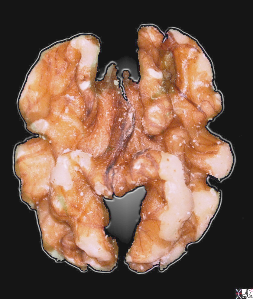
Form and Colors reminiscent of the Brain |
|
This image of a walnut has a few features that are reminiscent of a brain, including the symmetric hemispheric components, the pinkish color, and the undulations of the surface. However in reality it is about 1.8cms long, and only 5-8mms high, and is hard.
Courtesy Ashley Davidoff MD copyright 2010 all rights reserved 100286pb.93s
|
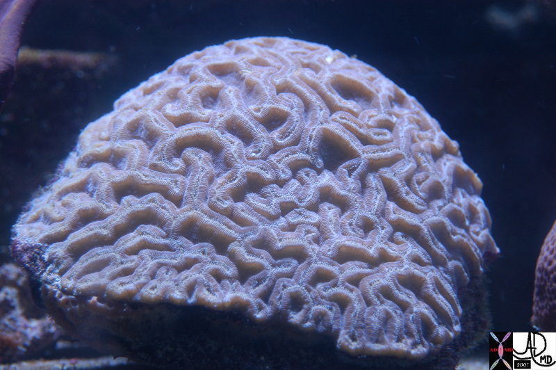
Brain Coral |
|
The brain coral
Courtesy Ashley Davidoff MD Copyright 2010 83018.800
|
Looking at the brain from above, it has, in its midline, a deep fissure that divides the organ in 2 halves called hemispheres. The external surface is also marked by serpiginous ridges and furrows called the gyri and sulci.
The gyri and fissures have characteristic form and position though there is variation among individuals. As we advance into the structure we will explore the detail of the gyri and sulci. As a a start we will exemplify the most central of the sulci and gyri in the diagram below.
|

Introduction to the Central Sulci and Gyri
Characteristic Location and Shape |
|
The lateral view of the brain shows the 4 important central sulci (aka fissures), 3 of which are vertically oriented and 1 of which is horizontal. The vertical sulci include the central sulcus, precentral sulcus and post central sulcus. The central sulcus is important because it separates the frontal lobe from the parietal lobe. The post central sulcus is important because together with the central sulcus it forms the borders of the post central gyrus (pink) which is the sensory cortex. The precentral sulcus together with the central sulcus forms the borders post central gyrus (blue) which is the motor cortex. The Sylvian fissure or lateral sulcus runs horizontally and separates the temporal lobe below from the frontal lobe and parietal lobe above.
Courtesy Ashley Davidoff copyright 2010 all rights reserved 83029b01b01b02g01L.8s |
Applied Biology
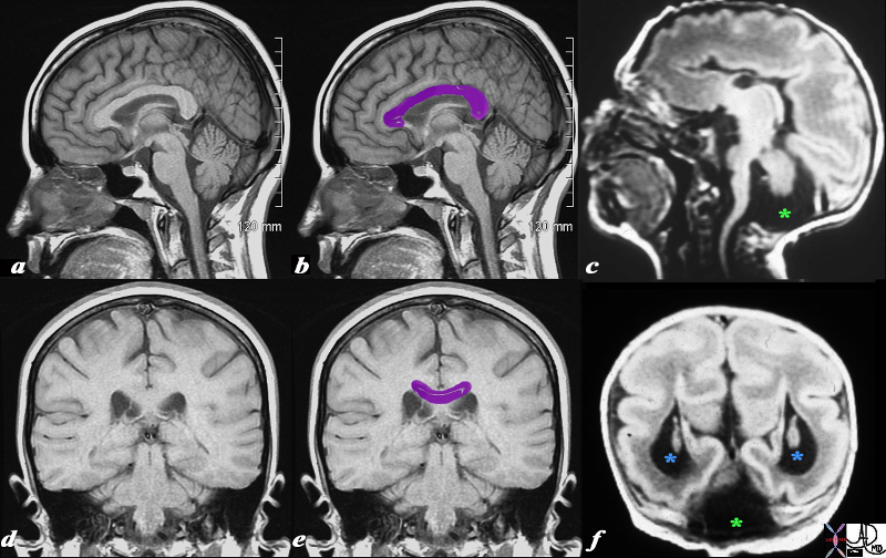
Normal (a,b,d,f) juxtaposed with Flattened, Shallow and Fewer Gyri – Lissencephaly (c,f) |
|
This T1 weighted MRI is from a normal patient juxtaposed on a T1 weighted MRI from a neonate with agenesis of the corpus callosum, congenital lissencephaly and Dandy Walker syndrome. The normal corpus callosum (purple overlay ) is seen above the ventricles between the ventricles and the supracallosul gyrus and cingulate gyrus. . In the neonate (c,f) the there is no white matter in the expected position of the corpus callosum. These findings are consistent with a diagnosis of agenesis of the corpus callosum In addition, the posterior fossa does not show a normal cerebellum. Instead it is mostly filled with CSF (dark T1 green asterisk) likely due to a dilated 4th ventricle and atrophy of the vermis. Only a small amount of posterior fossa soft tissue is seen. In addition the posterior horns are dilated (blue asterisk). This finding is consistent with a diagnosis of Dandy-Walker syndrome associated with hydrocephalus. Lastly the patient has fewer gyri and sulci than normal. The gyri that are present are also more shallow than normal. These findings are consistent with lissencephaly.
Courtesy James Donnelly MD Copyright 2010 All rights reserved 95466c01L03.8s
|
DOMElement Object
(
    [schemaTypeInfo] =>
    [tagName] => table
    [firstElementChild] => (object value omitted)
    [lastElementChild] => (object value omitted)
    [childElementCount] => 1
    [previousElementSibling] => (object value omitted)
    [nextElementSibling] =>
    [nodeName] => table
    [nodeValue] =>


Normal (a,b,d,f) juxtaposed with Flattened, Shallow and Fewer Gyri – Lissencephaly (c,f)


This T1 weighted MRI is from a normal patient juxtaposed on a T1 weighted MRI from a neonate with agenesis of the corpus callosum, congenital lissencephaly and Dandy Walker syndrome. The normal corpus callosum (purple overlay ) is seen above the ventricles between the ventricles and the supracallosul gyrus and cingulate gyrus. . In the neonate (c,f) the there is no white matter in the expected position of the corpus callosum. These findings are consistent with a diagnosis of agenesis of the corpus callosum In addition, the posterior fossa does not show a normal cerebellum. Instead it is mostly filled with CSF (dark T1 green asterisk) likely due to a dilated 4th ventricle and atrophy of the vermis. Only a small amount of posterior fossa soft tissue is seen. In addition the posterior horns are dilated (blue asterisk). This finding is consistent with a diagnosis of Dandy-Walker syndrome associated with hydrocephalus. Lastly the patient has fewer gyri and sulci than normal. The gyri that are present are also more shallow than normal. These findings are consistent with lissencephaly.
Courtesy James Donnelly MD Copyright 2010 All rights reserved 95466c01L03.8s


    [nodeType] => 1
    [parentNode] => (object value omitted)
    [childNodes] => (object value omitted)
    [firstChild] => (object value omitted)
    [lastChild] => (object value omitted)
    [previousSibling] => (object value omitted)
    [nextSibling] => (object value omitted)
    [attributes] => (object value omitted)
    [ownerDocument] => (object value omitted)
    [namespaceURI] =>
    [prefix] =>
    [localName] => table
    [baseURI] =>
    [textContent] =>


Normal (a,b,d,f) juxtaposed with Flattened, Shallow and Fewer Gyri – Lissencephaly (c,f)


This T1 weighted MRI is from a normal patient juxtaposed on a T1 weighted MRI from a neonate with agenesis of the corpus callosum, congenital lissencephaly and Dandy Walker syndrome. The normal corpus callosum (purple overlay ) is seen above the ventricles between the ventricles and the supracallosul gyrus and cingulate gyrus. . In the neonate (c,f) the there is no white matter in the expected position of the corpus callosum. These findings are consistent with a diagnosis of agenesis of the corpus callosum In addition, the posterior fossa does not show a normal cerebellum. Instead it is mostly filled with CSF (dark T1 green asterisk) likely due to a dilated 4th ventricle and atrophy of the vermis. Only a small amount of posterior fossa soft tissue is seen. In addition the posterior horns are dilated (blue asterisk). This finding is consistent with a diagnosis of Dandy-Walker syndrome associated with hydrocephalus. Lastly the patient has fewer gyri and sulci than normal. The gyri that are present are also more shallow than normal. These findings are consistent with lissencephaly.
Courtesy James Donnelly MD Copyright 2010 All rights reserved 95466c01L03.8s


)
DOMElement Object
(
    [schemaTypeInfo] =>
    [tagName] => td
    [firstElementChild] => (object value omitted)
    [lastElementChild] => (object value omitted)
    [childElementCount] => 2
    [previousElementSibling] =>
    [nextElementSibling] =>
    [nodeName] => td
    [nodeValue] =>
This T1 weighted MRI is from a normal patient juxtaposed on a T1 weighted MRI from a neonate with agenesis of the corpus callosum, congenital lissencephaly and Dandy Walker syndrome. The normal corpus callosum (purple overlay ) is seen above the ventricles between the ventricles and the supracallosul gyrus and cingulate gyrus. . In the neonate (c,f) the there is no white matter in the expected position of the corpus callosum. These findings are consistent with a diagnosis of agenesis of the corpus callosum In addition, the posterior fossa does not show a normal cerebellum. Instead it is mostly filled with CSF (dark T1 green asterisk) likely due to a dilated 4th ventricle and atrophy of the vermis. Only a small amount of posterior fossa soft tissue is seen. In addition the posterior horns are dilated (blue asterisk). This finding is consistent with a diagnosis of Dandy-Walker syndrome associated with hydrocephalus. Lastly the patient has fewer gyri and sulci than normal. The gyri that are present are also more shallow than normal. These findings are consistent with lissencephaly.
Courtesy James Donnelly MD Copyright 2010 All rights reserved 95466c01L03.8s

    [nodeType] => 1
    [parentNode] => (object value omitted)
    [childNodes] => (object value omitted)
    [firstChild] => (object value omitted)
    [lastChild] => (object value omitted)
    [previousSibling] => (object value omitted)
    [nextSibling] => (object value omitted)
    [attributes] => (object value omitted)
    [ownerDocument] => (object value omitted)
    [namespaceURI] =>
    [prefix] =>
    [localName] => td
    [baseURI] =>
    [textContent] =>
This T1 weighted MRI is from a normal patient juxtaposed on a T1 weighted MRI from a neonate with agenesis of the corpus callosum, congenital lissencephaly and Dandy Walker syndrome. The normal corpus callosum (purple overlay ) is seen above the ventricles between the ventricles and the supracallosul gyrus and cingulate gyrus. . In the neonate (c,f) the there is no white matter in the expected position of the corpus callosum. These findings are consistent with a diagnosis of agenesis of the corpus callosum In addition, the posterior fossa does not show a normal cerebellum. Instead it is mostly filled with CSF (dark T1 green asterisk) likely due to a dilated 4th ventricle and atrophy of the vermis. Only a small amount of posterior fossa soft tissue is seen. In addition the posterior horns are dilated (blue asterisk). This finding is consistent with a diagnosis of Dandy-Walker syndrome associated with hydrocephalus. Lastly the patient has fewer gyri and sulci than normal. The gyri that are present are also more shallow than normal. These findings are consistent with lissencephaly.
Courtesy James Donnelly MD Copyright 2010 All rights reserved 95466c01L03.8s

)
DOMElement Object
(
    [schemaTypeInfo] =>
    [tagName] => td
    [firstElementChild] => (object value omitted)
    [lastElementChild] => (object value omitted)
    [childElementCount] => 2
    [previousElementSibling] =>
    [nextElementSibling] =>
    [nodeName] => td
    [nodeValue] =>
Normal (a,b,d,f) juxtaposed with Flattened, Shallow and Fewer Gyri – Lissencephaly (c,f)
    [nodeType] => 1
    [parentNode] => (object value omitted)
    [childNodes] => (object value omitted)
    [firstChild] => (object value omitted)
    [lastChild] => (object value omitted)
    [previousSibling] => (object value omitted)
    [nextSibling] => (object value omitted)
    [attributes] => (object value omitted)
    [ownerDocument] => (object value omitted)
    [namespaceURI] =>
    [prefix] =>
    [localName] => td
    [baseURI] =>
    [textContent] =>
Normal (a,b,d,f) juxtaposed with Flattened, Shallow and Fewer Gyri – Lissencephaly (c,f)
)
DOMElement Object
(
    [schemaTypeInfo] =>
    [tagName] => table
    [firstElementChild] => (object value omitted)
    [lastElementChild] => (object value omitted)
    [childElementCount] => 1
    [previousElementSibling] => (object value omitted)
    [nextElementSibling] => (object value omitted)
    [nodeName] => table
    [nodeValue] =>


Introduction to the Central Sulci and Gyri
Characteristic Location and Shape


The lateral view of the brain shows the 4 important central sulci (aka fissures), 3 of which are vertically oriented and 1 of which is horizontal. The vertical sulci include the central sulcus, precentral sulcus and post central sulcus. The central sulcus is important because it separates the frontal lobe from the parietal lobe. The post central sulcus is important because together with the central sulcus it forms the borders of the post central gyrus (pink) which is the sensory cortex. The precentral sulcus together with the central sulcus forms the borders post central gyrus (blue) which is the motor cortex. The Sylvian fissure or lateral sulcus runs horizontally and separates the temporal lobe below from the frontal lobe and parietal lobe above.
Courtesy Ashley Davidoff copyright 2010 all rights reserved 83029b01b01b02g01L.8s


    [nodeType] => 1
    [parentNode] => (object value omitted)
    [childNodes] => (object value omitted)
    [firstChild] => (object value omitted)
    [lastChild] => (object value omitted)
    [previousSibling] => (object value omitted)
    [nextSibling] => (object value omitted)
    [attributes] => (object value omitted)
    [ownerDocument] => (object value omitted)
    [namespaceURI] =>
    [prefix] =>
    [localName] => table
    [baseURI] =>
    [textContent] =>


Introduction to the Central Sulci and Gyri
Characteristic Location and Shape


The lateral view of the brain shows the 4 important central sulci (aka fissures), 3 of which are vertically oriented and 1 of which is horizontal. The vertical sulci include the central sulcus, precentral sulcus and post central sulcus. The central sulcus is important because it separates the frontal lobe from the parietal lobe. The post central sulcus is important because together with the central sulcus it forms the borders of the post central gyrus (pink) which is the sensory cortex. The precentral sulcus together with the central sulcus forms the borders post central gyrus (blue) which is the motor cortex. The Sylvian fissure or lateral sulcus runs horizontally and separates the temporal lobe below from the frontal lobe and parietal lobe above.
Courtesy Ashley Davidoff copyright 2010 all rights reserved 83029b01b01b02g01L.8s


)
DOMElement Object
(
    [schemaTypeInfo] =>
    [tagName] => td
    [firstElementChild] => (object value omitted)
    [lastElementChild] => (object value omitted)
    [childElementCount] => 2
    [previousElementSibling] =>
    [nextElementSibling] =>
    [nodeName] => td
    [nodeValue] =>
The lateral view of the brain shows the 4 important central sulci (aka fissures), 3 of which are vertically oriented and 1 of which is horizontal. The vertical sulci include the central sulcus, precentral sulcus and post central sulcus. The central sulcus is important because it separates the frontal lobe from the parietal lobe. The post central sulcus is important because together with the central sulcus it forms the borders of the post central gyrus (pink) which is the sensory cortex. The precentral sulcus together with the central sulcus forms the borders post central gyrus (blue) which is the motor cortex. The Sylvian fissure or lateral sulcus runs horizontally and separates the temporal lobe below from the frontal lobe and parietal lobe above.
Courtesy Ashley Davidoff copyright 2010 all rights reserved 83029b01b01b02g01L.8s
    [nodeType] => 1
    [parentNode] => (object value omitted)
    [childNodes] => (object value omitted)
    [firstChild] => (object value omitted)
    [lastChild] => (object value omitted)
    [previousSibling] => (object value omitted)
    [nextSibling] => (object value omitted)
    [attributes] => (object value omitted)
    [ownerDocument] => (object value omitted)
    [namespaceURI] =>
    [prefix] =>
    [localName] => td
    [baseURI] =>
    [textContent] =>
The lateral view of the brain shows the 4 important central sulci (aka fissures), 3 of which are vertically oriented and 1 of which is horizontal. The vertical sulci include the central sulcus, precentral sulcus and post central sulcus. The central sulcus is important because it separates the frontal lobe from the parietal lobe. The post central sulcus is important because together with the central sulcus it forms the borders of the post central gyrus (pink) which is the sensory cortex. The precentral sulcus together with the central sulcus forms the borders post central gyrus (blue) which is the motor cortex. The Sylvian fissure or lateral sulcus runs horizontally and separates the temporal lobe below from the frontal lobe and parietal lobe above.
Courtesy Ashley Davidoff copyright 2010 all rights reserved 83029b01b01b02g01L.8s
)
DOMElement Object
(
    [schemaTypeInfo] =>
    [tagName] => td
    [firstElementChild] => (object value omitted)
    [lastElementChild] => (object value omitted)
    [childElementCount] => 3
    [previousElementSibling] =>
    [nextElementSibling] =>
    [nodeName] => td
    [nodeValue] =>

Introduction to the Central Sulci and Gyri
Characteristic Location and Shape
    [nodeType] => 1
    [parentNode] => (object value omitted)
    [childNodes] => (object value omitted)
    [firstChild] => (object value omitted)
    [lastChild] => (object value omitted)
    [previousSibling] => (object value omitted)
    [nextSibling] => (object value omitted)
    [attributes] => (object value omitted)
    [ownerDocument] => (object value omitted)
    [namespaceURI] =>
    [prefix] =>
    [localName] => td
    [baseURI] =>
    [textContent] =>

Introduction to the Central Sulci and Gyri
Characteristic Location and Shape
)
DOMElement Object
(
    [schemaTypeInfo] =>
    [tagName] => table
    [firstElementChild] => (object value omitted)
    [lastElementChild] => (object value omitted)
    [childElementCount] => 1
    [previousElementSibling] => (object value omitted)
    [nextElementSibling] => (object value omitted)
    [nodeName] => table
    [nodeValue] =>


Brain Coral


The brain coral
 Courtesy Ashley Davidoff MD Copyright 2010 83018.800


    [nodeType] => 1
    [parentNode] => (object value omitted)
    [childNodes] => (object value omitted)
    [firstChild] => (object value omitted)
    [lastChild] => (object value omitted)
    [previousSibling] => (object value omitted)
    [nextSibling] => (object value omitted)
    [attributes] => (object value omitted)
    [ownerDocument] => (object value omitted)
    [namespaceURI] =>
    [prefix] =>
    [localName] => table
    [baseURI] =>
    [textContent] =>


Brain Coral


The brain coral
 Courtesy Ashley Davidoff MD Copyright 2010 83018.800


)
DOMElement Object
(
    [schemaTypeInfo] =>
    [tagName] => td
    [firstElementChild] => (object value omitted)
    [lastElementChild] => (object value omitted)
    [childElementCount] => 2
    [previousElementSibling] =>
    [nextElementSibling] =>
    [nodeName] => td
    [nodeValue] =>
The brain coral
 Courtesy Ashley Davidoff MD Copyright 2010 83018.800

    [nodeType] => 1
    [parentNode] => (object value omitted)
    [childNodes] => (object value omitted)
    [firstChild] => (object value omitted)
    [lastChild] => (object value omitted)
    [previousSibling] => (object value omitted)
    [nextSibling] => (object value omitted)
    [attributes] => (object value omitted)
    [ownerDocument] => (object value omitted)
    [namespaceURI] =>
    [prefix] =>
    [localName] => td
    [baseURI] =>
    [textContent] =>
The brain coral
 Courtesy Ashley Davidoff MD Copyright 2010 83018.800

)
DOMElement Object
(
    [schemaTypeInfo] =>
    [tagName] => td
    [firstElementChild] => (object value omitted)
    [lastElementChild] => (object value omitted)
    [childElementCount] => 2
    [previousElementSibling] =>
    [nextElementSibling] =>
    [nodeName] => td
    [nodeValue] =>
Brain Coral
    [nodeType] => 1
    [parentNode] => (object value omitted)
    [childNodes] => (object value omitted)
    [firstChild] => (object value omitted)
    [lastChild] => (object value omitted)
    [previousSibling] => (object value omitted)
    [nextSibling] => (object value omitted)
    [attributes] => (object value omitted)
    [ownerDocument] => (object value omitted)
    [namespaceURI] =>
    [prefix] =>
    [localName] => td
    [baseURI] =>
    [textContent] =>
Brain Coral
)
DOMElement Object
(
    [schemaTypeInfo] =>
    [tagName] => table
    [firstElementChild] => (object value omitted)
    [lastElementChild] => (object value omitted)
    [childElementCount] => 1
    [previousElementSibling] => (object value omitted)
    [nextElementSibling] => (object value omitted)
    [nodeName] => table
    [nodeValue] =>


Form and Colors reminiscent of the Brain


This image of a walnut has a few features that are reminiscent of a brain, including the symmetric hemispheric components, the pinkish color, and the undulations of the surface. However in reality it is about 1.8cms long, and only 5-8mms high, and is hard.
Courtesy Ashley Davidoff MD copyright 2010 all rights reserved 100286pb.93s


    [nodeType] => 1
    [parentNode] => (object value omitted)
    [childNodes] => (object value omitted)
    [firstChild] => (object value omitted)
    [lastChild] => (object value omitted)
    [previousSibling] => (object value omitted)
    [nextSibling] => (object value omitted)
    [attributes] => (object value omitted)
    [ownerDocument] => (object value omitted)
    [namespaceURI] =>
    [prefix] =>
    [localName] => table
    [baseURI] =>
    [textContent] =>


Form and Colors reminiscent of the Brain


This image of a walnut has a few features that are reminiscent of a brain, including the symmetric hemispheric components, the pinkish color, and the undulations of the surface. However in reality it is about 1.8cms long, and only 5-8mms high, and is hard.
Courtesy Ashley Davidoff MD copyright 2010 all rights reserved 100286pb.93s


)
DOMElement Object
(
    [schemaTypeInfo] =>
    [tagName] => td
    [firstElementChild] => (object value omitted)
    [lastElementChild] => (object value omitted)
    [childElementCount] => 2
    [previousElementSibling] =>
    [nextElementSibling] =>
    [nodeName] => td
    [nodeValue] =>
This image of a walnut has a few features that are reminiscent of a brain, including the symmetric hemispheric components, the pinkish color, and the undulations of the surface. However in reality it is about 1.8cms long, and only 5-8mms high, and is hard.
Courtesy Ashley Davidoff MD copyright 2010 all rights reserved 100286pb.93s

    [nodeType] => 1
    [parentNode] => (object value omitted)
    [childNodes] => (object value omitted)
    [firstChild] => (object value omitted)
    [lastChild] => (object value omitted)
    [previousSibling] => (object value omitted)
    [nextSibling] => (object value omitted)
    [attributes] => (object value omitted)
    [ownerDocument] => (object value omitted)
    [namespaceURI] =>
    [prefix] =>
    [localName] => td
    [baseURI] =>
    [textContent] =>
This image of a walnut has a few features that are reminiscent of a brain, including the symmetric hemispheric components, the pinkish color, and the undulations of the surface. However in reality it is about 1.8cms long, and only 5-8mms high, and is hard.
Courtesy Ashley Davidoff MD copyright 2010 all rights reserved 100286pb.93s

)
DOMElement Object
(
    [schemaTypeInfo] =>
    [tagName] => td
    [firstElementChild] => (object value omitted)
    [lastElementChild] => (object value omitted)
    [childElementCount] => 2
    [previousElementSibling] =>
    [nextElementSibling] =>
    [nodeName] => td
    [nodeValue] =>

Form and Colors reminiscent of the Brain
    [nodeType] => 1
    [parentNode] => (object value omitted)
    [childNodes] => (object value omitted)
    [firstChild] => (object value omitted)
    [lastChild] => (object value omitted)
    [previousSibling] => (object value omitted)
    [nextSibling] => (object value omitted)
    [attributes] => (object value omitted)
    [ownerDocument] => (object value omitted)
    [namespaceURI] =>
    [prefix] =>
    [localName] => td
    [baseURI] =>
    [textContent] =>

Form and Colors reminiscent of the Brain
)
DOMElement Object
(
    [schemaTypeInfo] =>
    [tagName] => table
    [firstElementChild] => (object value omitted)
    [lastElementChild] => (object value omitted)
    [childElementCount] => 1
    [previousElementSibling] => (object value omitted)
    [nextElementSibling] => (object value omitted)
    [nodeName] => table
    [nodeValue] =>


Basic Shapes
Lateral Projection


When the external surface of the brain is viewed from its lateral aspect, , the ovoid shape of the conglomerate frontal parietal, occipital and part of the temporal lobe is still maintained. The part of the temporal lobe that projects into the middle cranial fossa adds a second oval directed slightly inferiorly. The medulla adds a new dimension having the shape somewhere between a half a sphere and the bottom of a top.
Davidoff art All Rights reserved copyright 2010 83029d03c01.9s


    [nodeType] => 1
    [parentNode] => (object value omitted)
    [childNodes] => (object value omitted)
    [firstChild] => (object value omitted)
    [lastChild] => (object value omitted)
    [previousSibling] => (object value omitted)
    [nextSibling] => (object value omitted)
    [attributes] => (object value omitted)
    [ownerDocument] => (object value omitted)
    [namespaceURI] =>
    [prefix] =>
    [localName] => table
    [baseURI] =>
    [textContent] =>


Basic Shapes
Lateral Projection


When the external surface of the brain is viewed from its lateral aspect, , the ovoid shape of the conglomerate frontal parietal, occipital and part of the temporal lobe is still maintained. The part of the temporal lobe that projects into the middle cranial fossa adds a second oval directed slightly inferiorly. The medulla adds a new dimension having the shape somewhere between a half a sphere and the bottom of a top.
Davidoff art All Rights reserved copyright 2010 83029d03c01.9s


)
DOMElement Object
(
    [schemaTypeInfo] =>
    [tagName] => td
    [firstElementChild] => (object value omitted)
    [lastElementChild] => (object value omitted)
    [childElementCount] => 2
    [previousElementSibling] =>
    [nextElementSibling] =>
    [nodeName] => td
    [nodeValue] =>
When the external surface of the brain is viewed from its lateral aspect, , the ovoid shape of the conglomerate frontal parietal, occipital and part of the temporal lobe is still maintained. The part of the temporal lobe that projects into the middle cranial fossa adds a second oval directed slightly inferiorly. The medulla adds a new dimension having the shape somewhere between a half a sphere and the bottom of a top.
Davidoff art All Rights reserved copyright 2010 83029d03c01.9s

    [nodeType] => 1
    [parentNode] => (object value omitted)
    [childNodes] => (object value omitted)
    [firstChild] => (object value omitted)
    [lastChild] => (object value omitted)
    [previousSibling] => (object value omitted)
    [nextSibling] => (object value omitted)
    [attributes] => (object value omitted)
    [ownerDocument] => (object value omitted)
    [namespaceURI] =>
    [prefix] =>
    [localName] => td
    [baseURI] =>
    [textContent] =>
When the external surface of the brain is viewed from its lateral aspect, , the ovoid shape of the conglomerate frontal parietal, occipital and part of the temporal lobe is still maintained. The part of the temporal lobe that projects into the middle cranial fossa adds a second oval directed slightly inferiorly. The medulla adds a new dimension having the shape somewhere between a half a sphere and the bottom of a top.
Davidoff art All Rights reserved copyright 2010 83029d03c01.9s

)
DOMElement Object
(
    [schemaTypeInfo] =>
    [tagName] => td
    [firstElementChild] => (object value omitted)
    [lastElementChild] => (object value omitted)
    [childElementCount] => 3
    [previousElementSibling] =>
    [nextElementSibling] =>
    [nodeName] => td
    [nodeValue] =>

Basic Shapes
Lateral Projection
    [nodeType] => 1
    [parentNode] => (object value omitted)
    [childNodes] => (object value omitted)
    [firstChild] => (object value omitted)
    [lastChild] => (object value omitted)
    [previousSibling] => (object value omitted)
    [nextSibling] => (object value omitted)
    [attributes] => (object value omitted)
    [ownerDocument] => (object value omitted)
    [namespaceURI] =>
    [prefix] =>
    [localName] => td
    [baseURI] =>
    [textContent] =>

Basic Shapes
Lateral Projection
)
DOMElement Object
(
    [schemaTypeInfo] =>
    [tagName] => table
    [firstElementChild] => (object value omitted)
    [lastElementChild] => (object value omitted)
    [childElementCount] => 1
    [previousElementSibling] => (object value omitted)
    [nextElementSibling] => (object value omitted)
    [nodeName] => table
    [nodeValue] =>


General Shape of the Brain
External Surface


The ovoid shape is easily appreciated when the external surface of the brain is viewed from above, and although it in general it has an ovoid shape when viewed from the side, more complex shapes start to appear, as the temporal and cerebellum start to appear.
source unknown modified by Davidoff 52981b05.8sd01


    [nodeType] => 1
    [parentNode] => (object value omitted)
    [childNodes] => (object value omitted)
    [firstChild] => (object value omitted)
    [lastChild] => (object value omitted)
    [previousSibling] => (object value omitted)
    [nextSibling] => (object value omitted)
    [attributes] => (object value omitted)
    [ownerDocument] => (object value omitted)
    [namespaceURI] =>
    [prefix] =>
    [localName] => table
    [baseURI] =>
    [textContent] =>


General Shape of the Brain
External Surface


The ovoid shape is easily appreciated when the external surface of the brain is viewed from above, and although it in general it has an ovoid shape when viewed from the side, more complex shapes start to appear, as the temporal and cerebellum start to appear.
source unknown modified by Davidoff 52981b05.8sd01


)
DOMElement Object
(
    [schemaTypeInfo] =>
    [tagName] => td
    [firstElementChild] => (object value omitted)
    [lastElementChild] => (object value omitted)
    [childElementCount] => 2
    [previousElementSibling] =>
    [nextElementSibling] =>
    [nodeName] => td
    [nodeValue] =>
The ovoid shape is easily appreciated when the external surface of the brain is viewed from above, and although it in general it has an ovoid shape when viewed from the side, more complex shapes start to appear, as the temporal and cerebellum start to appear.
source unknown modified by Davidoff 52981b05.8sd01

    [nodeType] => 1
    [parentNode] => (object value omitted)
    [childNodes] => (object value omitted)
    [firstChild] => (object value omitted)
    [lastChild] => (object value omitted)
    [previousSibling] => (object value omitted)
    [nextSibling] => (object value omitted)
    [attributes] => (object value omitted)
    [ownerDocument] => (object value omitted)
    [namespaceURI] =>
    [prefix] =>
    [localName] => td
    [baseURI] =>
    [textContent] =>
The ovoid shape is easily appreciated when the external surface of the brain is viewed from above, and although it in general it has an ovoid shape when viewed from the side, more complex shapes start to appear, as the temporal and cerebellum start to appear.
source unknown modified by Davidoff 52981b05.8sd01

)
DOMElement Object
(
    [schemaTypeInfo] =>
    [tagName] => td
    [firstElementChild] => (object value omitted)
    [lastElementChild] => (object value omitted)
    [childElementCount] => 3
    [previousElementSibling] =>
    [nextElementSibling] =>
    [nodeName] => td
    [nodeValue] =>

General Shape of the Brain
External Surface
    [nodeType] => 1
    [parentNode] => (object value omitted)
    [childNodes] => (object value omitted)
    [firstChild] => (object value omitted)
    [lastChild] => (object value omitted)
    [previousSibling] => (object value omitted)
    [nextSibling] => (object value omitted)
    [attributes] => (object value omitted)
    [ownerDocument] => (object value omitted)
    [namespaceURI] =>
    [prefix] =>
    [localName] => td
    [baseURI] =>
    [textContent] =>

General Shape of the Brain
External Surface
)
DOMElement Object
(
    [schemaTypeInfo] =>
    [tagName] => table
    [firstElementChild] => (object value omitted)
    [lastElementChild] => (object value omitted)
    [childElementCount] => 1
    [previousElementSibling] => (object value omitted)
    [nextElementSibling] => (object value omitted)
    [nodeName] => table
    [nodeValue] =>


Overview of the Gyri from the Lateral External View


The lateral view of the brain shows the frontal lobe (green) parietal lobe (light mauve), the occipital lobe (purple) and the temporal lobe (red) In this view the frontal lobe gyri that are visible are; superior frontal, middle frontal, inferior frontal and precentral gyri. The parietal gyri include the superior parietal and inferior parietal. The occipital gyrus that is visible is the lateral occipital. The temporal lobe gyri include the superior, middle and inferior temporal gyri.
Courtesy Ashley Davidoff MD Copyright 2010 83029d05g01.8s


    [nodeType] => 1
    [parentNode] => (object value omitted)
    [childNodes] => (object value omitted)
    [firstChild] => (object value omitted)
    [lastChild] => (object value omitted)
    [previousSibling] => (object value omitted)
    [nextSibling] => (object value omitted)
    [attributes] => (object value omitted)
    [ownerDocument] => (object value omitted)
    [namespaceURI] =>
    [prefix] =>
    [localName] => table
    [baseURI] =>
    [textContent] =>


Overview of the Gyri from the Lateral External View


The lateral view of the brain shows the frontal lobe (green) parietal lobe (light mauve), the occipital lobe (purple) and the temporal lobe (red) In this view the frontal lobe gyri that are visible are; superior frontal, middle frontal, inferior frontal and precentral gyri. The parietal gyri include the superior parietal and inferior parietal. The occipital gyrus that is visible is the lateral occipital. The temporal lobe gyri include the superior, middle and inferior temporal gyri.
Courtesy Ashley Davidoff MD Copyright 2010 83029d05g01.8s


)
DOMElement Object
(
    [schemaTypeInfo] =>
    [tagName] => td
    [firstElementChild] => (object value omitted)
    [lastElementChild] => (object value omitted)
    [childElementCount] => 2
    [previousElementSibling] =>
    [nextElementSibling] =>
    [nodeName] => td
    [nodeValue] =>
The lateral view of the brain shows the frontal lobe (green) parietal lobe (light mauve), the occipital lobe (purple) and the temporal lobe (red) In this view the frontal lobe gyri that are visible are; superior frontal, middle frontal, inferior frontal and precentral gyri. The parietal gyri include the superior parietal and inferior parietal. The occipital gyrus that is visible is the lateral occipital. The temporal lobe gyri include the superior, middle and inferior temporal gyri.
Courtesy Ashley Davidoff MD Copyright 2010 83029d05g01.8s

    [nodeType] => 1
    [parentNode] => (object value omitted)
    [childNodes] => (object value omitted)
    [firstChild] => (object value omitted)
    [lastChild] => (object value omitted)
    [previousSibling] => (object value omitted)
    [nextSibling] => (object value omitted)
    [attributes] => (object value omitted)
    [ownerDocument] => (object value omitted)
    [namespaceURI] =>
    [prefix] =>
    [localName] => td
    [baseURI] =>
    [textContent] =>
The lateral view of the brain shows the frontal lobe (green) parietal lobe (light mauve), the occipital lobe (purple) and the temporal lobe (red) In this view the frontal lobe gyri that are visible are; superior frontal, middle frontal, inferior frontal and precentral gyri. The parietal gyri include the superior parietal and inferior parietal. The occipital gyrus that is visible is the lateral occipital. The temporal lobe gyri include the superior, middle and inferior temporal gyri.
Courtesy Ashley Davidoff MD Copyright 2010 83029d05g01.8s

)
DOMElement Object
(
    [schemaTypeInfo] =>
    [tagName] => td
    [firstElementChild] => (object value omitted)
    [lastElementChild] => (object value omitted)
    [childElementCount] => 2
    [previousElementSibling] =>
    [nextElementSibling] =>
    [nodeName] => td
    [nodeValue] =>
Overview of the Gyri from the Lateral External View
    [nodeType] => 1
    [parentNode] => (object value omitted)
    [childNodes] => (object value omitted)
    [firstChild] => (object value omitted)
    [lastChild] => (object value omitted)
    [previousSibling] => (object value omitted)
    [nextSibling] => (object value omitted)
    [attributes] => (object value omitted)
    [ownerDocument] => (object value omitted)
    [namespaceURI] =>
    [prefix] =>
    [localName] => td
    [baseURI] =>
    [textContent] =>
Overview of the Gyri from the Lateral External View
)
DOMElement Object
(
    [schemaTypeInfo] =>
    [tagName] => table
    [firstElementChild] => (object value omitted)
    [lastElementChild] => (object value omitted)
    [childElementCount] => 1
    [previousElementSibling] => (object value omitted)
    [nextElementSibling] => (object value omitted)
    [nodeName] => table
    [nodeValue] =>


The Precentral Gyrus (blue) and Postcentral Gyrus (pink)


The lateral view of the external brain shows the most easily recognized gyri – the precentral gyrus motor cortex (blue) and the post central sensory gyrus (pink) surrounded by the sulci.  There are 4 important central sulci (aka fissures), 3 of which are vertically oriented and 1 of which is horizontal. The vertical sulci include the central sulcus, precentral sulcus and post central sulcus. The central sulcus is important because it separates the frontal lobe from the parietal lobe. The post central sulcus is important because together with the central sulcus it forms the borders of the post central gyrus (pink) which is the sensory cortex. The precentral sulcus together with the central sulcus forms the borders post central gyrus (blue) which is the motor cortex. The Sylvian fissure or lateral sulcus runs horizontally and separates the temporal lobe below from the frontal lobe and parietal lobe above.
Courtesy Ashley Davidoff copyright 2010 all rights reserved 83029b01b01b02g01L.8s


    [nodeType] => 1
    [parentNode] => (object value omitted)
    [childNodes] => (object value omitted)
    [firstChild] => (object value omitted)
    [lastChild] => (object value omitted)
    [previousSibling] => (object value omitted)
    [nextSibling] => (object value omitted)
    [attributes] => (object value omitted)
    [ownerDocument] => (object value omitted)
    [namespaceURI] =>
    [prefix] =>
    [localName] => table
    [baseURI] =>
    [textContent] =>


The Precentral Gyrus (blue) and Postcentral Gyrus (pink)


The lateral view of the external brain shows the most easily recognized gyri – the precentral gyrus motor cortex (blue) and the post central sensory gyrus (pink) surrounded by the sulci.  There are 4 important central sulci (aka fissures), 3 of which are vertically oriented and 1 of which is horizontal. The vertical sulci include the central sulcus, precentral sulcus and post central sulcus. The central sulcus is important because it separates the frontal lobe from the parietal lobe. The post central sulcus is important because together with the central sulcus it forms the borders of the post central gyrus (pink) which is the sensory cortex. The precentral sulcus together with the central sulcus forms the borders post central gyrus (blue) which is the motor cortex. The Sylvian fissure or lateral sulcus runs horizontally and separates the temporal lobe below from the frontal lobe and parietal lobe above.
Courtesy Ashley Davidoff copyright 2010 all rights reserved 83029b01b01b02g01L.8s


)
DOMElement Object
(
    [schemaTypeInfo] =>
    [tagName] => td
    [firstElementChild] => (object value omitted)
    [lastElementChild] => (object value omitted)
    [childElementCount] => 2
    [previousElementSibling] =>
    [nextElementSibling] =>
    [nodeName] => td
    [nodeValue] =>
The lateral view of the external brain shows the most easily recognized gyri – the precentral gyrus motor cortex (blue) and the post central sensory gyrus (pink) surrounded by the sulci.  There are 4 important central sulci (aka fissures), 3 of which are vertically oriented and 1 of which is horizontal. The vertical sulci include the central sulcus, precentral sulcus and post central sulcus. The central sulcus is important because it separates the frontal lobe from the parietal lobe. The post central sulcus is important because together with the central sulcus it forms the borders of the post central gyrus (pink) which is the sensory cortex. The precentral sulcus together with the central sulcus forms the borders post central gyrus (blue) which is the motor cortex. The Sylvian fissure or lateral sulcus runs horizontally and separates the temporal lobe below from the frontal lobe and parietal lobe above.
Courtesy Ashley Davidoff copyright 2010 all rights reserved 83029b01b01b02g01L.8s

    [nodeType] => 1
    [parentNode] => (object value omitted)
    [childNodes] => (object value omitted)
    [firstChild] => (object value omitted)
    [lastChild] => (object value omitted)
    [previousSibling] => (object value omitted)
    [nextSibling] => (object value omitted)
    [attributes] => (object value omitted)
    [ownerDocument] => (object value omitted)
    [namespaceURI] =>
    [prefix] =>
    [localName] => td
    [baseURI] =>
    [textContent] =>
The lateral view of the external brain shows the most easily recognized gyri – the precentral gyrus motor cortex (blue) and the post central sensory gyrus (pink) surrounded by the sulci.  There are 4 important central sulci (aka fissures), 3 of which are vertically oriented and 1 of which is horizontal. The vertical sulci include the central sulcus, precentral sulcus and post central sulcus. The central sulcus is important because it separates the frontal lobe from the parietal lobe. The post central sulcus is important because together with the central sulcus it forms the borders of the post central gyrus (pink) which is the sensory cortex. The precentral sulcus together with the central sulcus forms the borders post central gyrus (blue) which is the motor cortex. The Sylvian fissure or lateral sulcus runs horizontally and separates the temporal lobe below from the frontal lobe and parietal lobe above.
Courtesy Ashley Davidoff copyright 2010 all rights reserved 83029b01b01b02g01L.8s

)
DOMElement Object
(
    [schemaTypeInfo] =>
    [tagName] => td
    [firstElementChild] => (object value omitted)
    [lastElementChild] => (object value omitted)
    [childElementCount] => 2
    [previousElementSibling] =>
    [nextElementSibling] =>
    [nodeName] => td
    [nodeValue] =>
The Precentral Gyrus (blue) and Postcentral Gyrus (pink)
    [nodeType] => 1
    [parentNode] => (object value omitted)
    [childNodes] => (object value omitted)
    [firstChild] => (object value omitted)
    [lastChild] => (object value omitted)
    [previousSibling] => (object value omitted)
    [nextSibling] => (object value omitted)
    [attributes] => (object value omitted)
    [ownerDocument] => (object value omitted)
    [namespaceURI] =>
    [prefix] =>
    [localName] => td
    [baseURI] =>
    [textContent] =>
The Precentral Gyrus (blue) and Postcentral Gyrus (pink)
)
DOMElement Object
(
    [schemaTypeInfo] =>
    [tagName] => table
    [firstElementChild] => (object value omitted)
    [lastElementChild] => (object value omitted)
    [childElementCount] => 1
    [previousElementSibling] => (object value omitted)
    [nextElementSibling] => (object value omitted)
    [nodeName] => table
    [nodeValue] =>


Overall View of the Gyri


The MRI represents a sagittal image of the brain in an elderly patient where the associated atrophy accentuates the gyri. This general view reveals the gyri of the frontal lobe (light green, parietal lobe (mauve), and occipital lobe (darker purlple) with the inner cingulate gyrus seen in darker green) Which exemplirifies the gyra The specimen shows the approximate position of the 3 major frontal gyri in the anterior location including the superior, middle and inferior gyrus. The cingulate gyrus is not really part of the forebrain but part of the limbic system. It holds a medial deep and very central position.
Courtesy Ashley Davidoff MD Copyright 2010 92170b06b01.8sgL.8


    [nodeType] => 1
    [parentNode] => (object value omitted)
    [childNodes] => (object value omitted)
    [firstChild] => (object value omitted)
    [lastChild] => (object value omitted)
    [previousSibling] => (object value omitted)
    [nextSibling] => (object value omitted)
    [attributes] => (object value omitted)
    [ownerDocument] => (object value omitted)
    [namespaceURI] =>
    [prefix] =>
    [localName] => table
    [baseURI] =>
    [textContent] =>


Overall View of the Gyri


The MRI represents a sagittal image of the brain in an elderly patient where the associated atrophy accentuates the gyri. This general view reveals the gyri of the frontal lobe (light green, parietal lobe (mauve), and occipital lobe (darker purlple) with the inner cingulate gyrus seen in darker green) Which exemplirifies the gyra The specimen shows the approximate position of the 3 major frontal gyri in the anterior location including the superior, middle and inferior gyrus. The cingulate gyrus is not really part of the forebrain but part of the limbic system. It holds a medial deep and very central position.
Courtesy Ashley Davidoff MD Copyright 2010 92170b06b01.8sgL.8


)
DOMElement Object
(
    [schemaTypeInfo] =>
    [tagName] => td
    [firstElementChild] => (object value omitted)
    [lastElementChild] => (object value omitted)
    [childElementCount] => 2
    [previousElementSibling] =>
    [nextElementSibling] =>
    [nodeName] => td
    [nodeValue] =>
The MRI represents a sagittal image of the brain in an elderly patient where the associated atrophy accentuates the gyri. This general view reveals the gyri of the frontal lobe (light green, parietal lobe (mauve), and occipital lobe (darker purlple) with the inner cingulate gyrus seen in darker green) Which exemplirifies the gyra The specimen shows the approximate position of the 3 major frontal gyri in the anterior location including the superior, middle and inferior gyrus. The cingulate gyrus is not really part of the forebrain but part of the limbic system. It holds a medial deep and very central position.
Courtesy Ashley Davidoff MD Copyright 2010 92170b06b01.8sgL.8

    [nodeType] => 1
    [parentNode] => (object value omitted)
    [childNodes] => (object value omitted)
    [firstChild] => (object value omitted)
    [lastChild] => (object value omitted)
    [previousSibling] => (object value omitted)
    [nextSibling] => (object value omitted)
    [attributes] => (object value omitted)
    [ownerDocument] => (object value omitted)
    [namespaceURI] =>
    [prefix] =>
    [localName] => td
    [baseURI] =>
    [textContent] =>
The MRI represents a sagittal image of the brain in an elderly patient where the associated atrophy accentuates the gyri. This general view reveals the gyri of the frontal lobe (light green, parietal lobe (mauve), and occipital lobe (darker purlple) with the inner cingulate gyrus seen in darker green) Which exemplirifies the gyra The specimen shows the approximate position of the 3 major frontal gyri in the anterior location including the superior, middle and inferior gyrus. The cingulate gyrus is not really part of the forebrain but part of the limbic system. It holds a medial deep and very central position.
Courtesy Ashley Davidoff MD Copyright 2010 92170b06b01.8sgL.8

)
DOMElement Object
(
    [schemaTypeInfo] =>
    [tagName] => td
    [firstElementChild] => (object value omitted)
    [lastElementChild] => (object value omitted)
    [childElementCount] => 2
    [previousElementSibling] =>
    [nextElementSibling] =>
    [nodeName] => td
    [nodeValue] =>
Overall View of the Gyri
    [nodeType] => 1
    [parentNode] => (object value omitted)
    [childNodes] => (object value omitted)
    [firstChild] => (object value omitted)
    [lastChild] => (object value omitted)
    [previousSibling] => (object value omitted)
    [nextSibling] => (object value omitted)
    [attributes] => (object value omitted)
    [ownerDocument] => (object value omitted)
    [namespaceURI] =>
    [prefix] =>
    [localName] => td
    [baseURI] =>
    [textContent] =>
Overall View of the Gyri
)


