Hindbrain
Rhombencephalon
Ashley Davidoff MD
The Common Vein Copyright 2010
Definition
The hindbrain or rhombencephalon is that part of the brain that lies between the midbrain and the spimnal cord and consists of the pons, medulla, and cerebellum.
Embryologically it is divided into the metencephalon (pons and cerebellum) and the myelencephalon (medulla).
|
Concepts Major Parts of the Brain Hindbrain in Context |
|
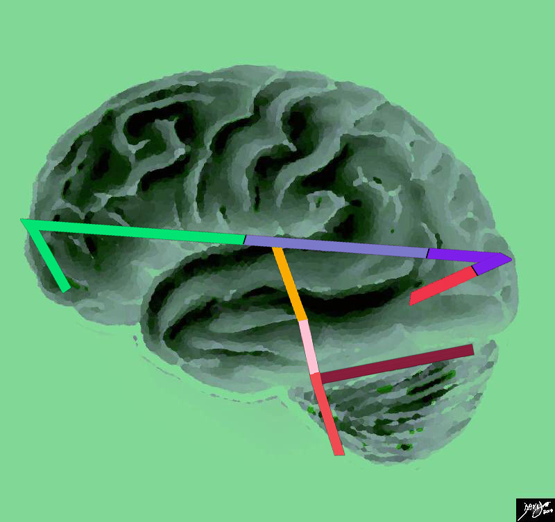
This artistic rendition of the brain reflects the vectors of the major parts of the brain with the stick diagram overlaid on a sagittal external view of the brain. In the stick diagram, the forebrain has now been divided into the frontal lobe (bright green), parietal lobe (light mauve) occipital lobe (purple) and temporal lobe (red). The midbrain is represented in orange, and the hind brain consists of the pons (pink) medulla (salmon) and the cerebellum (maroon) Courtesy Ashley Davidoff copyright 2010 all rights reserved 83029e04.83s |
Conceptual Framework
|
Conceptual Framework of the Hindbrain |
|
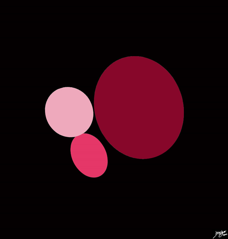
The hindbrain conceptually consists of 3 ovoids The pons is anterior and superior (light pink), the medulla is smaller and is anterior and inferior, and the cerebellum is the largest and is posterior. Courtesy Ashley Davidoff MD Copyright 2010 all rights reserved 92141.3kd03b03b01.8s |
|
The Concept In Vivo |
|
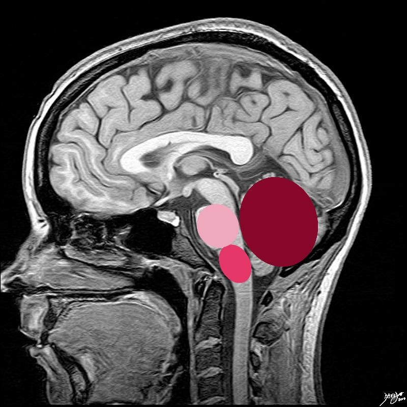
The 3 ovoids are situated in the posterior cranial fossa the pons is anterior and superior (light pink), the medulla is smaller and is anterior and inferior, and the cerebellum is the largest and is posterior. Courtesy Ashley Davidoff MD Copyright 2010 all rights reserved 92141.3kd03b03b.8s |
Gross Anatomy
DOMElement Object
(
    [schemaTypeInfo] =>
    [tagName] => table
    [firstElementChild] => (object value omitted)
    [lastElementChild] => (object value omitted)
    [childElementCount] => 1
    [previousElementSibling] => (object value omitted)
    [nextElementSibling] => (object value omitted)
    [nodeName] => table
    [nodeValue] =>


Hindbrain
Relation to Midbrain and Forebrain
Sagittal Section


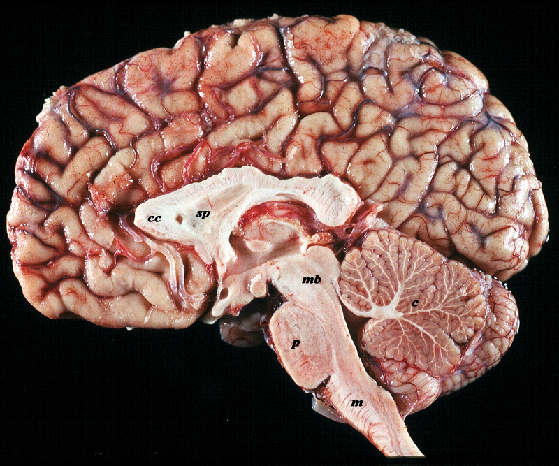
The midsagittal section view of brain reveals the distinctive shape position and character of the midline structures of the brain. The distinction between the character of the cerebral cortex which has a creamy color and the white matter exemplified by the corpus callosum (c) and septum pellucidum (sp) which are white, and the midbrain (mb) pons (p) and medulla (m) which are off white as opposed to the color of the cerebellum (c) which is light salmon pink is well demonstrated. The relative sizes of the forebrain, midbrain and hindbrain and their components are well appreciated in this section.
Image Courtesy of Thomas W.Smith, MD; Department of Pathology, University of Massachusetts Medical School. 97805b02


    [nodeType] => 1
    [parentNode] => (object value omitted)
    [childNodes] => (object value omitted)
    [firstChild] => (object value omitted)
    [lastChild] => (object value omitted)
    [previousSibling] => (object value omitted)
    [nextSibling] => (object value omitted)
    [attributes] => (object value omitted)
    [ownerDocument] => (object value omitted)
    [namespaceURI] =>
    [prefix] =>
    [localName] => table
    [baseURI] =>
    [textContent] =>


Hindbrain
Relation to Midbrain and Forebrain
Sagittal Section


The midsagittal section view of brain reveals the distinctive shape position and character of the midline structures of the brain. The distinction between the character of the cerebral cortex which has a creamy color and the white matter exemplified by the corpus callosum (c) and septum pellucidum (sp) which are white, and the midbrain (mb) pons (p) and medulla (m) which are off white as opposed to the color of the cerebellum (c) which is light salmon pink is well demonstrated. The relative sizes of the forebrain, midbrain and hindbrain and their components are well appreciated in this section.
Image Courtesy of Thomas W.Smith, MD; Department of Pathology, University of Massachusetts Medical School. 97805b02


)
DOMElement Object
(
    [schemaTypeInfo] =>
    [tagName] => td
    [firstElementChild] => (object value omitted)
    [lastElementChild] => (object value omitted)
    [childElementCount] => 2
    [previousElementSibling] =>
    [nextElementSibling] =>
    [nodeName] => td
    [nodeValue] =>
The midsagittal section view of brain reveals the distinctive shape position and character of the midline structures of the brain. The distinction between the character of the cerebral cortex which has a creamy color and the white matter exemplified by the corpus callosum (c) and septum pellucidum (sp) which are white, and the midbrain (mb) pons (p) and medulla (m) which are off white as opposed to the color of the cerebellum (c) which is light salmon pink is well demonstrated. The relative sizes of the forebrain, midbrain and hindbrain and their components are well appreciated in this section.
Image Courtesy of Thomas W.Smith, MD; Department of Pathology, University of Massachusetts Medical School. 97805b02

    [nodeType] => 1
    [parentNode] => (object value omitted)
    [childNodes] => (object value omitted)
    [firstChild] => (object value omitted)
    [lastChild] => (object value omitted)
    [previousSibling] => (object value omitted)
    [nextSibling] => (object value omitted)
    [attributes] => (object value omitted)
    [ownerDocument] => (object value omitted)
    [namespaceURI] =>
    [prefix] =>
    [localName] => td
    [baseURI] =>
    [textContent] =>
The midsagittal section view of brain reveals the distinctive shape position and character of the midline structures of the brain. The distinction between the character of the cerebral cortex which has a creamy color and the white matter exemplified by the corpus callosum (c) and septum pellucidum (sp) which are white, and the midbrain (mb) pons (p) and medulla (m) which are off white as opposed to the color of the cerebellum (c) which is light salmon pink is well demonstrated. The relative sizes of the forebrain, midbrain and hindbrain and their components are well appreciated in this section.
Image Courtesy of Thomas W.Smith, MD; Department of Pathology, University of Massachusetts Medical School. 97805b02

)
DOMElement Object
(
    [schemaTypeInfo] =>
    [tagName] => td
    [firstElementChild] => (object value omitted)
    [lastElementChild] => (object value omitted)
    [childElementCount] => 4
    [previousElementSibling] =>
    [nextElementSibling] =>
    [nodeName] => td
    [nodeValue] =>
Hindbrain
Relation to Midbrain and Forebrain
Sagittal Section
    [nodeType] => 1
    [parentNode] => (object value omitted)
    [childNodes] => (object value omitted)
    [firstChild] => (object value omitted)
    [lastChild] => (object value omitted)
    [previousSibling] => (object value omitted)
    [nextSibling] => (object value omitted)
    [attributes] => (object value omitted)
    [ownerDocument] => (object value omitted)
    [namespaceURI] =>
    [prefix] =>
    [localName] => td
    [baseURI] =>
    [textContent] =>
Hindbrain
Relation to Midbrain and Forebrain
Sagittal Section
)
DOMElement Object
(
    [schemaTypeInfo] =>
    [tagName] => table
    [firstElementChild] => (object value omitted)
    [lastElementChild] => (object value omitted)
    [childElementCount] => 1
    [previousElementSibling] => (object value omitted)
    [nextElementSibling] => (object value omitted)
    [nodeName] => table
    [nodeValue] =>


The Concept In Vivo


The 3 ovoids are situated in the posterior cranial fossa the pons is anterior and superior (light pink), the medulla is smaller and is anterior and inferior, and the cerebellum is the largest and is posterior.
Courtesy Ashley Davidoff MD Copyright 2010 all rights reserved 92141.3kd03b03b.8s


    [nodeType] => 1
    [parentNode] => (object value omitted)
    [childNodes] => (object value omitted)
    [firstChild] => (object value omitted)
    [lastChild] => (object value omitted)
    [previousSibling] => (object value omitted)
    [nextSibling] => (object value omitted)
    [attributes] => (object value omitted)
    [ownerDocument] => (object value omitted)
    [namespaceURI] =>
    [prefix] =>
    [localName] => table
    [baseURI] =>
    [textContent] =>


The Concept In Vivo


The 3 ovoids are situated in the posterior cranial fossa the pons is anterior and superior (light pink), the medulla is smaller and is anterior and inferior, and the cerebellum is the largest and is posterior.
Courtesy Ashley Davidoff MD Copyright 2010 all rights reserved 92141.3kd03b03b.8s


)
DOMElement Object
(
    [schemaTypeInfo] =>
    [tagName] => td
    [firstElementChild] => (object value omitted)
    [lastElementChild] => (object value omitted)
    [childElementCount] => 2
    [previousElementSibling] =>
    [nextElementSibling] =>
    [nodeName] => td
    [nodeValue] =>
The 3 ovoids are situated in the posterior cranial fossa the pons is anterior and superior (light pink), the medulla is smaller and is anterior and inferior, and the cerebellum is the largest and is posterior.
Courtesy Ashley Davidoff MD Copyright 2010 all rights reserved 92141.3kd03b03b.8s

    [nodeType] => 1
    [parentNode] => (object value omitted)
    [childNodes] => (object value omitted)
    [firstChild] => (object value omitted)
    [lastChild] => (object value omitted)
    [previousSibling] => (object value omitted)
    [nextSibling] => (object value omitted)
    [attributes] => (object value omitted)
    [ownerDocument] => (object value omitted)
    [namespaceURI] =>
    [prefix] =>
    [localName] => td
    [baseURI] =>
    [textContent] =>
The 3 ovoids are situated in the posterior cranial fossa the pons is anterior and superior (light pink), the medulla is smaller and is anterior and inferior, and the cerebellum is the largest and is posterior.
Courtesy Ashley Davidoff MD Copyright 2010 all rights reserved 92141.3kd03b03b.8s

)
DOMElement Object
(
    [schemaTypeInfo] =>
    [tagName] => td
    [firstElementChild] => (object value omitted)
    [lastElementChild] => (object value omitted)
    [childElementCount] => 2
    [previousElementSibling] =>
    [nextElementSibling] =>
    [nodeName] => td
    [nodeValue] =>

The Concept In Vivo
    [nodeType] => 1
    [parentNode] => (object value omitted)
    [childNodes] => (object value omitted)
    [firstChild] => (object value omitted)
    [lastChild] => (object value omitted)
    [previousSibling] => (object value omitted)
    [nextSibling] => (object value omitted)
    [attributes] => (object value omitted)
    [ownerDocument] => (object value omitted)
    [namespaceURI] =>
    [prefix] =>
    [localName] => td
    [baseURI] =>
    [textContent] =>

The Concept In Vivo
)
DOMElement Object
(
    [schemaTypeInfo] =>
    [tagName] => table
    [firstElementChild] => (object value omitted)
    [lastElementChild] => (object value omitted)
    [childElementCount] => 1
    [previousElementSibling] => (object value omitted)
    [nextElementSibling] => (object value omitted)
    [nodeName] => table
    [nodeValue] =>


Conceptual Framework of the Hindbrain


The hindbrain conceptually consists of 3 ovoids The pons is anterior and superior (light pink), the medulla is smaller and is anterior and inferior, and the cerebellum is the largest and is posterior.
Courtesy Ashley Davidoff MD Copyright 2010 all rights reserved 92141.3kd03b03b01.8s


    [nodeType] => 1
    [parentNode] => (object value omitted)
    [childNodes] => (object value omitted)
    [firstChild] => (object value omitted)
    [lastChild] => (object value omitted)
    [previousSibling] => (object value omitted)
    [nextSibling] => (object value omitted)
    [attributes] => (object value omitted)
    [ownerDocument] => (object value omitted)
    [namespaceURI] =>
    [prefix] =>
    [localName] => table
    [baseURI] =>
    [textContent] =>


Conceptual Framework of the Hindbrain


The hindbrain conceptually consists of 3 ovoids The pons is anterior and superior (light pink), the medulla is smaller and is anterior and inferior, and the cerebellum is the largest and is posterior.
Courtesy Ashley Davidoff MD Copyright 2010 all rights reserved 92141.3kd03b03b01.8s


)
DOMElement Object
(
    [schemaTypeInfo] =>
    [tagName] => td
    [firstElementChild] => (object value omitted)
    [lastElementChild] => (object value omitted)
    [childElementCount] => 2
    [previousElementSibling] =>
    [nextElementSibling] =>
    [nodeName] => td
    [nodeValue] =>
The hindbrain conceptually consists of 3 ovoids The pons is anterior and superior (light pink), the medulla is smaller and is anterior and inferior, and the cerebellum is the largest and is posterior.
Courtesy Ashley Davidoff MD Copyright 2010 all rights reserved 92141.3kd03b03b01.8s

    [nodeType] => 1
    [parentNode] => (object value omitted)
    [childNodes] => (object value omitted)
    [firstChild] => (object value omitted)
    [lastChild] => (object value omitted)
    [previousSibling] => (object value omitted)
    [nextSibling] => (object value omitted)
    [attributes] => (object value omitted)
    [ownerDocument] => (object value omitted)
    [namespaceURI] =>
    [prefix] =>
    [localName] => td
    [baseURI] =>
    [textContent] =>
The hindbrain conceptually consists of 3 ovoids The pons is anterior and superior (light pink), the medulla is smaller and is anterior and inferior, and the cerebellum is the largest and is posterior.
Courtesy Ashley Davidoff MD Copyright 2010 all rights reserved 92141.3kd03b03b01.8s

)
DOMElement Object
(
    [schemaTypeInfo] =>
    [tagName] => td
    [firstElementChild] => (object value omitted)
    [lastElementChild] => (object value omitted)
    [childElementCount] => 2
    [previousElementSibling] =>
    [nextElementSibling] =>
    [nodeName] => td
    [nodeValue] =>

Conceptual Framework of the Hindbrain
    [nodeType] => 1
    [parentNode] => (object value omitted)
    [childNodes] => (object value omitted)
    [firstChild] => (object value omitted)
    [lastChild] => (object value omitted)
    [previousSibling] => (object value omitted)
    [nextSibling] => (object value omitted)
    [attributes] => (object value omitted)
    [ownerDocument] => (object value omitted)
    [namespaceURI] =>
    [prefix] =>
    [localName] => td
    [baseURI] =>
    [textContent] =>

Conceptual Framework of the Hindbrain
)
DOMElement Object
(
    [schemaTypeInfo] =>
    [tagName] => table
    [firstElementChild] => (object value omitted)
    [lastElementChild] => (object value omitted)
    [childElementCount] => 1
    [previousElementSibling] => (object value omitted)
    [nextElementSibling] => (object value omitted)
    [nodeName] => table
    [nodeValue] =>


Concepts Major Parts of the Brain
Hindbrain in Context


This artistic rendition of the brain reflects the vectors of the major parts of the brain with the stick diagram overlaid on a sagittal external view of the brain. In the stick diagram, the forebrain has now been divided into the frontal lobe (bright green), parietal lobe (light mauve) occipital lobe (purple) and temporal lobe (red). The midbrain is represented in orange, and the hind brain consists of the pons (pink) medulla (salmon) and the cerebellum (maroon)
Courtesy Ashley Davidoff copyright 2010 all rights reserved 83029e04.83s


    [nodeType] => 1
    [parentNode] => (object value omitted)
    [childNodes] => (object value omitted)
    [firstChild] => (object value omitted)
    [lastChild] => (object value omitted)
    [previousSibling] => (object value omitted)
    [nextSibling] => (object value omitted)
    [attributes] => (object value omitted)
    [ownerDocument] => (object value omitted)
    [namespaceURI] =>
    [prefix] =>
    [localName] => table
    [baseURI] =>
    [textContent] =>


Concepts Major Parts of the Brain
Hindbrain in Context


This artistic rendition of the brain reflects the vectors of the major parts of the brain with the stick diagram overlaid on a sagittal external view of the brain. In the stick diagram, the forebrain has now been divided into the frontal lobe (bright green), parietal lobe (light mauve) occipital lobe (purple) and temporal lobe (red). The midbrain is represented in orange, and the hind brain consists of the pons (pink) medulla (salmon) and the cerebellum (maroon)
Courtesy Ashley Davidoff copyright 2010 all rights reserved 83029e04.83s


)
DOMElement Object
(
    [schemaTypeInfo] =>
    [tagName] => td
    [firstElementChild] => (object value omitted)
    [lastElementChild] => (object value omitted)
    [childElementCount] => 2
    [previousElementSibling] =>
    [nextElementSibling] =>
    [nodeName] => td
    [nodeValue] =>
This artistic rendition of the brain reflects the vectors of the major parts of the brain with the stick diagram overlaid on a sagittal external view of the brain. In the stick diagram, the forebrain has now been divided into the frontal lobe (bright green), parietal lobe (light mauve) occipital lobe (purple) and temporal lobe (red). The midbrain is represented in orange, and the hind brain consists of the pons (pink) medulla (salmon) and the cerebellum (maroon)
Courtesy Ashley Davidoff copyright 2010 all rights reserved 83029e04.83s

    [nodeType] => 1
    [parentNode] => (object value omitted)
    [childNodes] => (object value omitted)
    [firstChild] => (object value omitted)
    [lastChild] => (object value omitted)
    [previousSibling] => (object value omitted)
    [nextSibling] => (object value omitted)
    [attributes] => (object value omitted)
    [ownerDocument] => (object value omitted)
    [namespaceURI] =>
    [prefix] =>
    [localName] => td
    [baseURI] =>
    [textContent] =>
This artistic rendition of the brain reflects the vectors of the major parts of the brain with the stick diagram overlaid on a sagittal external view of the brain. In the stick diagram, the forebrain has now been divided into the frontal lobe (bright green), parietal lobe (light mauve) occipital lobe (purple) and temporal lobe (red). The midbrain is represented in orange, and the hind brain consists of the pons (pink) medulla (salmon) and the cerebellum (maroon)
Courtesy Ashley Davidoff copyright 2010 all rights reserved 83029e04.83s

)
DOMElement Object
(
    [schemaTypeInfo] =>
    [tagName] => td
    [firstElementChild] => (object value omitted)
    [lastElementChild] => (object value omitted)
    [childElementCount] => 3
    [previousElementSibling] =>
    [nextElementSibling] =>
    [nodeName] => td
    [nodeValue] =>

Concepts Major Parts of the Brain
Hindbrain in Context
    [nodeType] => 1
    [parentNode] => (object value omitted)
    [childNodes] => (object value omitted)
    [firstChild] => (object value omitted)
    [lastChild] => (object value omitted)
    [previousSibling] => (object value omitted)
    [nextSibling] => (object value omitted)
    [attributes] => (object value omitted)
    [ownerDocument] => (object value omitted)
    [namespaceURI] =>
    [prefix] =>
    [localName] => td
    [baseURI] =>
    [textContent] =>

Concepts Major Parts of the Brain
Hindbrain in Context
)
DOMElement Object
(
    [schemaTypeInfo] =>
    [tagName] => table
    [firstElementChild] => (object value omitted)
    [lastElementChild] => (object value omitted)
    [childElementCount] => 1
    [previousElementSibling] => (object value omitted)
    [nextElementSibling] => (object value omitted)
    [nodeName] => table
    [nodeValue] =>


The Hindbrain (rhombencephalon) in Context


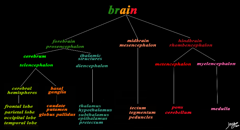
The basic and simplest classification of the brain into forebrain midbrain and hindbrain is shown in this diagram and advanced to a more complex tree using the embryological and evolutionary terminologies.
The hindbrain is shown in the right column and is colored in dark red.
The forebrain consists of the cerebrum also called the prosencephalon, which contains the more advanced form of the brain and the thalamic structures which contain more basic structures.
The cerebrum (telencephalon) itself consists of two cerebral hemispheres and paired basal ganglial structures. Each cerebral hemisphere will have gray and white matter distributed in the frontal parietal temporal and occipital lobe, with the basal ganglia being part of the gray matter deep in the cerebral hemispheres. The most important thalamic structures arising from the diencephalons include the thalamus itself and the hypothalamus.
The midbrain (mesencepaholon) consists of the tectum tegmentum and cerebral peduncles.
The hindbrain has two major branch points based on the evolutionary development classification. The pons and cerebellum(part of the metencephalon) are grouped and the medulla (part of the myelencephalon) s the second branch.
Courtesy Ashley Davidoff MD Copyright 2010 All rights reserved 97686.8s


    [nodeType] => 1
    [parentNode] => (object value omitted)
    [childNodes] => (object value omitted)
    [firstChild] => (object value omitted)
    [lastChild] => (object value omitted)
    [previousSibling] => (object value omitted)
    [nextSibling] => (object value omitted)
    [attributes] => (object value omitted)
    [ownerDocument] => (object value omitted)
    [namespaceURI] =>
    [prefix] =>
    [localName] => table
    [baseURI] =>
    [textContent] =>


The Hindbrain (rhombencephalon) in Context


The basic and simplest classification of the brain into forebrain midbrain and hindbrain is shown in this diagram and advanced to a more complex tree using the embryological and evolutionary terminologies.
The hindbrain is shown in the right column and is colored in dark red.
The forebrain consists of the cerebrum also called the prosencephalon, which contains the more advanced form of the brain and the thalamic structures which contain more basic structures.
The cerebrum (telencephalon) itself consists of two cerebral hemispheres and paired basal ganglial structures. Each cerebral hemisphere will have gray and white matter distributed in the frontal parietal temporal and occipital lobe, with the basal ganglia being part of the gray matter deep in the cerebral hemispheres. The most important thalamic structures arising from the diencephalons include the thalamus itself and the hypothalamus.
The midbrain (mesencepaholon) consists of the tectum tegmentum and cerebral peduncles.
The hindbrain has two major branch points based on the evolutionary development classification. The pons and cerebellum(part of the metencephalon) are grouped and the medulla (part of the myelencephalon) s the second branch.
Courtesy Ashley Davidoff MD Copyright 2010 All rights reserved 97686.8s


)
DOMElement Object
(
    [schemaTypeInfo] =>
    [tagName] => td
    [firstElementChild] => (object value omitted)
    [lastElementChild] => (object value omitted)
    [childElementCount] => 7
    [previousElementSibling] =>
    [nextElementSibling] =>
    [nodeName] => td
    [nodeValue] =>
The basic and simplest classification of the brain into forebrain midbrain and hindbrain is shown in this diagram and advanced to a more complex tree using the embryological and evolutionary terminologies.
The hindbrain is shown in the right column and is colored in dark red.
The forebrain consists of the cerebrum also called the prosencephalon, which contains the more advanced form of the brain and the thalamic structures which contain more basic structures.
The cerebrum (telencephalon) itself consists of two cerebral hemispheres and paired basal ganglial structures. Each cerebral hemisphere will have gray and white matter distributed in the frontal parietal temporal and occipital lobe, with the basal ganglia being part of the gray matter deep in the cerebral hemispheres. The most important thalamic structures arising from the diencephalons include the thalamus itself and the hypothalamus.
The midbrain (mesencepaholon) consists of the tectum tegmentum and cerebral peduncles.
The hindbrain has two major branch points based on the evolutionary development classification. The pons and cerebellum(part of the metencephalon) are grouped and the medulla (part of the myelencephalon) s the second branch.
Courtesy Ashley Davidoff MD Copyright 2010 All rights reserved 97686.8s

    [nodeType] => 1
    [parentNode] => (object value omitted)
    [childNodes] => (object value omitted)
    [firstChild] => (object value omitted)
    [lastChild] => (object value omitted)
    [previousSibling] => (object value omitted)
    [nextSibling] => (object value omitted)
    [attributes] => (object value omitted)
    [ownerDocument] => (object value omitted)
    [namespaceURI] =>
    [prefix] =>
    [localName] => td
    [baseURI] =>
    [textContent] =>
The basic and simplest classification of the brain into forebrain midbrain and hindbrain is shown in this diagram and advanced to a more complex tree using the embryological and evolutionary terminologies.
The hindbrain is shown in the right column and is colored in dark red.
The forebrain consists of the cerebrum also called the prosencephalon, which contains the more advanced form of the brain and the thalamic structures which contain more basic structures.
The cerebrum (telencephalon) itself consists of two cerebral hemispheres and paired basal ganglial structures. Each cerebral hemisphere will have gray and white matter distributed in the frontal parietal temporal and occipital lobe, with the basal ganglia being part of the gray matter deep in the cerebral hemispheres. The most important thalamic structures arising from the diencephalons include the thalamus itself and the hypothalamus.
The midbrain (mesencepaholon) consists of the tectum tegmentum and cerebral peduncles.
The hindbrain has two major branch points based on the evolutionary development classification. The pons and cerebellum(part of the metencephalon) are grouped and the medulla (part of the myelencephalon) s the second branch.
Courtesy Ashley Davidoff MD Copyright 2010 All rights reserved 97686.8s

)
DOMElement Object
(
    [schemaTypeInfo] =>
    [tagName] => td
    [firstElementChild] => (object value omitted)
    [lastElementChild] => (object value omitted)
    [childElementCount] => 2
    [previousElementSibling] =>
    [nextElementSibling] =>
    [nodeName] => td
    [nodeValue] =>
The Hindbrain (rhombencephalon) in Context
    [nodeType] => 1
    [parentNode] => (object value omitted)
    [childNodes] => (object value omitted)
    [firstChild] => (object value omitted)
    [lastChild] => (object value omitted)
    [previousSibling] => (object value omitted)
    [nextSibling] => (object value omitted)
    [attributes] => (object value omitted)
    [ownerDocument] => (object value omitted)
    [namespaceURI] =>
    [prefix] =>
    [localName] => td
    [baseURI] =>
    [textContent] =>
The Hindbrain (rhombencephalon) in Context
)

