Coronal View
Concepts
Ashley Davidoff MD
Copyright 2010
Introduction
The initial discussion will revolve around the relative positioning of the major parts of the forebrain and landmarks which allow us to recognise where we are
DOMElement Object
(
    [schemaTypeInfo] =>
    [tagName] => table
    [firstElementChild] => (object value omitted)
    [lastElementChild] => (object value omitted)
    [childElementCount] => 1
    [previousElementSibling] => (object value omitted)
    [nextElementSibling] =>
    [nodeName] => table
    [nodeValue] =>


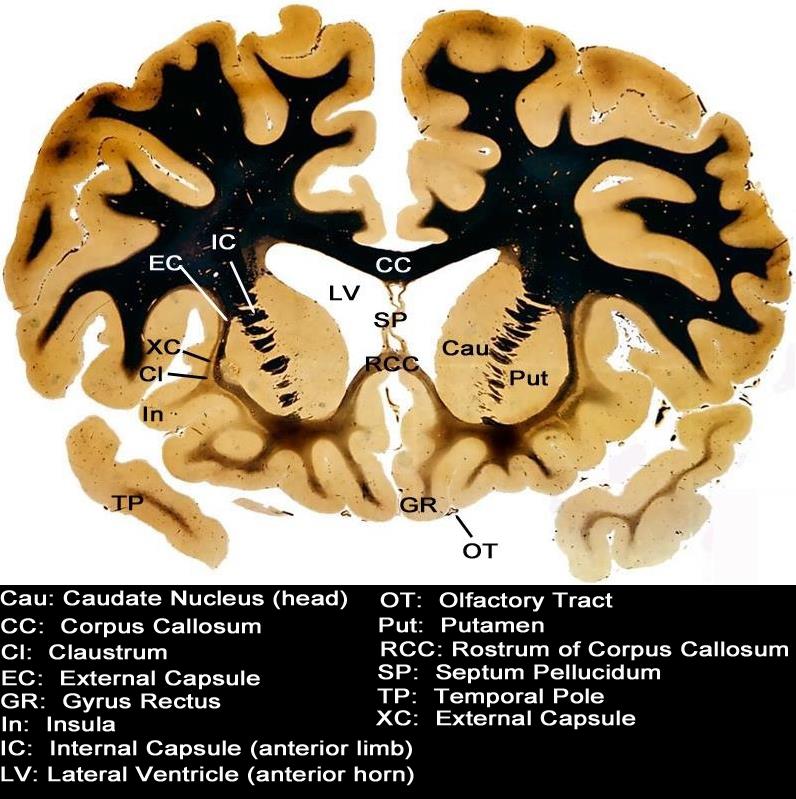
Anatomic Section through the Forebrain


This coronal section through the forebrain starts to introduce more complexity to the brain.  The cortical components at this level include the frontoparietal region and the temporal pole(TP).  Centrally the corpus callosum (CC) and septum  pellucidum SP are noted, and are surrounded by the next paracentral structures which include the lateral ventricles (LV).  The basal ganglia starting with the caudate nucleus  (Cau) and then the putamen follow, separated by the internal capsule (IC).  The external capsule (XC) and claustrum (Cl) follow as the two lateral structures till we return to the outer umbrella of the frontoparietal cortex again.
Courtesy Department of Anatomy and Neurobiology at Boston University School o


    [nodeType] => 1
    [parentNode] => (object value omitted)
    [childNodes] => (object value omitted)
    [firstChild] => (object value omitted)
    [lastChild] => (object value omitted)
    [previousSibling] => (object value omitted)
    [nextSibling] => (object value omitted)
    [attributes] => (object value omitted)
    [ownerDocument] => (object value omitted)
    [namespaceURI] =>
    [prefix] =>
    [localName] => table
    [baseURI] =>
    [textContent] =>


Anatomic Section through the Forebrain


This coronal section through the forebrain starts to introduce more complexity to the brain.  The cortical components at this level include the frontoparietal region and the temporal pole(TP).  Centrally the corpus callosum (CC) and septum  pellucidum SP are noted, and are surrounded by the next paracentral structures which include the lateral ventricles (LV).  The basal ganglia starting with the caudate nucleus  (Cau) and then the putamen follow, separated by the internal capsule (IC).  The external capsule (XC) and claustrum (Cl) follow as the two lateral structures till we return to the outer umbrella of the frontoparietal cortex again.
Courtesy Department of Anatomy and Neurobiology at Boston University School o


)
DOMElement Object
(
    [schemaTypeInfo] =>
    [tagName] => td
    [firstElementChild] => (object value omitted)
    [lastElementChild] => (object value omitted)
    [childElementCount] => 2
    [previousElementSibling] =>
    [nextElementSibling] =>
    [nodeName] => td
    [nodeValue] =>
This coronal section through the forebrain starts to introduce more complexity to the brain.  The cortical components at this level include the frontoparietal region and the temporal pole(TP).  Centrally the corpus callosum (CC) and septum  pellucidum SP are noted, and are surrounded by the next paracentral structures which include the lateral ventricles (LV).  The basal ganglia starting with the caudate nucleus  (Cau) and then the putamen follow, separated by the internal capsule (IC).  The external capsule (XC) and claustrum (Cl) follow as the two lateral structures till we return to the outer umbrella of the frontoparietal cortex again.
Courtesy Department of Anatomy and Neurobiology at Boston University School o

    [nodeType] => 1
    [parentNode] => (object value omitted)
    [childNodes] => (object value omitted)
    [firstChild] => (object value omitted)
    [lastChild] => (object value omitted)
    [previousSibling] => (object value omitted)
    [nextSibling] => (object value omitted)
    [attributes] => (object value omitted)
    [ownerDocument] => (object value omitted)
    [namespaceURI] =>
    [prefix] =>
    [localName] => td
    [baseURI] =>
    [textContent] =>
This coronal section through the forebrain starts to introduce more complexity to the brain.  The cortical components at this level include the frontoparietal region and the temporal pole(TP).  Centrally the corpus callosum (CC) and septum  pellucidum SP are noted, and are surrounded by the next paracentral structures which include the lateral ventricles (LV).  The basal ganglia starting with the caudate nucleus  (Cau) and then the putamen follow, separated by the internal capsule (IC).  The external capsule (XC) and claustrum (Cl) follow as the two lateral structures till we return to the outer umbrella of the frontoparietal cortex again.
Courtesy Department of Anatomy and Neurobiology at Boston University School o

)
DOMElement Object
(
    [schemaTypeInfo] =>
    [tagName] => td
    [firstElementChild] => (object value omitted)
    [lastElementChild] => (object value omitted)
    [childElementCount] => 2
    [previousElementSibling] =>
    [nextElementSibling] =>
    [nodeName] => td
    [nodeValue] =>
Anatomic Section through the Forebrain
    [nodeType] => 1
    [parentNode] => (object value omitted)
    [childNodes] => (object value omitted)
    [firstChild] => (object value omitted)
    [lastChild] => (object value omitted)
    [previousSibling] => (object value omitted)
    [nextSibling] => (object value omitted)
    [attributes] => (object value omitted)
    [ownerDocument] => (object value omitted)
    [namespaceURI] =>
    [prefix] =>
    [localName] => td
    [baseURI] =>
    [textContent] =>
Anatomic Section through the Forebrain
)
DOMElement Object
(
    [schemaTypeInfo] =>
    [tagName] => table
    [firstElementChild] => (object value omitted)
    [lastElementChild] => (object value omitted)
    [childElementCount] => 1
    [previousElementSibling] => (object value omitted)
    [nextElementSibling] => (object value omitted)
    [nodeName] => table
    [nodeValue] =>


The Parietal Lobes and the Temporal Lobes


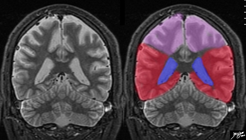
The coronal MRI shows the diversion of the ventriclesand their course inferiorly into the temporal horns allowing us to place the cut fairly posteriorly, and beyond the central sulcus, thus the superior aspect of the cut represents the parietal lobes, and the forebrain lying above the tentorium must represent the temporal lobes (red).
Courtesy Ashley Davidoff MD copyright 2010 all rights reserved 72246c02.8s


    [nodeType] => 1
    [parentNode] => (object value omitted)
    [childNodes] => (object value omitted)
    [firstChild] => (object value omitted)
    [lastChild] => (object value omitted)
    [previousSibling] => (object value omitted)
    [nextSibling] => (object value omitted)
    [attributes] => (object value omitted)
    [ownerDocument] => (object value omitted)
    [namespaceURI] =>
    [prefix] =>
    [localName] => table
    [baseURI] =>
    [textContent] =>


The Parietal Lobes and the Temporal Lobes


The coronal MRI shows the diversion of the ventriclesand their course inferiorly into the temporal horns allowing us to place the cut fairly posteriorly, and beyond the central sulcus, thus the superior aspect of the cut represents the parietal lobes, and the forebrain lying above the tentorium must represent the temporal lobes (red).
Courtesy Ashley Davidoff MD copyright 2010 all rights reserved 72246c02.8s


)
DOMElement Object
(
    [schemaTypeInfo] =>
    [tagName] => td
    [firstElementChild] => (object value omitted)
    [lastElementChild] => (object value omitted)
    [childElementCount] => 2
    [previousElementSibling] =>
    [nextElementSibling] =>
    [nodeName] => td
    [nodeValue] =>
The coronal MRI shows the diversion of the ventriclesand their course inferiorly into the temporal horns allowing us to place the cut fairly posteriorly, and beyond the central sulcus, thus the superior aspect of the cut represents the parietal lobes, and the forebrain lying above the tentorium must represent the temporal lobes (red).
Courtesy Ashley Davidoff MD copyright 2010 all rights reserved 72246c02.8s

    [nodeType] => 1
    [parentNode] => (object value omitted)
    [childNodes] => (object value omitted)
    [firstChild] => (object value omitted)
    [lastChild] => (object value omitted)
    [previousSibling] => (object value omitted)
    [nextSibling] => (object value omitted)
    [attributes] => (object value omitted)
    [ownerDocument] => (object value omitted)
    [namespaceURI] =>
    [prefix] =>
    [localName] => td
    [baseURI] =>
    [textContent] =>
The coronal MRI shows the diversion of the ventriclesand their course inferiorly into the temporal horns allowing us to place the cut fairly posteriorly, and beyond the central sulcus, thus the superior aspect of the cut represents the parietal lobes, and the forebrain lying above the tentorium must represent the temporal lobes (red).
Courtesy Ashley Davidoff MD copyright 2010 all rights reserved 72246c02.8s

)
DOMElement Object
(
    [schemaTypeInfo] =>
    [tagName] => td
    [firstElementChild] => (object value omitted)
    [lastElementChild] => (object value omitted)
    [childElementCount] => 2
    [previousElementSibling] =>
    [nextElementSibling] =>
    [nodeName] => td
    [nodeValue] =>
The Parietal Lobes and the Temporal Lobes
    [nodeType] => 1
    [parentNode] => (object value omitted)
    [childNodes] => (object value omitted)
    [firstChild] => (object value omitted)
    [lastChild] => (object value omitted)
    [previousSibling] => (object value omitted)
    [nextSibling] => (object value omitted)
    [attributes] => (object value omitted)
    [ownerDocument] => (object value omitted)
    [namespaceURI] =>
    [prefix] =>
    [localName] => td
    [baseURI] =>
    [textContent] =>
The Parietal Lobes and the Temporal Lobes
)
DOMElement Object
(
    [schemaTypeInfo] =>
    [tagName] => table
    [firstElementChild] => (object value omitted)
    [lastElementChild] => (object value omitted)
    [childElementCount] => 1
    [previousElementSibling] => (object value omitted)
    [nextElementSibling] => (object value omitted)
    [nodeName] => table
    [nodeValue] =>


Anterior Cut
The Frontal Lobe and the Temporal lobe


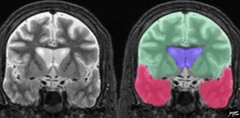
The series of coronal cuts uses the ventricles once again as a reference point. The relatively large size of the ventricles (and the presence of the caudate nuclii place the cut fairly anterior in front of the central sulcus, and so the top level in the anterior cranial fossa is the frontal lobe (light green) and the second level reveals the temporal lobes (red).
Courtesy Ashley Davidoff MD copyright 2010 all rights reserved 72233c03b.8s


    [nodeType] => 1
    [parentNode] => (object value omitted)
    [childNodes] => (object value omitted)
    [firstChild] => (object value omitted)
    [lastChild] => (object value omitted)
    [previousSibling] => (object value omitted)
    [nextSibling] => (object value omitted)
    [attributes] => (object value omitted)
    [ownerDocument] => (object value omitted)
    [namespaceURI] =>
    [prefix] =>
    [localName] => table
    [baseURI] =>
    [textContent] =>


Anterior Cut
The Frontal Lobe and the Temporal lobe


The series of coronal cuts uses the ventricles once again as a reference point. The relatively large size of the ventricles (and the presence of the caudate nuclii place the cut fairly anterior in front of the central sulcus, and so the top level in the anterior cranial fossa is the frontal lobe (light green) and the second level reveals the temporal lobes (red).
Courtesy Ashley Davidoff MD copyright 2010 all rights reserved 72233c03b.8s


)
DOMElement Object
(
    [schemaTypeInfo] =>
    [tagName] => td
    [firstElementChild] => (object value omitted)
    [lastElementChild] => (object value omitted)
    [childElementCount] => 2
    [previousElementSibling] =>
    [nextElementSibling] =>
    [nodeName] => td
    [nodeValue] =>
The series of coronal cuts uses the ventricles once again as a reference point. The relatively large size of the ventricles (and the presence of the caudate nuclii place the cut fairly anterior in front of the central sulcus, and so the top level in the anterior cranial fossa is the frontal lobe (light green) and the second level reveals the temporal lobes (red).
Courtesy Ashley Davidoff MD copyright 2010 all rights reserved 72233c03b.8s

    [nodeType] => 1
    [parentNode] => (object value omitted)
    [childNodes] => (object value omitted)
    [firstChild] => (object value omitted)
    [lastChild] => (object value omitted)
    [previousSibling] => (object value omitted)
    [nextSibling] => (object value omitted)
    [attributes] => (object value omitted)
    [ownerDocument] => (object value omitted)
    [namespaceURI] =>
    [prefix] =>
    [localName] => td
    [baseURI] =>
    [textContent] =>
The series of coronal cuts uses the ventricles once again as a reference point. The relatively large size of the ventricles (and the presence of the caudate nuclii place the cut fairly anterior in front of the central sulcus, and so the top level in the anterior cranial fossa is the frontal lobe (light green) and the second level reveals the temporal lobes (red).
Courtesy Ashley Davidoff MD copyright 2010 all rights reserved 72233c03b.8s

)
DOMElement Object
(
    [schemaTypeInfo] =>
    [tagName] => td
    [firstElementChild] => (object value omitted)
    [lastElementChild] => (object value omitted)
    [childElementCount] => 3
    [previousElementSibling] =>
    [nextElementSibling] =>
    [nodeName] => td
    [nodeValue] =>
Anterior Cut
The Frontal Lobe and the Temporal lobe
    [nodeType] => 1
    [parentNode] => (object value omitted)
    [childNodes] => (object value omitted)
    [firstChild] => (object value omitted)
    [lastChild] => (object value omitted)
    [previousSibling] => (object value omitted)
    [nextSibling] => (object value omitted)
    [attributes] => (object value omitted)
    [ownerDocument] => (object value omitted)
    [namespaceURI] =>
    [prefix] =>
    [localName] => td
    [baseURI] =>
    [textContent] =>
Anterior Cut
The Frontal Lobe and the Temporal lobe
)
DOMElement Object
(
    [schemaTypeInfo] =>
    [tagName] => table
    [firstElementChild] => (object value omitted)
    [lastElementChild] => (object value omitted)
    [childElementCount] => 1
    [previousElementSibling] => (object value omitted)
    [nextElementSibling] => (object value omitted)
    [nodeName] => table
    [nodeValue] =>


Forebrain


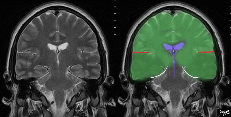
In this T2 weighted MRI image – the forebrain (green is seen centered aroubnd the ventriclar system. The red linees indicate the Sylvian fissures which in this instance in a relatively posterior cut reflect the parietal lobe superiorly and the temporal lobes inferiorly (darker green) The temporal lobes rest of the tentorium (white curvilinear convex lines). below the tenntorium is the midbrain and hind brain
Courtesy Ashley Davidoff MD copyright 2010 all rights reserved 89721c02b03.8s


    [nodeType] => 1
    [parentNode] => (object value omitted)
    [childNodes] => (object value omitted)
    [firstChild] => (object value omitted)
    [lastChild] => (object value omitted)
    [previousSibling] => (object value omitted)
    [nextSibling] => (object value omitted)
    [attributes] => (object value omitted)
    [ownerDocument] => (object value omitted)
    [namespaceURI] =>
    [prefix] =>
    [localName] => table
    [baseURI] =>
    [textContent] =>


Forebrain


In this T2 weighted MRI image – the forebrain (green is seen centered aroubnd the ventriclar system. The red linees indicate the Sylvian fissures which in this instance in a relatively posterior cut reflect the parietal lobe superiorly and the temporal lobes inferiorly (darker green) The temporal lobes rest of the tentorium (white curvilinear convex lines). below the tenntorium is the midbrain and hind brain
Courtesy Ashley Davidoff MD copyright 2010 all rights reserved 89721c02b03.8s


)
DOMElement Object
(
    [schemaTypeInfo] =>
    [tagName] => td
    [firstElementChild] => (object value omitted)
    [lastElementChild] => (object value omitted)
    [childElementCount] => 2
    [previousElementSibling] =>
    [nextElementSibling] =>
    [nodeName] => td
    [nodeValue] =>
In this T2 weighted MRI image – the forebrain (green is seen centered aroubnd the ventriclar system. The red linees indicate the Sylvian fissures which in this instance in a relatively posterior cut reflect the parietal lobe superiorly and the temporal lobes inferiorly (darker green) The temporal lobes rest of the tentorium (white curvilinear convex lines). below the tenntorium is the midbrain and hind brain
Courtesy Ashley Davidoff MD copyright 2010 all rights reserved 89721c02b03.8s

    [nodeType] => 1
    [parentNode] => (object value omitted)
    [childNodes] => (object value omitted)
    [firstChild] => (object value omitted)
    [lastChild] => (object value omitted)
    [previousSibling] => (object value omitted)
    [nextSibling] => (object value omitted)
    [attributes] => (object value omitted)
    [ownerDocument] => (object value omitted)
    [namespaceURI] =>
    [prefix] =>
    [localName] => td
    [baseURI] =>
    [textContent] =>
In this T2 weighted MRI image – the forebrain (green is seen centered aroubnd the ventriclar system. The red linees indicate the Sylvian fissures which in this instance in a relatively posterior cut reflect the parietal lobe superiorly and the temporal lobes inferiorly (darker green) The temporal lobes rest of the tentorium (white curvilinear convex lines). below the tenntorium is the midbrain and hind brain
Courtesy Ashley Davidoff MD copyright 2010 all rights reserved 89721c02b03.8s

)
DOMElement Object
(
    [schemaTypeInfo] =>
    [tagName] => td
    [firstElementChild] => (object value omitted)
    [lastElementChild] => (object value omitted)
    [childElementCount] => 2
    [previousElementSibling] =>
    [nextElementSibling] =>
    [nodeName] => td
    [nodeValue] =>
Forebrain

    [nodeType] => 1
    [parentNode] => (object value omitted)
    [childNodes] => (object value omitted)
    [firstChild] => (object value omitted)
    [lastChild] => (object value omitted)
    [previousSibling] => (object value omitted)
    [nextSibling] => (object value omitted)
    [attributes] => (object value omitted)
    [ownerDocument] => (object value omitted)
    [namespaceURI] =>
    [prefix] =>
    [localName] => td
    [baseURI] =>
    [textContent] =>
Forebrain

)
DOMElement Object
(
    [schemaTypeInfo] =>
    [tagName] => table
    [firstElementChild] => (object value omitted)
    [lastElementChild] => (object value omitted)
    [childElementCount] => 1
    [previousElementSibling] => (object value omitted)
    [nextElementSibling] => (object value omitted)
    [nodeName] => table
    [nodeValue] =>


The Ventricles
Reference Points


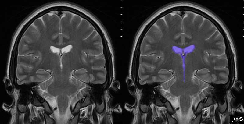
The MRI T2 weighted image is taken through the most revealing portions of the ventricles, imcluding from superior the paired lateral ventricles, the keyhole third ventricle just below, and then to the even smaller aqueduct of Sylvius. The structures of the brain are most easily recognized by using the ventricles as a reference point At this level all the major components of the brain can be identified as well.
Courtesy Ashley Davidoff MD copyright 2010 all rights reserved 89721c02.8s


    [nodeType] => 1
    [parentNode] => (object value omitted)
    [childNodes] => (object value omitted)
    [firstChild] => (object value omitted)
    [lastChild] => (object value omitted)
    [previousSibling] => (object value omitted)
    [nextSibling] => (object value omitted)
    [attributes] => (object value omitted)
    [ownerDocument] => (object value omitted)
    [namespaceURI] =>
    [prefix] =>
    [localName] => table
    [baseURI] =>
    [textContent] =>


The Ventricles
Reference Points


The MRI T2 weighted image is taken through the most revealing portions of the ventricles, imcluding from superior the paired lateral ventricles, the keyhole third ventricle just below, and then to the even smaller aqueduct of Sylvius. The structures of the brain are most easily recognized by using the ventricles as a reference point At this level all the major components of the brain can be identified as well.
Courtesy Ashley Davidoff MD copyright 2010 all rights reserved 89721c02.8s


)
DOMElement Object
(
    [schemaTypeInfo] =>
    [tagName] => td
    [firstElementChild] => (object value omitted)
    [lastElementChild] => (object value omitted)
    [childElementCount] => 2
    [previousElementSibling] =>
    [nextElementSibling] =>
    [nodeName] => td
    [nodeValue] =>
The MRI T2 weighted image is taken through the most revealing portions of the ventricles, imcluding from superior the paired lateral ventricles, the keyhole third ventricle just below, and then to the even smaller aqueduct of Sylvius. The structures of the brain are most easily recognized by using the ventricles as a reference point At this level all the major components of the brain can be identified as well.
Courtesy Ashley Davidoff MD copyright 2010 all rights reserved 89721c02.8s

    [nodeType] => 1
    [parentNode] => (object value omitted)
    [childNodes] => (object value omitted)
    [firstChild] => (object value omitted)
    [lastChild] => (object value omitted)
    [previousSibling] => (object value omitted)
    [nextSibling] => (object value omitted)
    [attributes] => (object value omitted)
    [ownerDocument] => (object value omitted)
    [namespaceURI] =>
    [prefix] =>
    [localName] => td
    [baseURI] =>
    [textContent] =>
The MRI T2 weighted image is taken through the most revealing portions of the ventricles, imcluding from superior the paired lateral ventricles, the keyhole third ventricle just below, and then to the even smaller aqueduct of Sylvius. The structures of the brain are most easily recognized by using the ventricles as a reference point At this level all the major components of the brain can be identified as well.
Courtesy Ashley Davidoff MD copyright 2010 all rights reserved 89721c02.8s

)
DOMElement Object
(
    [schemaTypeInfo] =>
    [tagName] => td
    [firstElementChild] => (object value omitted)
    [lastElementChild] => (object value omitted)
    [childElementCount] => 3
    [previousElementSibling] =>
    [nextElementSibling] =>
    [nodeName] => td
    [nodeValue] =>
The Ventricles
Reference Points

    [nodeType] => 1
    [parentNode] => (object value omitted)
    [childNodes] => (object value omitted)
    [firstChild] => (object value omitted)
    [lastChild] => (object value omitted)
    [previousSibling] => (object value omitted)
    [nextSibling] => (object value omitted)
    [attributes] => (object value omitted)
    [ownerDocument] => (object value omitted)
    [namespaceURI] =>
    [prefix] =>
    [localName] => td
    [baseURI] =>
    [textContent] =>
The Ventricles
Reference Points

)

