Temporal Lobe
Ashley Davidoff MD
Copyright 2010
Definition
The temporal lobe is part of the forebrain and is located at the side of the brain beneath the Sylvian fissure.
The temporal lobe is an important area for many cognitive functions, including hearing, speech and memory and vision of complex shapes.
Structure
The temporal lobe is located in the middle and inferior part of the hemisphere. Behind, it is limited by an imaginary line continuing the parieto-occipital sulcus. Anteriorly and superiorly by the Sylvian fissure. It contains 3 convolutions (superior, middle and inferior temporal gyri) divided by 2 fissures (superior and middle temporal sulci).
Function
The temporal lobes process the immediate events in recent and remote memory. They receive and interpret sounds and images, store events in the form of memory and recall those already memorized the roads and generate emotional.
The superior temporal gyrus contains Wernicke’s area, which is invovled in the incorporation and comprehension of spoken language. From the lateral gyri, several short folds are apparent, which form the gyri of Heschl, which represents the primary auditory area. The hippocampus is located within the temporal lobe.
Disease
Memory of sounds and shapes tends to be affected; the understanding of language come from external or internal sources is dramatically impaired and typically prevents the individual to express himself through language. Individuals with may also develop personality changes, for example, lack of humor, unusual degrees of religiosity, obsessions, and loss of libido.
Dysfunction in the temporal lobe leads to memory impairment as well as difficulties producing and understanding speech.
Dysfunction is diagnosed through the observation of imapairment, cognitive and speech related tests, and neuroimaging to identify the cause. Treatment includes speech therapy as well as pharmocological agents to combat personality changes.
|
Conceptual Framework of the Brain |
|
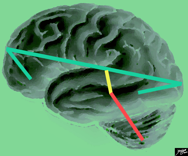
The vector of the forebrain (green) is folded at its anterior aspect as a projection of the frontal lobe and the posterior fold forms the temporal lobe. The midbrain (yellow has its axis just tipped slightly anteriorly and the hindbrain (salmon red) is tipped more posteriorly Courtesy Ashley Davidoff MD copyright 2010 all rights reserved |
|
Axial Projection MRI |
|
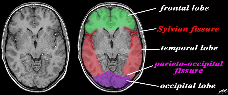
The axial image is intended to demonstrate ttwo important fissures; the Sylvian fissure (in red) the parieto-occipital fissure (pink) in order to demonstrate the border between the frontal and temporal lobe in the axial plane and the junction of the temporal and occipital lobe MRI SENSE Courtesy Philips medicaL systems rendred by Davidoff art 92142c06b01 label.8s |
DOMElement Object
(
    [schemaTypeInfo] =>
    [tagName] => table
    [firstElementChild] => (object value omitted)
    [lastElementChild] => (object value omitted)
    [childElementCount] => 1
    [previousElementSibling] => (object value omitted)
    [nextElementSibling] =>
    [nodeName] => table
    [nodeValue] =>


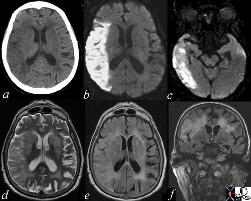
Right Parietal and Temporal Lobe Acute Infarction


71275c01.800 brain cerebrum acute right mca infarct parietal lobe temporal lobe right middle cerebral artery infarction a= CTscan b = DWI diffusion weighted imaging bright c= DWI d= T2 weighted MRI e = FLAIR


    [nodeType] => 1
    [parentNode] => (object value omitted)
    [childNodes] => (object value omitted)
    [firstChild] => (object value omitted)
    [lastChild] => (object value omitted)
    [previousSibling] => (object value omitted)
    [nextSibling] => (object value omitted)
    [attributes] => (object value omitted)
    [ownerDocument] => (object value omitted)
    [namespaceURI] =>
    [prefix] =>
    [localName] => table
    [baseURI] =>
    [textContent] =>


Right Parietal and Temporal Lobe Acute Infarction


71275c01.800 brain cerebrum acute right mca infarct parietal lobe temporal lobe right middle cerebral artery infarction a= CTscan b = DWI diffusion weighted imaging bright c= DWI d= T2 weighted MRI e = FLAIR


)
DOMElement Object
(
    [schemaTypeInfo] =>
    [tagName] => td
    [firstElementChild] =>
    [lastElementChild] =>
    [childElementCount] => 0
    [previousElementSibling] =>
    [nextElementSibling] =>
    [nodeName] => td
    [nodeValue] => 71275c01.800 brain cerebrum acute right mca infarct parietal lobe temporal lobe right middle cerebral artery infarction a= CTscan b = DWI diffusion weighted imaging bright c= DWI d= T2 weighted MRI e = FLAIR
    [nodeType] => 1
    [parentNode] => (object value omitted)
    [childNodes] => (object value omitted)
    [firstChild] => (object value omitted)
    [lastChild] => (object value omitted)
    [previousSibling] => (object value omitted)
    [nextSibling] => (object value omitted)
    [attributes] => (object value omitted)
    [ownerDocument] => (object value omitted)
    [namespaceURI] =>
    [prefix] =>
    [localName] => td
    [baseURI] =>
    [textContent] => 71275c01.800 brain cerebrum acute right mca infarct parietal lobe temporal lobe right middle cerebral artery infarction a= CTscan b = DWI diffusion weighted imaging bright c= DWI d= T2 weighted MRI e = FLAIR
)
DOMElement Object
(
    [schemaTypeInfo] =>
    [tagName] => td
    [firstElementChild] => (object value omitted)
    [lastElementChild] => (object value omitted)
    [childElementCount] => 2
    [previousElementSibling] =>
    [nextElementSibling] =>
    [nodeName] => td
    [nodeValue] =>
Right Parietal and Temporal Lobe Acute Infarction
    [nodeType] => 1
    [parentNode] => (object value omitted)
    [childNodes] => (object value omitted)
    [firstChild] => (object value omitted)
    [lastChild] => (object value omitted)
    [previousSibling] => (object value omitted)
    [nextSibling] => (object value omitted)
    [attributes] => (object value omitted)
    [ownerDocument] => (object value omitted)
    [namespaceURI] =>
    [prefix] =>
    [localName] => td
    [baseURI] =>
    [textContent] =>
Right Parietal and Temporal Lobe Acute Infarction
)
DOMElement Object
(
    [schemaTypeInfo] =>
    [tagName] => table
    [firstElementChild] => (object value omitted)
    [lastElementChild] => (object value omitted)
    [childElementCount] => 1
    [previousElementSibling] => (object value omitted)
    [nextElementSibling] => (object value omitted)
    [nodeName] => table
    [nodeValue] =>


Posterior Coroanl Cut
Temporal and Parietal Lobes


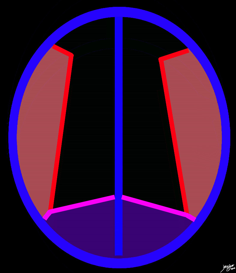
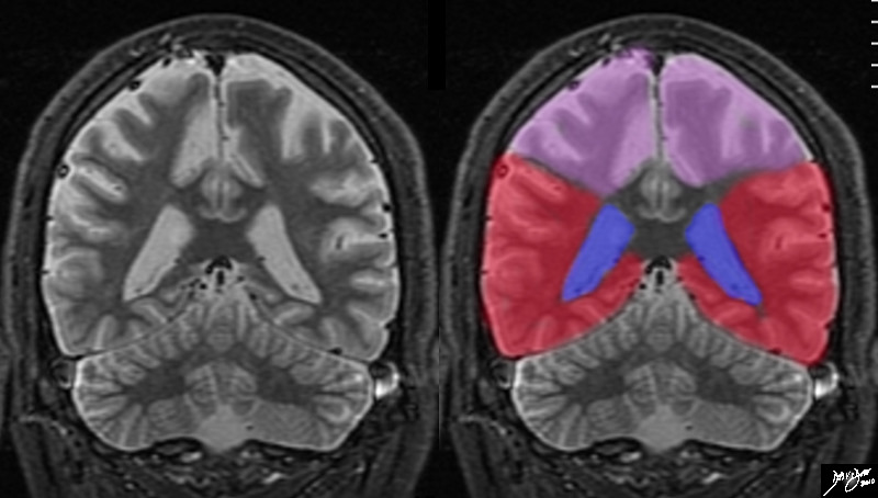
The coronal MRI shows the diversion of the ventriclesand their course inferiorly into the temporal horns allowing us to place the cut fairly posteriorly, and beyond the central sulcus, thus the superior aspect of the cut represents the parietal lobes, and the forebrain lying above the tentorium must represent the temporal lobes (red).
Courtesy Ashley Davidoff MD copyright 2010 all rights reserved 72246c02.8s


    [nodeType] => 1
    [parentNode] => (object value omitted)
    [childNodes] => (object value omitted)
    [firstChild] => (object value omitted)
    [lastChild] => (object value omitted)
    [previousSibling] => (object value omitted)
    [nextSibling] => (object value omitted)
    [attributes] => (object value omitted)
    [ownerDocument] => (object value omitted)
    [namespaceURI] =>
    [prefix] =>
    [localName] => table
    [baseURI] =>
    [textContent] =>


Posterior Coroanl Cut
Temporal and Parietal Lobes


The coronal MRI shows the diversion of the ventriclesand their course inferiorly into the temporal horns allowing us to place the cut fairly posteriorly, and beyond the central sulcus, thus the superior aspect of the cut represents the parietal lobes, and the forebrain lying above the tentorium must represent the temporal lobes (red).
Courtesy Ashley Davidoff MD copyright 2010 all rights reserved 72246c02.8s


)
DOMElement Object
(
    [schemaTypeInfo] =>
    [tagName] => td
    [firstElementChild] => (object value omitted)
    [lastElementChild] => (object value omitted)
    [childElementCount] => 2
    [previousElementSibling] =>
    [nextElementSibling] =>
    [nodeName] => td
    [nodeValue] =>
The coronal MRI shows the diversion of the ventriclesand their course inferiorly into the temporal horns allowing us to place the cut fairly posteriorly, and beyond the central sulcus, thus the superior aspect of the cut represents the parietal lobes, and the forebrain lying above the tentorium must represent the temporal lobes (red).
Courtesy Ashley Davidoff MD copyright 2010 all rights reserved 72246c02.8s

    [nodeType] => 1
    [parentNode] => (object value omitted)
    [childNodes] => (object value omitted)
    [firstChild] => (object value omitted)
    [lastChild] => (object value omitted)
    [previousSibling] => (object value omitted)
    [nextSibling] => (object value omitted)
    [attributes] => (object value omitted)
    [ownerDocument] => (object value omitted)
    [namespaceURI] =>
    [prefix] =>
    [localName] => td
    [baseURI] =>
    [textContent] =>
The coronal MRI shows the diversion of the ventriclesand their course inferiorly into the temporal horns allowing us to place the cut fairly posteriorly, and beyond the central sulcus, thus the superior aspect of the cut represents the parietal lobes, and the forebrain lying above the tentorium must represent the temporal lobes (red).
Courtesy Ashley Davidoff MD copyright 2010 all rights reserved 72246c02.8s

)
DOMElement Object
(
    [schemaTypeInfo] =>
    [tagName] => td
    [firstElementChild] => (object value omitted)
    [lastElementChild] => (object value omitted)
    [childElementCount] => 3
    [previousElementSibling] =>
    [nextElementSibling] =>
    [nodeName] => td
    [nodeValue] =>
Posterior Coroanl Cut
Temporal and Parietal Lobes
    [nodeType] => 1
    [parentNode] => (object value omitted)
    [childNodes] => (object value omitted)
    [firstChild] => (object value omitted)
    [lastChild] => (object value omitted)
    [previousSibling] => (object value omitted)
    [nextSibling] => (object value omitted)
    [attributes] => (object value omitted)
    [ownerDocument] => (object value omitted)
    [namespaceURI] =>
    [prefix] =>
    [localName] => td
    [baseURI] =>
    [textContent] =>
Posterior Coroanl Cut
Temporal and Parietal Lobes
)
DOMElement Object
(
    [schemaTypeInfo] =>
    [tagName] => table
    [firstElementChild] => (object value omitted)
    [lastElementChild] => (object value omitted)
    [childElementCount] => 1
    [previousElementSibling] => (object value omitted)
    [nextElementSibling] => (object value omitted)
    [nodeName] => table
    [nodeValue] =>


Coronal Projection Sylvian Fissure Parietal Lobe and Temporal Lobe


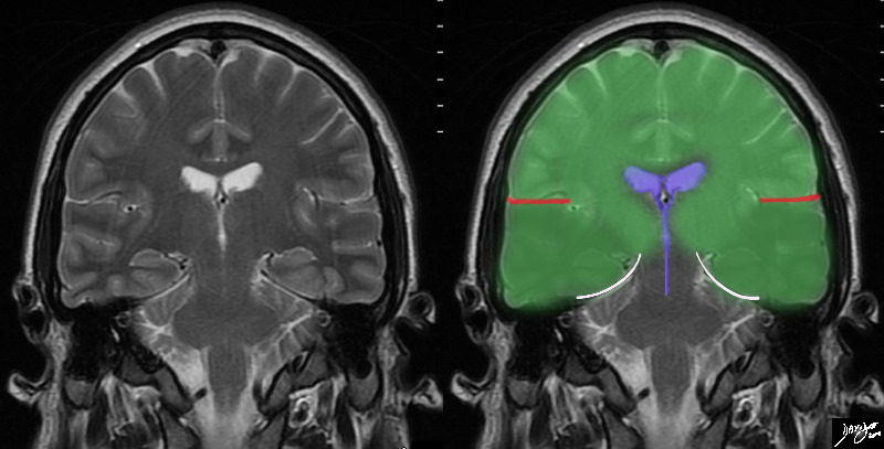
In this T2 weighted MRI image – the forebrain (green is seen centered aroubnd the ventriclar system. The red linees indicate the Sylvian fissures which in this instance in a relatively posterior cut reflect the parietal lobe superiorly and the temporal lobes inferiorly (darker green) The temporal lobes rest of the tentorium (white curvilinear convex lines). below the tenntorium is the midbrain and hind brain
Courtesy Ashley Davidoff MD copyright 2010 all rights reserved 89721c02b03.8s 89721c02b03


    [nodeType] => 1
    [parentNode] => (object value omitted)
    [childNodes] => (object value omitted)
    [firstChild] => (object value omitted)
    [lastChild] => (object value omitted)
    [previousSibling] => (object value omitted)
    [nextSibling] => (object value omitted)
    [attributes] => (object value omitted)
    [ownerDocument] => (object value omitted)
    [namespaceURI] =>
    [prefix] =>
    [localName] => table
    [baseURI] =>
    [textContent] =>


Coronal Projection Sylvian Fissure Parietal Lobe and Temporal Lobe


In this T2 weighted MRI image – the forebrain (green is seen centered aroubnd the ventriclar system. The red linees indicate the Sylvian fissures which in this instance in a relatively posterior cut reflect the parietal lobe superiorly and the temporal lobes inferiorly (darker green) The temporal lobes rest of the tentorium (white curvilinear convex lines). below the tenntorium is the midbrain and hind brain
Courtesy Ashley Davidoff MD copyright 2010 all rights reserved 89721c02b03.8s 89721c02b03


)
DOMElement Object
(
    [schemaTypeInfo] =>
    [tagName] => td
    [firstElementChild] => (object value omitted)
    [lastElementChild] => (object value omitted)
    [childElementCount] => 2
    [previousElementSibling] =>
    [nextElementSibling] =>
    [nodeName] => td
    [nodeValue] =>
In this T2 weighted MRI image – the forebrain (green is seen centered aroubnd the ventriclar system. The red linees indicate the Sylvian fissures which in this instance in a relatively posterior cut reflect the parietal lobe superiorly and the temporal lobes inferiorly (darker green) The temporal lobes rest of the tentorium (white curvilinear convex lines). below the tenntorium is the midbrain and hind brain
Courtesy Ashley Davidoff MD copyright 2010 all rights reserved 89721c02b03.8s 89721c02b03

    [nodeType] => 1
    [parentNode] => (object value omitted)
    [childNodes] => (object value omitted)
    [firstChild] => (object value omitted)
    [lastChild] => (object value omitted)
    [previousSibling] => (object value omitted)
    [nextSibling] => (object value omitted)
    [attributes] => (object value omitted)
    [ownerDocument] => (object value omitted)
    [namespaceURI] =>
    [prefix] =>
    [localName] => td
    [baseURI] =>
    [textContent] =>
In this T2 weighted MRI image – the forebrain (green is seen centered aroubnd the ventriclar system. The red linees indicate the Sylvian fissures which in this instance in a relatively posterior cut reflect the parietal lobe superiorly and the temporal lobes inferiorly (darker green) The temporal lobes rest of the tentorium (white curvilinear convex lines). below the tenntorium is the midbrain and hind brain
Courtesy Ashley Davidoff MD copyright 2010 all rights reserved 89721c02b03.8s 89721c02b03

)
DOMElement Object
(
    [schemaTypeInfo] =>
    [tagName] => td
    [firstElementChild] => (object value omitted)
    [lastElementChild] => (object value omitted)
    [childElementCount] => 2
    [previousElementSibling] =>
    [nextElementSibling] =>
    [nodeName] => td
    [nodeValue] =>
Coronal Projection Sylvian Fissure Parietal Lobe and Temporal Lobe
    [nodeType] => 1
    [parentNode] => (object value omitted)
    [childNodes] => (object value omitted)
    [firstChild] => (object value omitted)
    [lastChild] => (object value omitted)
    [previousSibling] => (object value omitted)
    [nextSibling] => (object value omitted)
    [attributes] => (object value omitted)
    [ownerDocument] => (object value omitted)
    [namespaceURI] =>
    [prefix] =>
    [localName] => td
    [baseURI] =>
    [textContent] =>
Coronal Projection Sylvian Fissure Parietal Lobe and Temporal Lobe
)
DOMElement Object
(
    [schemaTypeInfo] =>
    [tagName] => table
    [firstElementChild] => (object value omitted)
    [lastElementChild] => (object value omitted)
    [childElementCount] => 1
    [previousElementSibling] => (object value omitted)
    [nextElementSibling] => (object value omitted)
    [nodeName] => table
    [nodeValue] =>


Axial Projection MRI


The axial image is intended to demonstrate ttwo important fissures; the Sylvian fissure (in red) the parieto-occipital fissure (pink) in order to demonstrate the border between the frontal and temporal lobe in the axial plane and the junction of the temporal and occipital lobe
MRI SENSE Courtesy Philips medicaL systems rendred by Davidoff art 92142c06b01 label.8s


    [nodeType] => 1
    [parentNode] => (object value omitted)
    [childNodes] => (object value omitted)
    [firstChild] => (object value omitted)
    [lastChild] => (object value omitted)
    [previousSibling] => (object value omitted)
    [nextSibling] => (object value omitted)
    [attributes] => (object value omitted)
    [ownerDocument] => (object value omitted)
    [namespaceURI] =>
    [prefix] =>
    [localName] => table
    [baseURI] =>
    [textContent] =>


Axial Projection MRI


The axial image is intended to demonstrate ttwo important fissures; the Sylvian fissure (in red) the parieto-occipital fissure (pink) in order to demonstrate the border between the frontal and temporal lobe in the axial plane and the junction of the temporal and occipital lobe
MRI SENSE Courtesy Philips medicaL systems rendred by Davidoff art 92142c06b01 label.8s


)
DOMElement Object
(
    [schemaTypeInfo] =>
    [tagName] => td
    [firstElementChild] => (object value omitted)
    [lastElementChild] => (object value omitted)
    [childElementCount] => 2
    [previousElementSibling] =>
    [nextElementSibling] =>
    [nodeName] => td
    [nodeValue] =>
The axial image is intended to demonstrate ttwo important fissures; the Sylvian fissure (in red) the parieto-occipital fissure (pink) in order to demonstrate the border between the frontal and temporal lobe in the axial plane and the junction of the temporal and occipital lobe
MRI SENSE Courtesy Philips medicaL systems rendred by Davidoff art 92142c06b01 label.8s

    [nodeType] => 1
    [parentNode] => (object value omitted)
    [childNodes] => (object value omitted)
    [firstChild] => (object value omitted)
    [lastChild] => (object value omitted)
    [previousSibling] => (object value omitted)
    [nextSibling] => (object value omitted)
    [attributes] => (object value omitted)
    [ownerDocument] => (object value omitted)
    [namespaceURI] =>
    [prefix] =>
    [localName] => td
    [baseURI] =>
    [textContent] =>
The axial image is intended to demonstrate ttwo important fissures; the Sylvian fissure (in red) the parieto-occipital fissure (pink) in order to demonstrate the border between the frontal and temporal lobe in the axial plane and the junction of the temporal and occipital lobe
MRI SENSE Courtesy Philips medicaL systems rendred by Davidoff art 92142c06b01 label.8s

)
DOMElement Object
(
    [schemaTypeInfo] =>
    [tagName] => td
    [firstElementChild] => (object value omitted)
    [lastElementChild] => (object value omitted)
    [childElementCount] => 2
    [previousElementSibling] =>
    [nextElementSibling] =>
    [nodeName] => td
    [nodeValue] =>

Axial Projection MRI
    [nodeType] => 1
    [parentNode] => (object value omitted)
    [childNodes] => (object value omitted)
    [firstChild] => (object value omitted)
    [lastChild] => (object value omitted)
    [previousSibling] => (object value omitted)
    [nextSibling] => (object value omitted)
    [attributes] => (object value omitted)
    [ownerDocument] => (object value omitted)
    [namespaceURI] =>
    [prefix] =>
    [localName] => td
    [baseURI] =>
    [textContent] =>

Axial Projection MRI
)
DOMElement Object
(
    [schemaTypeInfo] =>
    [tagName] => table
    [firstElementChild] => (object value omitted)
    [lastElementChild] => (object value omitted)
    [childElementCount] => 1
    [previousElementSibling] => (object value omitted)
    [nextElementSibling] => (object value omitted)
    [nodeName] => table
    [nodeValue] =>


Axial at a Lower Level


At a lower level the parietooccipital fissure (pink) defines the anterior edge of the occipital lobe posteriorly (purple). The Sylvian fissure (bright red lines) defines the temporal lobes (light red)
Copyright 2010 all rights reserved 93914.36k.81s


    [nodeType] => 1
    [parentNode] => (object value omitted)
    [childNodes] => (object value omitted)
    [firstChild] => (object value omitted)
    [lastChild] => (object value omitted)
    [previousSibling] => (object value omitted)
    [nextSibling] => (object value omitted)
    [attributes] => (object value omitted)
    [ownerDocument] => (object value omitted)
    [namespaceURI] =>
    [prefix] =>
    [localName] => table
    [baseURI] =>
    [textContent] =>


Axial at a Lower Level


At a lower level the parietooccipital fissure (pink) defines the anterior edge of the occipital lobe posteriorly (purple). The Sylvian fissure (bright red lines) defines the temporal lobes (light red)
Copyright 2010 all rights reserved 93914.36k.81s


)
DOMElement Object
(
    [schemaTypeInfo] =>
    [tagName] => td
    [firstElementChild] => (object value omitted)
    [lastElementChild] => (object value omitted)
    [childElementCount] => 2
    [previousElementSibling] =>
    [nextElementSibling] =>
    [nodeName] => td
    [nodeValue] =>
At a lower level the parietooccipital fissure (pink) defines the anterior edge of the occipital lobe posteriorly (purple). The Sylvian fissure (bright red lines) defines the temporal lobes (light red)
Copyright 2010 all rights reserved 93914.36k.81s

    [nodeType] => 1
    [parentNode] => (object value omitted)
    [childNodes] => (object value omitted)
    [firstChild] => (object value omitted)
    [lastChild] => (object value omitted)
    [previousSibling] => (object value omitted)
    [nextSibling] => (object value omitted)
    [attributes] => (object value omitted)
    [ownerDocument] => (object value omitted)
    [namespaceURI] =>
    [prefix] =>
    [localName] => td
    [baseURI] =>
    [textContent] =>
At a lower level the parietooccipital fissure (pink) defines the anterior edge of the occipital lobe posteriorly (purple). The Sylvian fissure (bright red lines) defines the temporal lobes (light red)
Copyright 2010 all rights reserved 93914.36k.81s

)
DOMElement Object
(
    [schemaTypeInfo] =>
    [tagName] => td
    [firstElementChild] => (object value omitted)
    [lastElementChild] => (object value omitted)
    [childElementCount] => 2
    [previousElementSibling] =>
    [nextElementSibling] =>
    [nodeName] => td
    [nodeValue] =>
Axial at a Lower Level
    [nodeType] => 1
    [parentNode] => (object value omitted)
    [childNodes] => (object value omitted)
    [firstChild] => (object value omitted)
    [lastChild] => (object value omitted)
    [previousSibling] => (object value omitted)
    [nextSibling] => (object value omitted)
    [attributes] => (object value omitted)
    [ownerDocument] => (object value omitted)
    [namespaceURI] =>
    [prefix] =>
    [localName] => td
    [baseURI] =>
    [textContent] =>
Axial at a Lower Level
)
DOMElement Object
(
    [schemaTypeInfo] =>
    [tagName] => table
    [firstElementChild] => (object value omitted)
    [lastElementChild] => (object value omitted)
    [childElementCount] => 1
    [previousElementSibling] => (object value omitted)
    [nextElementSibling] => (object value omitted)
    [nodeName] => table
    [nodeValue] =>


Somatosensory Region
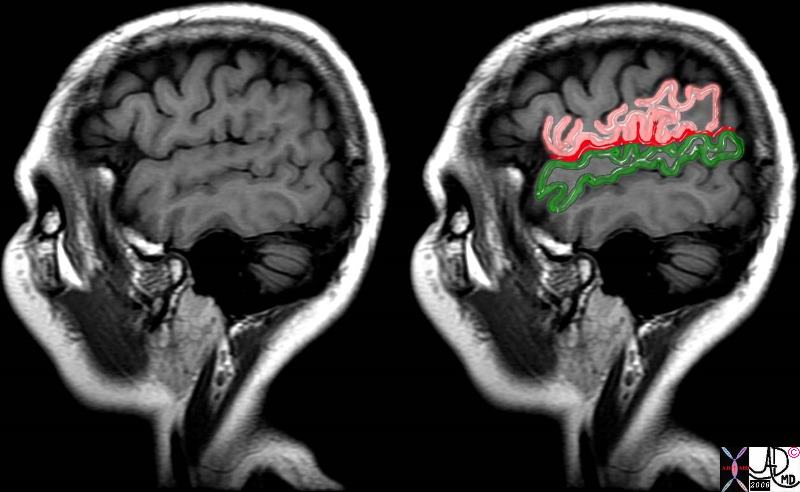
Parietal Lobe Above the Sylvian Fissure (red) and Temporal Lobe Below


71060c07.8s brain pain pathway somotosensory cortex S 2 SII operculum parietal lobe temporal lobe frontal lobe lateral sulcus Sylvian fissure MRI T1 weighted Courtesy Ashley Davidoff MD copyright 2008


    [nodeType] => 1
    [parentNode] => (object value omitted)
    [childNodes] => (object value omitted)
    [firstChild] => (object value omitted)
    [lastChild] => (object value omitted)
    [previousSibling] => (object value omitted)
    [nextSibling] => (object value omitted)
    [attributes] => (object value omitted)
    [ownerDocument] => (object value omitted)
    [namespaceURI] =>
    [prefix] =>
    [localName] => table
    [baseURI] =>
    [textContent] =>


Somatosensory Region
Parietal Lobe Above the Sylvian Fissure (red) and Temporal Lobe Below


71060c07.8s brain pain pathway somotosensory cortex S 2 SII operculum parietal lobe temporal lobe frontal lobe lateral sulcus Sylvian fissure MRI T1 weighted Courtesy Ashley Davidoff MD copyright 2008


)
DOMElement Object
(
    [schemaTypeInfo] =>
    [tagName] => td
    [firstElementChild] =>
    [lastElementChild] =>
    [childElementCount] => 0
    [previousElementSibling] =>
    [nextElementSibling] =>
    [nodeName] => td
    [nodeValue] => 71060c07.8s brain pain pathway somotosensory cortex S 2 SII operculum parietal lobe temporal lobe frontal lobe lateral sulcus Sylvian fissure MRI T1 weighted Courtesy Ashley Davidoff MD copyright 2008
    [nodeType] => 1
    [parentNode] => (object value omitted)
    [childNodes] => (object value omitted)
    [firstChild] => (object value omitted)
    [lastChild] => (object value omitted)
    [previousSibling] => (object value omitted)
    [nextSibling] => (object value omitted)
    [attributes] => (object value omitted)
    [ownerDocument] => (object value omitted)
    [namespaceURI] =>
    [prefix] =>
    [localName] => td
    [baseURI] =>
    [textContent] => 71060c07.8s brain pain pathway somotosensory cortex S 2 SII operculum parietal lobe temporal lobe frontal lobe lateral sulcus Sylvian fissure MRI T1 weighted Courtesy Ashley Davidoff MD copyright 2008
)
DOMElement Object
(
    [schemaTypeInfo] =>
    [tagName] => td
    [firstElementChild] => (object value omitted)
    [lastElementChild] => (object value omitted)
    [childElementCount] => 3
    [previousElementSibling] =>
    [nextElementSibling] =>
    [nodeName] => td
    [nodeValue] =>
Somatosensory Region
Parietal Lobe Above the Sylvian Fissure (red) and Temporal Lobe Below
    [nodeType] => 1
    [parentNode] => (object value omitted)
    [childNodes] => (object value omitted)
    [firstChild] => (object value omitted)
    [lastChild] => (object value omitted)
    [previousSibling] => (object value omitted)
    [nextSibling] => (object value omitted)
    [attributes] => (object value omitted)
    [ownerDocument] => (object value omitted)
    [namespaceURI] =>
    [prefix] =>
    [localName] => td
    [baseURI] =>
    [textContent] =>
Somatosensory Region
Parietal Lobe Above the Sylvian Fissure (red) and Temporal Lobe Below
)
DOMElement Object
(
    [schemaTypeInfo] =>
    [tagName] => table
    [firstElementChild] => (object value omitted)
    [lastElementChild] => (object value omitted)
    [childElementCount] => 1
    [previousElementSibling] => (object value omitted)
    [nextElementSibling] => (object value omitted)
    [nodeName] => table
    [nodeValue] =>


83029d13.8s


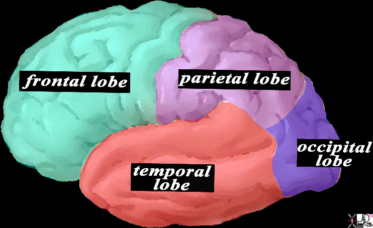
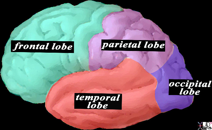
This is a diagram looking at the forebrain from the side showing the anteriorly placed frontal lobe, inferiorly placed temporal lobe posteriorly placed occipital lobe and superiorly placed occipital lobe. In this view the size of the frontal lobe dominates and in fact it is the largest portion of the forebrain.
Courtesy Ashley DAvidoff MD copyright 2010 all rights reserved 83029d13.8s


    [nodeType] => 1
    [parentNode] => (object value omitted)
    [childNodes] => (object value omitted)
    [firstChild] => (object value omitted)
    [lastChild] => (object value omitted)
    [previousSibling] => (object value omitted)
    [nextSibling] => (object value omitted)
    [attributes] => (object value omitted)
    [ownerDocument] => (object value omitted)
    [namespaceURI] =>
    [prefix] =>
    [localName] => table
    [baseURI] =>
    [textContent] =>


83029d13.8s


This is a diagram looking at the forebrain from the side showing the anteriorly placed frontal lobe, inferiorly placed temporal lobe posteriorly placed occipital lobe and superiorly placed occipital lobe. In this view the size of the frontal lobe dominates and in fact it is the largest portion of the forebrain.
Courtesy Ashley DAvidoff MD copyright 2010 all rights reserved 83029d13.8s


)
DOMElement Object
(
    [schemaTypeInfo] =>
    [tagName] => td
    [firstElementChild] => (object value omitted)
    [lastElementChild] => (object value omitted)
    [childElementCount] => 2
    [previousElementSibling] =>
    [nextElementSibling] =>
    [nodeName] => td
    [nodeValue] =>
This is a diagram looking at the forebrain from the side showing the anteriorly placed frontal lobe, inferiorly placed temporal lobe posteriorly placed occipital lobe and superiorly placed occipital lobe. In this view the size of the frontal lobe dominates and in fact it is the largest portion of the forebrain.
Courtesy Ashley DAvidoff MD copyright 2010 all rights reserved 83029d13.8s

    [nodeType] => 1
    [parentNode] => (object value omitted)
    [childNodes] => (object value omitted)
    [firstChild] => (object value omitted)
    [lastChild] => (object value omitted)
    [previousSibling] => (object value omitted)
    [nextSibling] => (object value omitted)
    [attributes] => (object value omitted)
    [ownerDocument] => (object value omitted)
    [namespaceURI] =>
    [prefix] =>
    [localName] => td
    [baseURI] =>
    [textContent] =>
This is a diagram looking at the forebrain from the side showing the anteriorly placed frontal lobe, inferiorly placed temporal lobe posteriorly placed occipital lobe and superiorly placed occipital lobe. In this view the size of the frontal lobe dominates and in fact it is the largest portion of the forebrain.
Courtesy Ashley DAvidoff MD copyright 2010 all rights reserved 83029d13.8s

)
DOMElement Object
(
    [schemaTypeInfo] =>
    [tagName] => td
    [firstElementChild] => (object value omitted)
    [lastElementChild] => (object value omitted)
    [childElementCount] => 2
    [previousElementSibling] =>
    [nextElementSibling] =>
    [nodeName] => td
    [nodeValue] =>

83029d13.8s
    [nodeType] => 1
    [parentNode] => (object value omitted)
    [childNodes] => (object value omitted)
    [firstChild] => (object value omitted)
    [lastChild] => (object value omitted)
    [previousSibling] => (object value omitted)
    [nextSibling] => (object value omitted)
    [attributes] => (object value omitted)
    [ownerDocument] => (object value omitted)
    [namespaceURI] =>
    [prefix] =>
    [localName] => td
    [baseURI] =>
    [textContent] =>

83029d13.8s
)
DOMElement Object
(
    [schemaTypeInfo] =>
    [tagName] => table
    [firstElementChild] => (object value omitted)
    [lastElementChild] => (object value omitted)
    [childElementCount] => 1
    [previousElementSibling] => (object value omitted)
    [nextElementSibling] => (object value omitted)
    [nodeName] => table
    [nodeValue] =>


The Central Sulcus is the Border between the Frontal Lobe and the Parietal Lobe


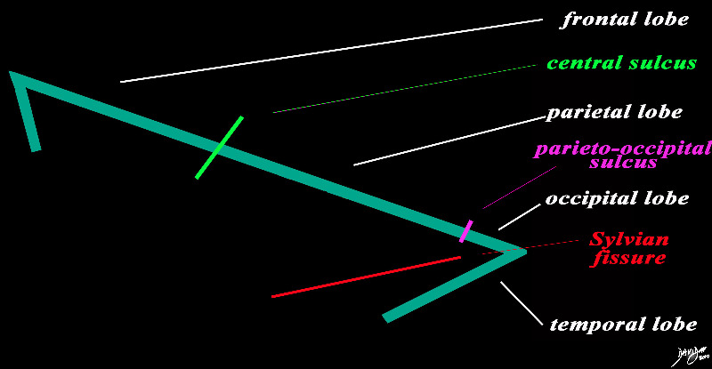
The forebrain is divided into the frontal lobe anetriorly, separated by the central sulcus (bright green), the parietal lobe separated from the occipital lobe by the parieto-occipital fissure (pink), and the temporal lobe separated from the frontal and occip[ital lobe by the Sylvian fissure (lateral sulcus) (red). The distinction between the occipital and temporal lobe is more of a functional division than a structural division
Courtesy Ashley Davidoff MD copyright 2010 93887b03b05.8s


    [nodeType] => 1
    [parentNode] => (object value omitted)
    [childNodes] => (object value omitted)
    [firstChild] => (object value omitted)
    [lastChild] => (object value omitted)
    [previousSibling] => (object value omitted)
    [nextSibling] => (object value omitted)
    [attributes] => (object value omitted)
    [ownerDocument] => (object value omitted)
    [namespaceURI] =>
    [prefix] =>
    [localName] => table
    [baseURI] =>
    [textContent] =>


The Central Sulcus is the Border between the Frontal Lobe and the Parietal Lobe


The forebrain is divided into the frontal lobe anetriorly, separated by the central sulcus (bright green), the parietal lobe separated from the occipital lobe by the parieto-occipital fissure (pink), and the temporal lobe separated from the frontal and occip[ital lobe by the Sylvian fissure (lateral sulcus) (red). The distinction between the occipital and temporal lobe is more of a functional division than a structural division
Courtesy Ashley Davidoff MD copyright 2010 93887b03b05.8s


)
DOMElement Object
(
    [schemaTypeInfo] =>
    [tagName] => td
    [firstElementChild] => (object value omitted)
    [lastElementChild] => (object value omitted)
    [childElementCount] => 2
    [previousElementSibling] =>
    [nextElementSibling] =>
    [nodeName] => td
    [nodeValue] =>
The forebrain is divided into the frontal lobe anetriorly, separated by the central sulcus (bright green), the parietal lobe separated from the occipital lobe by the parieto-occipital fissure (pink), and the temporal lobe separated from the frontal and occip[ital lobe by the Sylvian fissure (lateral sulcus) (red). The distinction between the occipital and temporal lobe is more of a functional division than a structural division
Courtesy Ashley Davidoff MD copyright 2010 93887b03b05.8s
    [nodeType] => 1
    [parentNode] => (object value omitted)
    [childNodes] => (object value omitted)
    [firstChild] => (object value omitted)
    [lastChild] => (object value omitted)
    [previousSibling] => (object value omitted)
    [nextSibling] => (object value omitted)
    [attributes] => (object value omitted)
    [ownerDocument] => (object value omitted)
    [namespaceURI] =>
    [prefix] =>
    [localName] => td
    [baseURI] =>
    [textContent] =>
The forebrain is divided into the frontal lobe anetriorly, separated by the central sulcus (bright green), the parietal lobe separated from the occipital lobe by the parieto-occipital fissure (pink), and the temporal lobe separated from the frontal and occip[ital lobe by the Sylvian fissure (lateral sulcus) (red). The distinction between the occipital and temporal lobe is more of a functional division than a structural division
Courtesy Ashley Davidoff MD copyright 2010 93887b03b05.8s
)
DOMElement Object
(
    [schemaTypeInfo] =>
    [tagName] => td
    [firstElementChild] => (object value omitted)
    [lastElementChild] => (object value omitted)
    [childElementCount] => 2
    [previousElementSibling] =>
    [nextElementSibling] =>
    [nodeName] => td
    [nodeValue] =>
The Central Sulcus is the Border between the Frontal Lobe and the Parietal Lobe
    [nodeType] => 1
    [parentNode] => (object value omitted)
    [childNodes] => (object value omitted)
    [firstChild] => (object value omitted)
    [lastChild] => (object value omitted)
    [previousSibling] => (object value omitted)
    [nextSibling] => (object value omitted)
    [attributes] => (object value omitted)
    [ownerDocument] => (object value omitted)
    [namespaceURI] =>
    [prefix] =>
    [localName] => td
    [baseURI] =>
    [textContent] =>
The Central Sulcus is the Border between the Frontal Lobe and the Parietal Lobe
)
DOMElement Object
(
    [schemaTypeInfo] =>
    [tagName] => table
    [firstElementChild] => (object value omitted)
    [lastElementChild] => (object value omitted)
    [childElementCount] => 1
    [previousElementSibling] => (object value omitted)
    [nextElementSibling] => (object value omitted)
    [nodeName] => table
    [nodeValue] =>


Conceptual Framework of the Brain


The vector of the forebrain (green) is folded at its anterior aspect as a projection of the frontal lobe and the posterior fold forms the temporal lobe. The midbrain (yellow has its axis just tipped slightly anteriorly and the hindbrain (salmon red) is tipped more posteriorly
Courtesy Ashley Davidoff MD copyright 2010 all rights reserved


    [nodeType] => 1
    [parentNode] => (object value omitted)
    [childNodes] => (object value omitted)
    [firstChild] => (object value omitted)
    [lastChild] => (object value omitted)
    [previousSibling] => (object value omitted)
    [nextSibling] => (object value omitted)
    [attributes] => (object value omitted)
    [ownerDocument] => (object value omitted)
    [namespaceURI] =>
    [prefix] =>
    [localName] => table
    [baseURI] =>
    [textContent] =>


Conceptual Framework of the Brain


The vector of the forebrain (green) is folded at its anterior aspect as a projection of the frontal lobe and the posterior fold forms the temporal lobe. The midbrain (yellow has its axis just tipped slightly anteriorly and the hindbrain (salmon red) is tipped more posteriorly
Courtesy Ashley Davidoff MD copyright 2010 all rights reserved


)
DOMElement Object
(
    [schemaTypeInfo] =>
    [tagName] => td
    [firstElementChild] => (object value omitted)
    [lastElementChild] => (object value omitted)
    [childElementCount] => 2
    [previousElementSibling] =>
    [nextElementSibling] =>
    [nodeName] => td
    [nodeValue] =>
The vector of the forebrain (green) is folded at its anterior aspect as a projection of the frontal lobe and the posterior fold forms the temporal lobe. The midbrain (yellow has its axis just tipped slightly anteriorly and the hindbrain (salmon red) is tipped more posteriorly
Courtesy Ashley Davidoff MD copyright 2010 all rights reserved

    [nodeType] => 1
    [parentNode] => (object value omitted)
    [childNodes] => (object value omitted)
    [firstChild] => (object value omitted)
    [lastChild] => (object value omitted)
    [previousSibling] => (object value omitted)
    [nextSibling] => (object value omitted)
    [attributes] => (object value omitted)
    [ownerDocument] => (object value omitted)
    [namespaceURI] =>
    [prefix] =>
    [localName] => td
    [baseURI] =>
    [textContent] =>
The vector of the forebrain (green) is folded at its anterior aspect as a projection of the frontal lobe and the posterior fold forms the temporal lobe. The midbrain (yellow has its axis just tipped slightly anteriorly and the hindbrain (salmon red) is tipped more posteriorly
Courtesy Ashley Davidoff MD copyright 2010 all rights reserved

)
DOMElement Object
(
    [schemaTypeInfo] =>
    [tagName] => td
    [firstElementChild] => (object value omitted)
    [lastElementChild] => (object value omitted)
    [childElementCount] => 2
    [previousElementSibling] =>
    [nextElementSibling] =>
    [nodeName] => td
    [nodeValue] =>

Conceptual Framework of the Brain
    [nodeType] => 1
    [parentNode] => (object value omitted)
    [childNodes] => (object value omitted)
    [firstChild] => (object value omitted)
    [lastChild] => (object value omitted)
    [previousSibling] => (object value omitted)
    [nextSibling] => (object value omitted)
    [attributes] => (object value omitted)
    [ownerDocument] => (object value omitted)
    [namespaceURI] =>
    [prefix] =>
    [localName] => td
    [baseURI] =>
    [textContent] =>

Conceptual Framework of the Brain
)
DOMElement Object
(
    [schemaTypeInfo] =>
    [tagName] => table
    [firstElementChild] => (object value omitted)
    [lastElementChild] => (object value omitted)
    [childElementCount] => 1
    [previousElementSibling] => (object value omitted)
    [nextElementSibling] => (object value omitted)
    [nodeName] => table
    [nodeValue] =>


The Occipital Lobe (lime green)
Part of the Forebrain (Prosencephalon)
Member of the Cerebrum (Telencephalon)
Member of the Cerebral Hemispheres


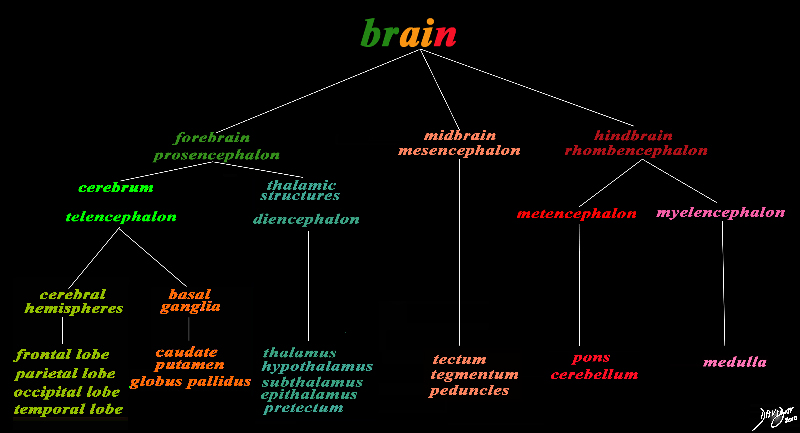
The basic and simplest classification of the brain into forebrain midbrain and hindbrain is shown in this diagram and advanced to a more complex tree using the embryological and evolutionary terminologies. The forebrain consists of the cerebrum also called the prosencephalon, which contains the more advanced form of the brain and the thalamic structures which contain more basic structures. The cerebrum (telencephalon) itself consists of two cerebral hemispheres and paired basal ganglial structures. Each cerebral hemisphere will have gray and white matter distributed in the frontal parietal temporal and occipital lobe, with the basal ganglia being part of the gray matter deep in the cerebral hemispheres. The most important thalamic structures arising from the diencephalons include the thalamus itself and the hypothalamus. The midbrain (mesencepaholon) consists of the tectum tegmentum and cerebral peduncles. The hindbrain has two major branch points based on the evolutionary development. The pons and cerebellum(part of the metencephalon) are grouped and the medulla (part of the myelencephalon is the second branch.
Courtesy Ashley Davidoff MD Copyright 2010 All rights reserved 97686.8s


    [nodeType] => 1
    [parentNode] => (object value omitted)
    [childNodes] => (object value omitted)
    [firstChild] => (object value omitted)
    [lastChild] => (object value omitted)
    [previousSibling] => (object value omitted)
    [nextSibling] => (object value omitted)
    [attributes] => (object value omitted)
    [ownerDocument] => (object value omitted)
    [namespaceURI] =>
    [prefix] =>
    [localName] => table
    [baseURI] =>
    [textContent] =>


The Occipital Lobe (lime green)
Part of the Forebrain (Prosencephalon)
Member of the Cerebrum (Telencephalon)
Member of the Cerebral Hemispheres


The basic and simplest classification of the brain into forebrain midbrain and hindbrain is shown in this diagram and advanced to a more complex tree using the embryological and evolutionary terminologies. The forebrain consists of the cerebrum also called the prosencephalon, which contains the more advanced form of the brain and the thalamic structures which contain more basic structures. The cerebrum (telencephalon) itself consists of two cerebral hemispheres and paired basal ganglial structures. Each cerebral hemisphere will have gray and white matter distributed in the frontal parietal temporal and occipital lobe, with the basal ganglia being part of the gray matter deep in the cerebral hemispheres. The most important thalamic structures arising from the diencephalons include the thalamus itself and the hypothalamus. The midbrain (mesencepaholon) consists of the tectum tegmentum and cerebral peduncles. The hindbrain has two major branch points based on the evolutionary development. The pons and cerebellum(part of the metencephalon) are grouped and the medulla (part of the myelencephalon is the second branch.
Courtesy Ashley Davidoff MD Copyright 2010 All rights reserved 97686.8s


)
DOMElement Object
(
    [schemaTypeInfo] =>
    [tagName] => td
    [firstElementChild] => (object value omitted)
    [lastElementChild] => (object value omitted)
    [childElementCount] => 2
    [previousElementSibling] =>
    [nextElementSibling] =>
    [nodeName] => td
    [nodeValue] =>
The basic and simplest classification of the brain into forebrain midbrain and hindbrain is shown in this diagram and advanced to a more complex tree using the embryological and evolutionary terminologies. The forebrain consists of the cerebrum also called the prosencephalon, which contains the more advanced form of the brain and the thalamic structures which contain more basic structures. The cerebrum (telencephalon) itself consists of two cerebral hemispheres and paired basal ganglial structures. Each cerebral hemisphere will have gray and white matter distributed in the frontal parietal temporal and occipital lobe, with the basal ganglia being part of the gray matter deep in the cerebral hemispheres. The most important thalamic structures arising from the diencephalons include the thalamus itself and the hypothalamus. The midbrain (mesencepaholon) consists of the tectum tegmentum and cerebral peduncles. The hindbrain has two major branch points based on the evolutionary development. The pons and cerebellum(part of the metencephalon) are grouped and the medulla (part of the myelencephalon is the second branch.
Courtesy Ashley Davidoff MD Copyright 2010 All rights reserved 97686.8s

    [nodeType] => 1
    [parentNode] => (object value omitted)
    [childNodes] => (object value omitted)
    [firstChild] => (object value omitted)
    [lastChild] => (object value omitted)
    [previousSibling] => (object value omitted)
    [nextSibling] => (object value omitted)
    [attributes] => (object value omitted)
    [ownerDocument] => (object value omitted)
    [namespaceURI] =>
    [prefix] =>
    [localName] => td
    [baseURI] =>
    [textContent] =>
The basic and simplest classification of the brain into forebrain midbrain and hindbrain is shown in this diagram and advanced to a more complex tree using the embryological and evolutionary terminologies. The forebrain consists of the cerebrum also called the prosencephalon, which contains the more advanced form of the brain and the thalamic structures which contain more basic structures. The cerebrum (telencephalon) itself consists of two cerebral hemispheres and paired basal ganglial structures. Each cerebral hemisphere will have gray and white matter distributed in the frontal parietal temporal and occipital lobe, with the basal ganglia being part of the gray matter deep in the cerebral hemispheres. The most important thalamic structures arising from the diencephalons include the thalamus itself and the hypothalamus. The midbrain (mesencepaholon) consists of the tectum tegmentum and cerebral peduncles. The hindbrain has two major branch points based on the evolutionary development. The pons and cerebellum(part of the metencephalon) are grouped and the medulla (part of the myelencephalon is the second branch.
Courtesy Ashley Davidoff MD Copyright 2010 All rights reserved 97686.8s

)
DOMElement Object
(
    [schemaTypeInfo] =>
    [tagName] => td
    [firstElementChild] => (object value omitted)
    [lastElementChild] => (object value omitted)
    [childElementCount] => 5
    [previousElementSibling] =>
    [nextElementSibling] =>
    [nodeName] => td
    [nodeValue] =>
The Occipital Lobe (lime green)
Part of the Forebrain (Prosencephalon)
Member of the Cerebrum (Telencephalon)
Member of the Cerebral Hemispheres
    [nodeType] => 1
    [parentNode] => (object value omitted)
    [childNodes] => (object value omitted)
    [firstChild] => (object value omitted)
    [lastChild] => (object value omitted)
    [previousSibling] => (object value omitted)
    [nextSibling] => (object value omitted)
    [attributes] => (object value omitted)
    [ownerDocument] => (object value omitted)
    [namespaceURI] =>
    [prefix] =>
    [localName] => td
    [baseURI] =>
    [textContent] =>
The Occipital Lobe (lime green)
Part of the Forebrain (Prosencephalon)
Member of the Cerebrum (Telencephalon)
Member of the Cerebral Hemispheres
)

