Sylvian Fissure or Lateral Sulcus
Ashley Davidoff MD
The Common Vein Copyright 2010
Definition
The Sylvian fissure or lateral sulcus is one the most prominent and most easily recognized fissures of the brain contributes to the defining and well recognized shape of the external brain. It lies between the temporal lobe inferiorly amd the frontal and parieta lobe superiorly.
Applied Biology – Diseases Around the Fissure
DOMElement Object
(
    [schemaTypeInfo] =>
    [tagName] => table
    [firstElementChild] => (object value omitted)
    [lastElementChild] => (object value omitted)
    [childElementCount] => 1
    [previousElementSibling] => (object value omitted)
    [nextElementSibling] =>
    [nodeName] => table
    [nodeValue] =>


Chronic Infarction in the Region of the Sylvian Fissure


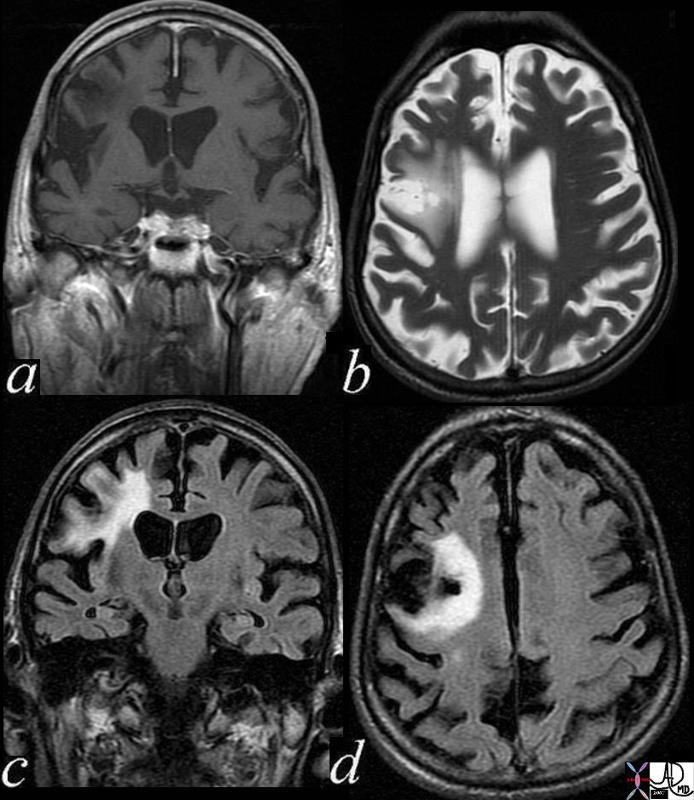
The MRI from a 91 year old male patient with a chronic infarction in the region of the Sylvian fissure. Image a shows a coronal T1 weighted image with an expanded Sylvian fissure due to volume loss in the region of the insular cortex (insula) with secondary enlargement of the Sylvian fissure as a result of loss of tissue, caused by gliosis and encephalomalacia of the insular cortex. In image b an axial T2 weighted image shows the hyperintense expanded Sylvian fissure surrounded by the less intense chronic infarct in the cerebral tissue. The border between the expanded fissure and the infarcted tissue is ill defined. Image c is a coronal FLAIR image showing the process of gliosis and FLAIR intensity of chronic infarction, and this is seen again in the FLAIR image in axial projection 9D) where the distinction between the expanded Sylvian fissure and the diseased cerebral tissue is quite clear. The deformity of the lateral ventricle indicates ex vacuo change and encephalomalacia male brain cerebrum right frontal lobe extending between the Sylvian fissure and precentral gyrus white matter and medially to the surface of the lateral ventricle
Courtesy Ashley Davidoff MD Copyright 2010 71559c04


    [nodeType] => 1
    [parentNode] => (object value omitted)
    [childNodes] => (object value omitted)
    [firstChild] => (object value omitted)
    [lastChild] => (object value omitted)
    [previousSibling] => (object value omitted)
    [nextSibling] => (object value omitted)
    [attributes] => (object value omitted)
    [ownerDocument] => (object value omitted)
    [namespaceURI] =>
    [prefix] =>
    [localName] => table
    [baseURI] =>
    [textContent] =>


Chronic Infarction in the Region of the Sylvian Fissure


The MRI from a 91 year old male patient with a chronic infarction in the region of the Sylvian fissure. Image a shows a coronal T1 weighted image with an expanded Sylvian fissure due to volume loss in the region of the insular cortex (insula) with secondary enlargement of the Sylvian fissure as a result of loss of tissue, caused by gliosis and encephalomalacia of the insular cortex. In image b an axial T2 weighted image shows the hyperintense expanded Sylvian fissure surrounded by the less intense chronic infarct in the cerebral tissue. The border between the expanded fissure and the infarcted tissue is ill defined. Image c is a coronal FLAIR image showing the process of gliosis and FLAIR intensity of chronic infarction, and this is seen again in the FLAIR image in axial projection 9D) where the distinction between the expanded Sylvian fissure and the diseased cerebral tissue is quite clear. The deformity of the lateral ventricle indicates ex vacuo change and encephalomalacia male brain cerebrum right frontal lobe extending between the Sylvian fissure and precentral gyrus white matter and medially to the surface of the lateral ventricle
Courtesy Ashley Davidoff MD Copyright 2010 71559c04


)
DOMElement Object
(
    [schemaTypeInfo] =>
    [tagName] => td
    [firstElementChild] => (object value omitted)
    [lastElementChild] => (object value omitted)
    [childElementCount] => 2
    [previousElementSibling] =>
    [nextElementSibling] =>
    [nodeName] => td
    [nodeValue] =>
The MRI from a 91 year old male patient with a chronic infarction in the region of the Sylvian fissure. Image a shows a coronal T1 weighted image with an expanded Sylvian fissure due to volume loss in the region of the insular cortex (insula) with secondary enlargement of the Sylvian fissure as a result of loss of tissue, caused by gliosis and encephalomalacia of the insular cortex. In image b an axial T2 weighted image shows the hyperintense expanded Sylvian fissure surrounded by the less intense chronic infarct in the cerebral tissue. The border between the expanded fissure and the infarcted tissue is ill defined. Image c is a coronal FLAIR image showing the process of gliosis and FLAIR intensity of chronic infarction, and this is seen again in the FLAIR image in axial projection 9D) where the distinction between the expanded Sylvian fissure and the diseased cerebral tissue is quite clear. The deformity of the lateral ventricle indicates ex vacuo change and encephalomalacia male brain cerebrum right frontal lobe extending between the Sylvian fissure and precentral gyrus white matter and medially to the surface of the lateral ventricle
Courtesy Ashley Davidoff MD Copyright 2010 71559c04
    [nodeType] => 1
    [parentNode] => (object value omitted)
    [childNodes] => (object value omitted)
    [firstChild] => (object value omitted)
    [lastChild] => (object value omitted)
    [previousSibling] => (object value omitted)
    [nextSibling] => (object value omitted)
    [attributes] => (object value omitted)
    [ownerDocument] => (object value omitted)
    [namespaceURI] =>
    [prefix] =>
    [localName] => td
    [baseURI] =>
    [textContent] =>
The MRI from a 91 year old male patient with a chronic infarction in the region of the Sylvian fissure. Image a shows a coronal T1 weighted image with an expanded Sylvian fissure due to volume loss in the region of the insular cortex (insula) with secondary enlargement of the Sylvian fissure as a result of loss of tissue, caused by gliosis and encephalomalacia of the insular cortex. In image b an axial T2 weighted image shows the hyperintense expanded Sylvian fissure surrounded by the less intense chronic infarct in the cerebral tissue. The border between the expanded fissure and the infarcted tissue is ill defined. Image c is a coronal FLAIR image showing the process of gliosis and FLAIR intensity of chronic infarction, and this is seen again in the FLAIR image in axial projection 9D) where the distinction between the expanded Sylvian fissure and the diseased cerebral tissue is quite clear. The deformity of the lateral ventricle indicates ex vacuo change and encephalomalacia male brain cerebrum right frontal lobe extending between the Sylvian fissure and precentral gyrus white matter and medially to the surface of the lateral ventricle
Courtesy Ashley Davidoff MD Copyright 2010 71559c04
)
DOMElement Object
(
    [schemaTypeInfo] =>
    [tagName] => td
    [firstElementChild] => (object value omitted)
    [lastElementChild] => (object value omitted)
    [childElementCount] => 2
    [previousElementSibling] =>
    [nextElementSibling] =>
    [nodeName] => td
    [nodeValue] =>
Chronic Infarction in the Region of the Sylvian Fissure
    [nodeType] => 1
    [parentNode] => (object value omitted)
    [childNodes] => (object value omitted)
    [firstChild] => (object value omitted)
    [lastChild] => (object value omitted)
    [previousSibling] => (object value omitted)
    [nextSibling] => (object value omitted)
    [attributes] => (object value omitted)
    [ownerDocument] => (object value omitted)
    [namespaceURI] =>
    [prefix] =>
    [localName] => td
    [baseURI] =>
    [textContent] =>
Chronic Infarction in the Region of the Sylvian Fissure
)
DOMElement Object
(
    [schemaTypeInfo] =>
    [tagName] => table
    [firstElementChild] => (object value omitted)
    [lastElementChild] => (object value omitted)
    [childElementCount] => 1
    [previousElementSibling] => (object value omitted)
    [nextElementSibling] => (object value omitted)
    [nodeName] => table
    [nodeValue] =>


Chric Infarction of the Insula Cortex with Secondary Enlargement of the Sylvian Fissure


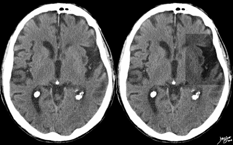
The non contrast CTscan is from elderly patient and reveals volume loss in the region of the insular cortex (insula) with secondary enlargement of the Sylvian fissure as a result of loss of tissue, caused by gliosis and encephalomalacia of the insular cortex. The process may involve the claustrum as well A low density lesion in the left caudate nucleus represents an old infarct. (lacune)
Courtesy Ashley Davidoff MD Copyright 2010 All rights reserved 89384.8s


    [nodeType] => 1
    [parentNode] => (object value omitted)
    [childNodes] => (object value omitted)
    [firstChild] => (object value omitted)
    [lastChild] => (object value omitted)
    [previousSibling] => (object value omitted)
    [nextSibling] => (object value omitted)
    [attributes] => (object value omitted)
    [ownerDocument] => (object value omitted)
    [namespaceURI] =>
    [prefix] =>
    [localName] => table
    [baseURI] =>
    [textContent] =>


Chric Infarction of the Insula Cortex with Secondary Enlargement of the Sylvian Fissure


The non contrast CTscan is from elderly patient and reveals volume loss in the region of the insular cortex (insula) with secondary enlargement of the Sylvian fissure as a result of loss of tissue, caused by gliosis and encephalomalacia of the insular cortex. The process may involve the claustrum as well A low density lesion in the left caudate nucleus represents an old infarct. (lacune)
Courtesy Ashley Davidoff MD Copyright 2010 All rights reserved 89384.8s


)
DOMElement Object
(
    [schemaTypeInfo] =>
    [tagName] => td
    [firstElementChild] => (object value omitted)
    [lastElementChild] => (object value omitted)
    [childElementCount] => 2
    [previousElementSibling] =>
    [nextElementSibling] =>
    [nodeName] => td
    [nodeValue] =>
The non contrast CTscan is from elderly patient and reveals volume loss in the region of the insular cortex (insula) with secondary enlargement of the Sylvian fissure as a result of loss of tissue, caused by gliosis and encephalomalacia of the insular cortex. The process may involve the claustrum as well A low density lesion in the left caudate nucleus represents an old infarct. (lacune)
Courtesy Ashley Davidoff MD Copyright 2010 All rights reserved 89384.8s

    [nodeType] => 1
    [parentNode] => (object value omitted)
    [childNodes] => (object value omitted)
    [firstChild] => (object value omitted)
    [lastChild] => (object value omitted)
    [previousSibling] => (object value omitted)
    [nextSibling] => (object value omitted)
    [attributes] => (object value omitted)
    [ownerDocument] => (object value omitted)
    [namespaceURI] =>
    [prefix] =>
    [localName] => td
    [baseURI] =>
    [textContent] =>
The non contrast CTscan is from elderly patient and reveals volume loss in the region of the insular cortex (insula) with secondary enlargement of the Sylvian fissure as a result of loss of tissue, caused by gliosis and encephalomalacia of the insular cortex. The process may involve the claustrum as well A low density lesion in the left caudate nucleus represents an old infarct. (lacune)
Courtesy Ashley Davidoff MD Copyright 2010 All rights reserved 89384.8s

)
DOMElement Object
(
    [schemaTypeInfo] =>
    [tagName] => td
    [firstElementChild] => (object value omitted)
    [lastElementChild] => (object value omitted)
    [childElementCount] => 2
    [previousElementSibling] =>
    [nextElementSibling] =>
    [nodeName] => td
    [nodeValue] =>
Chric Infarction of the Insula Cortex with Secondary Enlargement of the Sylvian Fissure
    [nodeType] => 1
    [parentNode] => (object value omitted)
    [childNodes] => (object value omitted)
    [firstChild] => (object value omitted)
    [lastChild] => (object value omitted)
    [previousSibling] => (object value omitted)
    [nextSibling] => (object value omitted)
    [attributes] => (object value omitted)
    [ownerDocument] => (object value omitted)
    [namespaceURI] =>
    [prefix] =>
    [localName] => td
    [baseURI] =>
    [textContent] =>
Chric Infarction of the Insula Cortex with Secondary Enlargement of the Sylvian Fissure
)
DOMElement Object
(
    [schemaTypeInfo] =>
    [tagName] => table
    [firstElementChild] => (object value omitted)
    [lastElementChild] => (object value omitted)
    [childElementCount] => 1
    [previousElementSibling] => (object value omitted)
    [nextElementSibling] => (object value omitted)
    [nodeName] => table
    [nodeValue] =>


Acute Lenticulostriate Infarction – Subinsular and Deep to the Sylvian Fissure


The CT and MRI images are from an 82 year old make with acute neurological deficit. An acute infarction is shown that involves territory within and beyond the basal ganglia. In the CTscan two low density regions are seen medial to the Sylvian fissure and medial to the insular cortex, likely involving the putamen and the part of the right caudate nucleus as well as some white matter in right middle cerebra;l territory. In the second image a high intensity region in the putamen and part of the caudate nucleus is shown together with an area that does not involve the basal ganglia more posteriorly, likely part of the white matter of the right parietal lobe shown in an axial projection on DWI consistent with an acute infarction. The third image is an axial FLAIR sequence and shows the area of parenchymal edema with presumed central necrosis, while the image at low right shows the periventricular disease to better advantage. The constellation of findings is consistent with an acute infarction with distribution of the lenticulostriate vessels arising from the right middle cerebral territory
Courtesy Ashley Davidoff MD Copyright 2010 All rights reserved 89367c01.8s


    [nodeType] => 1
    [parentNode] => (object value omitted)
    [childNodes] => (object value omitted)
    [firstChild] => (object value omitted)
    [lastChild] => (object value omitted)
    [previousSibling] => (object value omitted)
    [nextSibling] => (object value omitted)
    [attributes] => (object value omitted)
    [ownerDocument] => (object value omitted)
    [namespaceURI] =>
    [prefix] =>
    [localName] => table
    [baseURI] =>
    [textContent] =>


Acute Lenticulostriate Infarction – Subinsular and Deep to the Sylvian Fissure


The CT and MRI images are from an 82 year old make with acute neurological deficit. An acute infarction is shown that involves territory within and beyond the basal ganglia. In the CTscan two low density regions are seen medial to the Sylvian fissure and medial to the insular cortex, likely involving the putamen and the part of the right caudate nucleus as well as some white matter in right middle cerebra;l territory. In the second image a high intensity region in the putamen and part of the caudate nucleus is shown together with an area that does not involve the basal ganglia more posteriorly, likely part of the white matter of the right parietal lobe shown in an axial projection on DWI consistent with an acute infarction. The third image is an axial FLAIR sequence and shows the area of parenchymal edema with presumed central necrosis, while the image at low right shows the periventricular disease to better advantage. The constellation of findings is consistent with an acute infarction with distribution of the lenticulostriate vessels arising from the right middle cerebral territory
Courtesy Ashley Davidoff MD Copyright 2010 All rights reserved 89367c01.8s


)
DOMElement Object
(
    [schemaTypeInfo] =>
    [tagName] => td
    [firstElementChild] => (object value omitted)
    [lastElementChild] => (object value omitted)
    [childElementCount] => 2
    [previousElementSibling] =>
    [nextElementSibling] =>
    [nodeName] => td
    [nodeValue] =>
The CT and MRI images are from an 82 year old make with acute neurological deficit. An acute infarction is shown that involves territory within and beyond the basal ganglia. In the CTscan two low density regions are seen medial to the Sylvian fissure and medial to the insular cortex, likely involving the putamen and the part of the right caudate nucleus as well as some white matter in right middle cerebra;l territory. In the second image a high intensity region in the putamen and part of the caudate nucleus is shown together with an area that does not involve the basal ganglia more posteriorly, likely part of the white matter of the right parietal lobe shown in an axial projection on DWI consistent with an acute infarction. The third image is an axial FLAIR sequence and shows the area of parenchymal edema with presumed central necrosis, while the image at low right shows the periventricular disease to better advantage. The constellation of findings is consistent with an acute infarction with distribution of the lenticulostriate vessels arising from the right middle cerebral territory
Courtesy Ashley Davidoff MD Copyright 2010 All rights reserved 89367c01.8s

    [nodeType] => 1
    [parentNode] => (object value omitted)
    [childNodes] => (object value omitted)
    [firstChild] => (object value omitted)
    [lastChild] => (object value omitted)
    [previousSibling] => (object value omitted)
    [nextSibling] => (object value omitted)
    [attributes] => (object value omitted)
    [ownerDocument] => (object value omitted)
    [namespaceURI] =>
    [prefix] =>
    [localName] => td
    [baseURI] =>
    [textContent] =>
The CT and MRI images are from an 82 year old make with acute neurological deficit. An acute infarction is shown that involves territory within and beyond the basal ganglia. In the CTscan two low density regions are seen medial to the Sylvian fissure and medial to the insular cortex, likely involving the putamen and the part of the right caudate nucleus as well as some white matter in right middle cerebra;l territory. In the second image a high intensity region in the putamen and part of the caudate nucleus is shown together with an area that does not involve the basal ganglia more posteriorly, likely part of the white matter of the right parietal lobe shown in an axial projection on DWI consistent with an acute infarction. The third image is an axial FLAIR sequence and shows the area of parenchymal edema with presumed central necrosis, while the image at low right shows the periventricular disease to better advantage. The constellation of findings is consistent with an acute infarction with distribution of the lenticulostriate vessels arising from the right middle cerebral territory
Courtesy Ashley Davidoff MD Copyright 2010 All rights reserved 89367c01.8s

)
DOMElement Object
(
    [schemaTypeInfo] =>
    [tagName] => td
    [firstElementChild] => (object value omitted)
    [lastElementChild] => (object value omitted)
    [childElementCount] => 2
    [previousElementSibling] =>
    [nextElementSibling] =>
    [nodeName] => td
    [nodeValue] =>
Acute Lenticulostriate Infarction – Subinsular and Deep to the Sylvian Fissure
    [nodeType] => 1
    [parentNode] => (object value omitted)
    [childNodes] => (object value omitted)
    [firstChild] => (object value omitted)
    [lastChild] => (object value omitted)
    [previousSibling] => (object value omitted)
    [nextSibling] => (object value omitted)
    [attributes] => (object value omitted)
    [ownerDocument] => (object value omitted)
    [namespaceURI] =>
    [prefix] =>
    [localName] => td
    [baseURI] =>
    [textContent] =>
Acute Lenticulostriate Infarction – Subinsular and Deep to the Sylvian Fissure
)
DOMElement Object
(
    [schemaTypeInfo] =>
    [tagName] => table
    [firstElementChild] => (object value omitted)
    [lastElementChild] => (object value omitted)
    [childElementCount] => 1
    [previousElementSibling] => (object value omitted)
    [nextElementSibling] => (object value omitted)
    [nodeName] => table
    [nodeValue] =>


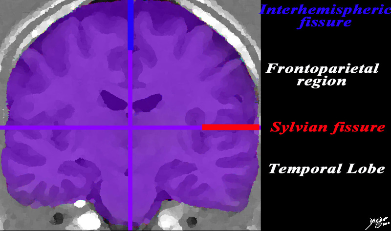
The Sylvian Fissure as a Border between the Frontoparietal region and the Temporal Lobe


In the same cut the fissural landmarks enable us to divide the brain into the two hemispheres (blue interhemispheric fissure) and the Sylvian fissure (red) divides the frontoparietal region in the anterior craial fossa from the temporal lobe in the middle cranial fossa.
Courtesy Ashley Davidoff MD Copyright 2010 All rights reserved 92167..84slabel


    [nodeType] => 1
    [parentNode] => (object value omitted)
    [childNodes] => (object value omitted)
    [firstChild] => (object value omitted)
    [lastChild] => (object value omitted)
    [previousSibling] => (object value omitted)
    [nextSibling] => (object value omitted)
    [attributes] => (object value omitted)
    [ownerDocument] => (object value omitted)
    [namespaceURI] =>
    [prefix] =>
    [localName] => table
    [baseURI] =>
    [textContent] =>


The Sylvian Fissure as a Border between the Frontoparietal region and the Temporal Lobe


In the same cut the fissural landmarks enable us to divide the brain into the two hemispheres (blue interhemispheric fissure) and the Sylvian fissure (red) divides the frontoparietal region in the anterior craial fossa from the temporal lobe in the middle cranial fossa.
Courtesy Ashley Davidoff MD Copyright 2010 All rights reserved 92167..84slabel


)
DOMElement Object
(
    [schemaTypeInfo] =>
    [tagName] => td
    [firstElementChild] => (object value omitted)
    [lastElementChild] => (object value omitted)
    [childElementCount] => 2
    [previousElementSibling] =>
    [nextElementSibling] =>
    [nodeName] => td
    [nodeValue] =>
In the same cut the fissural landmarks enable us to divide the brain into the two hemispheres (blue interhemispheric fissure) and the Sylvian fissure (red) divides the frontoparietal region in the anterior craial fossa from the temporal lobe in the middle cranial fossa.
Courtesy Ashley Davidoff MD Copyright 2010 All rights reserved 92167..84slabel

    [nodeType] => 1
    [parentNode] => (object value omitted)
    [childNodes] => (object value omitted)
    [firstChild] => (object value omitted)
    [lastChild] => (object value omitted)
    [previousSibling] => (object value omitted)
    [nextSibling] => (object value omitted)
    [attributes] => (object value omitted)
    [ownerDocument] => (object value omitted)
    [namespaceURI] =>
    [prefix] =>
    [localName] => td
    [baseURI] =>
    [textContent] =>
In the same cut the fissural landmarks enable us to divide the brain into the two hemispheres (blue interhemispheric fissure) and the Sylvian fissure (red) divides the frontoparietal region in the anterior craial fossa from the temporal lobe in the middle cranial fossa.
Courtesy Ashley Davidoff MD Copyright 2010 All rights reserved 92167..84slabel

)
DOMElement Object
(
    [schemaTypeInfo] =>
    [tagName] => td
    [firstElementChild] => (object value omitted)
    [lastElementChild] => (object value omitted)
    [childElementCount] => 2
    [previousElementSibling] =>
    [nextElementSibling] =>
    [nodeName] => td
    [nodeValue] =>
The Sylvian Fissure as a Border between the Frontoparietal region and the Temporal Lobe
    [nodeType] => 1
    [parentNode] => (object value omitted)
    [childNodes] => (object value omitted)
    [firstChild] => (object value omitted)
    [lastChild] => (object value omitted)
    [previousSibling] => (object value omitted)
    [nextSibling] => (object value omitted)
    [attributes] => (object value omitted)
    [ownerDocument] => (object value omitted)
    [namespaceURI] =>
    [prefix] =>
    [localName] => td
    [baseURI] =>
    [textContent] =>
The Sylvian Fissure as a Border between the Frontoparietal region and the Temporal Lobe
)
DOMElement Object
(
    [schemaTypeInfo] =>
    [tagName] => table
    [firstElementChild] => (object value omitted)
    [lastElementChild] => (object value omitted)
    [childElementCount] => 1
    [previousElementSibling] => (object value omitted)
    [nextElementSibling] => (object value omitted)
    [nodeName] => table
    [nodeValue] =>


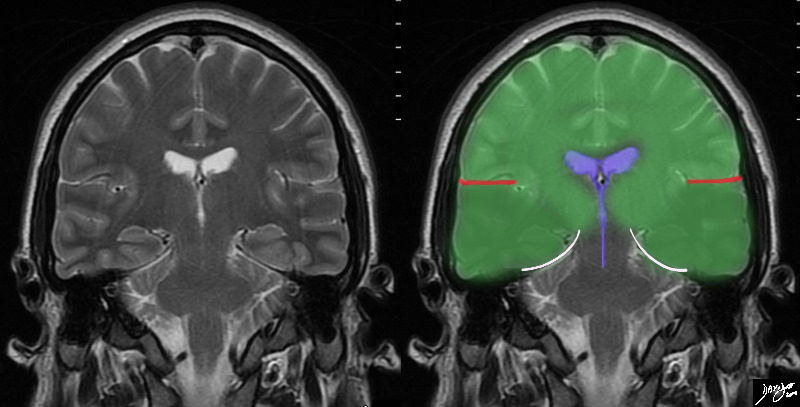
Sylvian Fissure in Coronal Projection


In this T2 weighted MRI image – the forebrain (green is seen centered aroubnd the ventriclar system. The red linees indicate the Sylvian fissures which in this instance in a relatively posterior cut reflect the parietal lobe superiorly and the temporal lobes inferiorly (darker green) The temporal lobes rest of the tentorium (white curvilinear convex lines). below the tenntorium is the midbrain and hind brain
Courtesy Ashley Davidoff MD copyright 2010 all rights reserved 89721c02b03.8s


    [nodeType] => 1
    [parentNode] => (object value omitted)
    [childNodes] => (object value omitted)
    [firstChild] => (object value omitted)
    [lastChild] => (object value omitted)
    [previousSibling] => (object value omitted)
    [nextSibling] => (object value omitted)
    [attributes] => (object value omitted)
    [ownerDocument] => (object value omitted)
    [namespaceURI] =>
    [prefix] =>
    [localName] => table
    [baseURI] =>
    [textContent] =>


Sylvian Fissure in Coronal Projection


In this T2 weighted MRI image – the forebrain (green is seen centered aroubnd the ventriclar system. The red linees indicate the Sylvian fissures which in this instance in a relatively posterior cut reflect the parietal lobe superiorly and the temporal lobes inferiorly (darker green) The temporal lobes rest of the tentorium (white curvilinear convex lines). below the tenntorium is the midbrain and hind brain
Courtesy Ashley Davidoff MD copyright 2010 all rights reserved 89721c02b03.8s


)
DOMElement Object
(
    [schemaTypeInfo] =>
    [tagName] => td
    [firstElementChild] => (object value omitted)
    [lastElementChild] => (object value omitted)
    [childElementCount] => 2
    [previousElementSibling] =>
    [nextElementSibling] =>
    [nodeName] => td
    [nodeValue] =>
In this T2 weighted MRI image – the forebrain (green is seen centered aroubnd the ventriclar system. The red linees indicate the Sylvian fissures which in this instance in a relatively posterior cut reflect the parietal lobe superiorly and the temporal lobes inferiorly (darker green) The temporal lobes rest of the tentorium (white curvilinear convex lines). below the tenntorium is the midbrain and hind brain
Courtesy Ashley Davidoff MD copyright 2010 all rights reserved 89721c02b03.8s

    [nodeType] => 1
    [parentNode] => (object value omitted)
    [childNodes] => (object value omitted)
    [firstChild] => (object value omitted)
    [lastChild] => (object value omitted)
    [previousSibling] => (object value omitted)
    [nextSibling] => (object value omitted)
    [attributes] => (object value omitted)
    [ownerDocument] => (object value omitted)
    [namespaceURI] =>
    [prefix] =>
    [localName] => td
    [baseURI] =>
    [textContent] =>
In this T2 weighted MRI image – the forebrain (green is seen centered aroubnd the ventriclar system. The red linees indicate the Sylvian fissures which in this instance in a relatively posterior cut reflect the parietal lobe superiorly and the temporal lobes inferiorly (darker green) The temporal lobes rest of the tentorium (white curvilinear convex lines). below the tenntorium is the midbrain and hind brain
Courtesy Ashley Davidoff MD copyright 2010 all rights reserved 89721c02b03.8s

)
DOMElement Object
(
    [schemaTypeInfo] =>
    [tagName] => td
    [firstElementChild] => (object value omitted)
    [lastElementChild] => (object value omitted)
    [childElementCount] => 2
    [previousElementSibling] =>
    [nextElementSibling] =>
    [nodeName] => td
    [nodeValue] =>
Sylvian Fissure in Coronal Projection
    [nodeType] => 1
    [parentNode] => (object value omitted)
    [childNodes] => (object value omitted)
    [firstChild] => (object value omitted)
    [lastChild] => (object value omitted)
    [previousSibling] => (object value omitted)
    [nextSibling] => (object value omitted)
    [attributes] => (object value omitted)
    [ownerDocument] => (object value omitted)
    [namespaceURI] =>
    [prefix] =>
    [localName] => td
    [baseURI] =>
    [textContent] =>
Sylvian Fissure in Coronal Projection
)
DOMElement Object
(
    [schemaTypeInfo] =>
    [tagName] => table
    [firstElementChild] => (object value omitted)
    [lastElementChild] => (object value omitted)
    [childElementCount] => 1
    [previousElementSibling] => (object value omitted)
    [nextElementSibling] => (object value omitted)
    [nodeName] => table
    [nodeValue] =>


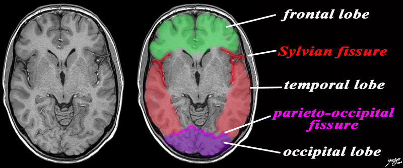
Sylvian Fissure in Axial Projection


The axial image is intended to demonstrate ttwo important fissures; the Sylvian fissure (in red) the parieto-occipital fissure (pink) in order to demonstrate the border between the frontal and temporal lobe in the axial plane and the junction of the temporal and occipital lobe
Courtesy Philips medicaL systems rendred by Davidoff art 92142c06b01 label.8s


    [nodeType] => 1
    [parentNode] => (object value omitted)
    [childNodes] => (object value omitted)
    [firstChild] => (object value omitted)
    [lastChild] => (object value omitted)
    [previousSibling] => (object value omitted)
    [nextSibling] => (object value omitted)
    [attributes] => (object value omitted)
    [ownerDocument] => (object value omitted)
    [namespaceURI] =>
    [prefix] =>
    [localName] => table
    [baseURI] =>
    [textContent] =>


Sylvian Fissure in Axial Projection


The axial image is intended to demonstrate ttwo important fissures; the Sylvian fissure (in red) the parieto-occipital fissure (pink) in order to demonstrate the border between the frontal and temporal lobe in the axial plane and the junction of the temporal and occipital lobe
Courtesy Philips medicaL systems rendred by Davidoff art 92142c06b01 label.8s


)
DOMElement Object
(
    [schemaTypeInfo] =>
    [tagName] => td
    [firstElementChild] => (object value omitted)
    [lastElementChild] => (object value omitted)
    [childElementCount] => 2
    [previousElementSibling] =>
    [nextElementSibling] =>
    [nodeName] => td
    [nodeValue] =>
The axial image is intended to demonstrate ttwo important fissures; the Sylvian fissure (in red) the parieto-occipital fissure (pink) in order to demonstrate the border between the frontal and temporal lobe in the axial plane and the junction of the temporal and occipital lobe
Courtesy Philips medicaL systems rendred by Davidoff art 92142c06b01 label.8s

    [nodeType] => 1
    [parentNode] => (object value omitted)
    [childNodes] => (object value omitted)
    [firstChild] => (object value omitted)
    [lastChild] => (object value omitted)
    [previousSibling] => (object value omitted)
    [nextSibling] => (object value omitted)
    [attributes] => (object value omitted)
    [ownerDocument] => (object value omitted)
    [namespaceURI] =>
    [prefix] =>
    [localName] => td
    [baseURI] =>
    [textContent] =>
The axial image is intended to demonstrate ttwo important fissures; the Sylvian fissure (in red) the parieto-occipital fissure (pink) in order to demonstrate the border between the frontal and temporal lobe in the axial plane and the junction of the temporal and occipital lobe
Courtesy Philips medicaL systems rendred by Davidoff art 92142c06b01 label.8s

)
DOMElement Object
(
    [schemaTypeInfo] =>
    [tagName] => td
    [firstElementChild] => (object value omitted)
    [lastElementChild] => (object value omitted)
    [childElementCount] => 2
    [previousElementSibling] =>
    [nextElementSibling] =>
    [nodeName] => td
    [nodeValue] =>
Sylvian Fissure in Axial Projection
    [nodeType] => 1
    [parentNode] => (object value omitted)
    [childNodes] => (object value omitted)
    [firstChild] => (object value omitted)
    [lastChild] => (object value omitted)
    [previousSibling] => (object value omitted)
    [nextSibling] => (object value omitted)
    [attributes] => (object value omitted)
    [ownerDocument] => (object value omitted)
    [namespaceURI] =>
    [prefix] =>
    [localName] => td
    [baseURI] =>
    [textContent] =>
Sylvian Fissure in Axial Projection
)
DOMElement Object
(
    [schemaTypeInfo] =>
    [tagName] => table
    [firstElementChild] => (object value omitted)
    [lastElementChild] => (object value omitted)
    [childElementCount] => 1
    [previousElementSibling] => (object value omitted)
    [nextElementSibling] => (object value omitted)
    [nodeName] => table
    [nodeValue] =>


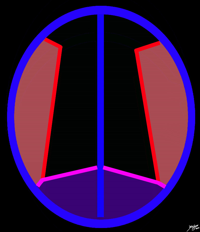
Conceptual Drawing of the Sylvian Fissure in Axial Projection


At this level the parietooccipital fissure (pink) defines the anterior edge of the occipital lobe posteriorly (purple). The Sylvian fissure (bright red lines) defines the temporal lobes (light red)
Courtesy Ashley Davidoff MDCopyright 2010 all rights reserved 93914.36k.81s


    [nodeType] => 1
    [parentNode] => (object value omitted)
    [childNodes] => (object value omitted)
    [firstChild] => (object value omitted)
    [lastChild] => (object value omitted)
    [previousSibling] => (object value omitted)
    [nextSibling] => (object value omitted)
    [attributes] => (object value omitted)
    [ownerDocument] => (object value omitted)
    [namespaceURI] =>
    [prefix] =>
    [localName] => table
    [baseURI] =>
    [textContent] =>


Conceptual Drawing of the Sylvian Fissure in Axial Projection


At this level the parietooccipital fissure (pink) defines the anterior edge of the occipital lobe posteriorly (purple). The Sylvian fissure (bright red lines) defines the temporal lobes (light red)
Courtesy Ashley Davidoff MDCopyright 2010 all rights reserved 93914.36k.81s


)
DOMElement Object
(
    [schemaTypeInfo] =>
    [tagName] => td
    [firstElementChild] => (object value omitted)
    [lastElementChild] => (object value omitted)
    [childElementCount] => 2
    [previousElementSibling] =>
    [nextElementSibling] =>
    [nodeName] => td
    [nodeValue] =>
At this level the parietooccipital fissure (pink) defines the anterior edge of the occipital lobe posteriorly (purple). The Sylvian fissure (bright red lines) defines the temporal lobes (light red)
Courtesy Ashley Davidoff MDCopyright 2010 all rights reserved 93914.36k.81s

    [nodeType] => 1
    [parentNode] => (object value omitted)
    [childNodes] => (object value omitted)
    [firstChild] => (object value omitted)
    [lastChild] => (object value omitted)
    [previousSibling] => (object value omitted)
    [nextSibling] => (object value omitted)
    [attributes] => (object value omitted)
    [ownerDocument] => (object value omitted)
    [namespaceURI] =>
    [prefix] =>
    [localName] => td
    [baseURI] =>
    [textContent] =>
At this level the parietooccipital fissure (pink) defines the anterior edge of the occipital lobe posteriorly (purple). The Sylvian fissure (bright red lines) defines the temporal lobes (light red)
Courtesy Ashley Davidoff MDCopyright 2010 all rights reserved 93914.36k.81s

)
DOMElement Object
(
    [schemaTypeInfo] =>
    [tagName] => td
    [firstElementChild] => (object value omitted)
    [lastElementChild] => (object value omitted)
    [childElementCount] => 2
    [previousElementSibling] =>
    [nextElementSibling] =>
    [nodeName] => td
    [nodeValue] =>
Conceptual Drawing of the Sylvian Fissure in Axial Projection
    [nodeType] => 1
    [parentNode] => (object value omitted)
    [childNodes] => (object value omitted)
    [firstChild] => (object value omitted)
    [lastChild] => (object value omitted)
    [previousSibling] => (object value omitted)
    [nextSibling] => (object value omitted)
    [attributes] => (object value omitted)
    [ownerDocument] => (object value omitted)
    [namespaceURI] =>
    [prefix] =>
    [localName] => td
    [baseURI] =>
    [textContent] =>
Conceptual Drawing of the Sylvian Fissure in Axial Projection
)
DOMElement Object
(
    [schemaTypeInfo] =>
    [tagName] => table
    [firstElementChild] => (object value omitted)
    [lastElementChild] => (object value omitted)
    [childElementCount] => 1
    [previousElementSibling] => (object value omitted)
    [nextElementSibling] => (object value omitted)
    [nodeName] => table
    [nodeValue] =>


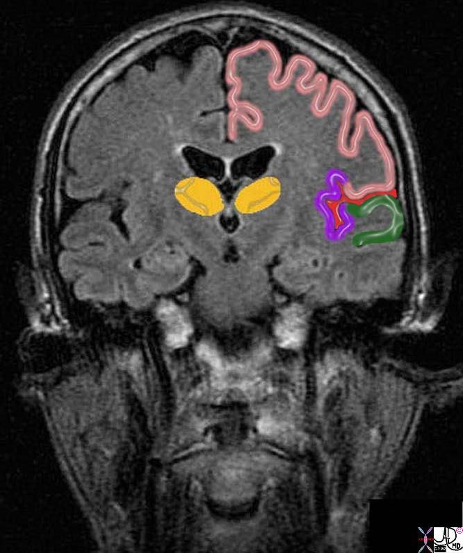
The Sylvian Fissure, the Frontoparietal region and the Temporal Lobe


The coronal T1 weighted image of the brain reveals the relationship of the Sylvian Fissure to the insular cortex.   The lateral somatosensory cortex is centered around the Sylvian fissure (red) It incorporates the insula (purple) the upper lid of the operculum which is part of the parietal cortex (pink) as well as parts of the frontal cortex, and the lower lid of the operculum (green) which is part of the temporal lobe.
Courtesy Ashley Davidoff MD copyright 2010  38610c06b06.8s


    [nodeType] => 1
    [parentNode] => (object value omitted)
    [childNodes] => (object value omitted)
    [firstChild] => (object value omitted)
    [lastChild] => (object value omitted)
    [previousSibling] => (object value omitted)
    [nextSibling] => (object value omitted)
    [attributes] => (object value omitted)
    [ownerDocument] => (object value omitted)
    [namespaceURI] =>
    [prefix] =>
    [localName] => table
    [baseURI] =>
    [textContent] =>


The Sylvian Fissure, the Frontoparietal region and the Temporal Lobe


The coronal T1 weighted image of the brain reveals the relationship of the Sylvian Fissure to the insular cortex.   The lateral somatosensory cortex is centered around the Sylvian fissure (red) It incorporates the insula (purple) the upper lid of the operculum which is part of the parietal cortex (pink) as well as parts of the frontal cortex, and the lower lid of the operculum (green) which is part of the temporal lobe.
Courtesy Ashley Davidoff MD copyright 2010  38610c06b06.8s


)
DOMElement Object
(
    [schemaTypeInfo] =>
    [tagName] => td
    [firstElementChild] => (object value omitted)
    [lastElementChild] => (object value omitted)
    [childElementCount] => 2
    [previousElementSibling] =>
    [nextElementSibling] =>
    [nodeName] => td
    [nodeValue] =>
The coronal T1 weighted image of the brain reveals the relationship of the Sylvian Fissure to the insular cortex.   The lateral somatosensory cortex is centered around the Sylvian fissure (red) It incorporates the insula (purple) the upper lid of the operculum which is part of the parietal cortex (pink) as well as parts of the frontal cortex, and the lower lid of the operculum (green) which is part of the temporal lobe.
Courtesy Ashley Davidoff MD copyright 2010  38610c06b06.8s

    [nodeType] => 1
    [parentNode] => (object value omitted)
    [childNodes] => (object value omitted)
    [firstChild] => (object value omitted)
    [lastChild] => (object value omitted)
    [previousSibling] => (object value omitted)
    [nextSibling] => (object value omitted)
    [attributes] => (object value omitted)
    [ownerDocument] => (object value omitted)
    [namespaceURI] =>
    [prefix] =>
    [localName] => td
    [baseURI] =>
    [textContent] =>
The coronal T1 weighted image of the brain reveals the relationship of the Sylvian Fissure to the insular cortex.   The lateral somatosensory cortex is centered around the Sylvian fissure (red) It incorporates the insula (purple) the upper lid of the operculum which is part of the parietal cortex (pink) as well as parts of the frontal cortex, and the lower lid of the operculum (green) which is part of the temporal lobe.
Courtesy Ashley Davidoff MD copyright 2010  38610c06b06.8s

)
DOMElement Object
(
    [schemaTypeInfo] =>
    [tagName] => td
    [firstElementChild] => (object value omitted)
    [lastElementChild] => (object value omitted)
    [childElementCount] => 2
    [previousElementSibling] =>
    [nextElementSibling] =>
    [nodeName] => td
    [nodeValue] =>
The Sylvian Fissure, the Frontoparietal region and the Temporal Lobe
    [nodeType] => 1
    [parentNode] => (object value omitted)
    [childNodes] => (object value omitted)
    [firstChild] => (object value omitted)
    [lastChild] => (object value omitted)
    [previousSibling] => (object value omitted)
    [nextSibling] => (object value omitted)
    [attributes] => (object value omitted)
    [ownerDocument] => (object value omitted)
    [namespaceURI] =>
    [prefix] =>
    [localName] => td
    [baseURI] =>
    [textContent] =>
The Sylvian Fissure, the Frontoparietal region and the Temporal Lobe
)
DOMElement Object
(
    [schemaTypeInfo] =>
    [tagName] => table
    [firstElementChild] => (object value omitted)
    [lastElementChild] => (object value omitted)
    [childElementCount] => 1
    [previousElementSibling] => (object value omitted)
    [nextElementSibling] => (object value omitted)
    [nodeName] => table
    [nodeValue] =>


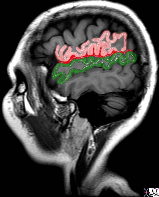
The Sylvian Fissure and the Operculum


The Sylvian fissure (red) lies in close association to the the insula (purple).  The upper lid of the operculum is part of the parietal cortex (pink) as well as parts of the frontal cortex, and the lower lid of the operculum (green) is part of the temporal lobe.
Courtesy Ashley Davidoff MD copyright 2010 71060c07.81s


    [nodeType] => 1
    [parentNode] => (object value omitted)
    [childNodes] => (object value omitted)
    [firstChild] => (object value omitted)
    [lastChild] => (object value omitted)
    [previousSibling] => (object value omitted)
    [nextSibling] => (object value omitted)
    [attributes] => (object value omitted)
    [ownerDocument] => (object value omitted)
    [namespaceURI] =>
    [prefix] =>
    [localName] => table
    [baseURI] =>
    [textContent] =>


The Sylvian Fissure and the Operculum


The Sylvian fissure (red) lies in close association to the the insula (purple).  The upper lid of the operculum is part of the parietal cortex (pink) as well as parts of the frontal cortex, and the lower lid of the operculum (green) is part of the temporal lobe.
Courtesy Ashley Davidoff MD copyright 2010 71060c07.81s


)
DOMElement Object
(
    [schemaTypeInfo] =>
    [tagName] => td
    [firstElementChild] => (object value omitted)
    [lastElementChild] => (object value omitted)
    [childElementCount] => 2
    [previousElementSibling] =>
    [nextElementSibling] =>
    [nodeName] => td
    [nodeValue] =>
The Sylvian fissure (red) lies in close association to the the insula (purple).  The upper lid of the operculum is part of the parietal cortex (pink) as well as parts of the frontal cortex, and the lower lid of the operculum (green) is part of the temporal lobe.
Courtesy Ashley Davidoff MD copyright 2010 71060c07.81s
    [nodeType] => 1
    [parentNode] => (object value omitted)
    [childNodes] => (object value omitted)
    [firstChild] => (object value omitted)
    [lastChild] => (object value omitted)
    [previousSibling] => (object value omitted)
    [nextSibling] => (object value omitted)
    [attributes] => (object value omitted)
    [ownerDocument] => (object value omitted)
    [namespaceURI] =>
    [prefix] =>
    [localName] => td
    [baseURI] =>
    [textContent] =>
The Sylvian fissure (red) lies in close association to the the insula (purple).  The upper lid of the operculum is part of the parietal cortex (pink) as well as parts of the frontal cortex, and the lower lid of the operculum (green) is part of the temporal lobe.
Courtesy Ashley Davidoff MD copyright 2010 71060c07.81s
)
DOMElement Object
(
    [schemaTypeInfo] =>
    [tagName] => td
    [firstElementChild] => (object value omitted)
    [lastElementChild] => (object value omitted)
    [childElementCount] => 2
    [previousElementSibling] =>
    [nextElementSibling] =>
    [nodeName] => td
    [nodeValue] =>
The Sylvian Fissure and the Operculum
    [nodeType] => 1
    [parentNode] => (object value omitted)
    [childNodes] => (object value omitted)
    [firstChild] => (object value omitted)
    [lastChild] => (object value omitted)
    [previousSibling] => (object value omitted)
    [nextSibling] => (object value omitted)
    [attributes] => (object value omitted)
    [ownerDocument] => (object value omitted)
    [namespaceURI] =>
    [prefix] =>
    [localName] => td
    [baseURI] =>
    [textContent] =>
The Sylvian Fissure and the Operculum
)
DOMElement Object
(
    [schemaTypeInfo] =>
    [tagName] => table
    [firstElementChild] => (object value omitted)
    [lastElementChild] => (object value omitted)
    [childElementCount] => 1
    [previousElementSibling] => (object value omitted)
    [nextElementSibling] => (object value omitted)
    [nodeName] => table
    [nodeValue] =>


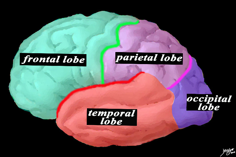
External Sagittal View
Sylvian Fissure in Red


This is a diagram looking at the forebrain from the side showing the anteriorly placed frontal lobe, separated from the inferiorly placed temporal lobe by the Sylvian fissure (red), and separated from the parietal lobe by the central sulcus (green). The parietal lobe is also separated from the temporal lobe by the Sylvian fissure and from the occipital lobe by the parieto-occipital fissure (pink).
Courtesy Ashley DAvidoff MD copyright 2010 83029d13b01.8s


    [nodeType] => 1
    [parentNode] => (object value omitted)
    [childNodes] => (object value omitted)
    [firstChild] => (object value omitted)
    [lastChild] => (object value omitted)
    [previousSibling] => (object value omitted)
    [nextSibling] => (object value omitted)
    [attributes] => (object value omitted)
    [ownerDocument] => (object value omitted)
    [namespaceURI] =>
    [prefix] =>
    [localName] => table
    [baseURI] =>
    [textContent] =>


External Sagittal View
Sylvian Fissure in Red


This is a diagram looking at the forebrain from the side showing the anteriorly placed frontal lobe, separated from the inferiorly placed temporal lobe by the Sylvian fissure (red), and separated from the parietal lobe by the central sulcus (green). The parietal lobe is also separated from the temporal lobe by the Sylvian fissure and from the occipital lobe by the parieto-occipital fissure (pink).
Courtesy Ashley DAvidoff MD copyright 2010 83029d13b01.8s


)
DOMElement Object
(
    [schemaTypeInfo] =>
    [tagName] => td
    [firstElementChild] => (object value omitted)
    [lastElementChild] => (object value omitted)
    [childElementCount] => 2
    [previousElementSibling] =>
    [nextElementSibling] =>
    [nodeName] => td
    [nodeValue] =>
This is a diagram looking at the forebrain from the side showing the anteriorly placed frontal lobe, separated from the inferiorly placed temporal lobe by the Sylvian fissure (red), and separated from the parietal lobe by the central sulcus (green). The parietal lobe is also separated from the temporal lobe by the Sylvian fissure and from the occipital lobe by the parieto-occipital fissure (pink).
Courtesy Ashley DAvidoff MD copyright 2010 83029d13b01.8s

    [nodeType] => 1
    [parentNode] => (object value omitted)
    [childNodes] => (object value omitted)
    [firstChild] => (object value omitted)
    [lastChild] => (object value omitted)
    [previousSibling] => (object value omitted)
    [nextSibling] => (object value omitted)
    [attributes] => (object value omitted)
    [ownerDocument] => (object value omitted)
    [namespaceURI] =>
    [prefix] =>
    [localName] => td
    [baseURI] =>
    [textContent] =>
This is a diagram looking at the forebrain from the side showing the anteriorly placed frontal lobe, separated from the inferiorly placed temporal lobe by the Sylvian fissure (red), and separated from the parietal lobe by the central sulcus (green). The parietal lobe is also separated from the temporal lobe by the Sylvian fissure and from the occipital lobe by the parieto-occipital fissure (pink).
Courtesy Ashley DAvidoff MD copyright 2010 83029d13b01.8s

)
DOMElement Object
(
    [schemaTypeInfo] =>
    [tagName] => td
    [firstElementChild] => (object value omitted)
    [lastElementChild] => (object value omitted)
    [childElementCount] => 3
    [previousElementSibling] =>
    [nextElementSibling] =>
    [nodeName] => td
    [nodeValue] =>
External Sagittal View
Sylvian Fissure in Red
    [nodeType] => 1
    [parentNode] => (object value omitted)
    [childNodes] => (object value omitted)
    [firstChild] => (object value omitted)
    [lastChild] => (object value omitted)
    [previousSibling] => (object value omitted)
    [nextSibling] => (object value omitted)
    [attributes] => (object value omitted)
    [ownerDocument] => (object value omitted)
    [namespaceURI] =>
    [prefix] =>
    [localName] => td
    [baseURI] =>
    [textContent] =>
External Sagittal View
Sylvian Fissure in Red
)
DOMElement Object
(
    [schemaTypeInfo] =>
    [tagName] => table
    [firstElementChild] => (object value omitted)
    [lastElementChild] => (object value omitted)
    [childElementCount] => 1
    [previousElementSibling] => (object value omitted)
    [nextElementSibling] => (object value omitted)
    [nodeName] => table
    [nodeValue] =>


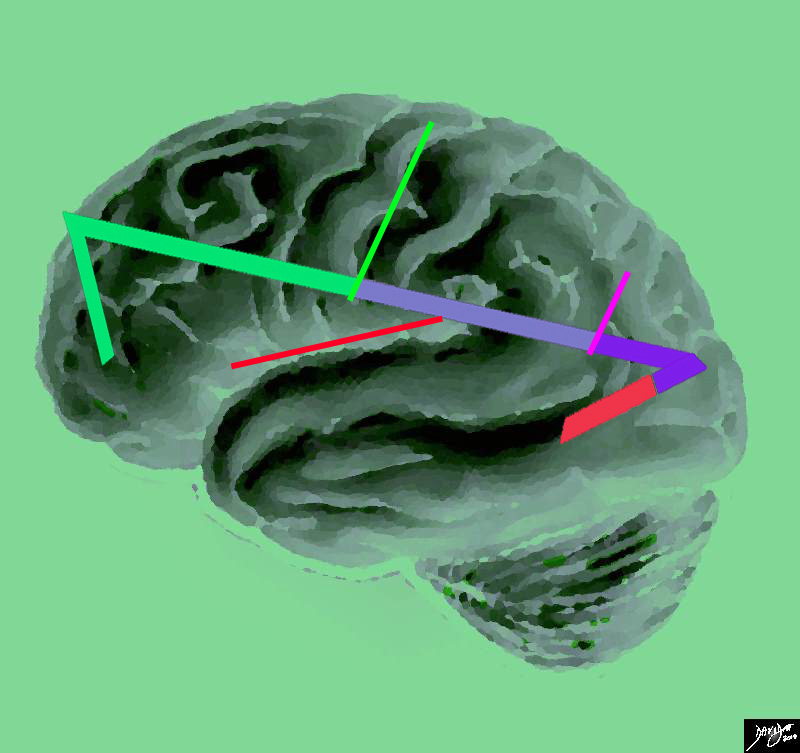
Basic Concept
Sylvian Fissure (thin red line)


This artistic rendition of the brain reflects the vectors of the major parts of the brain revealing the major border forming fissures and sulci. The central sulcus (bright green line) divides the frontal lobe from the parietal lobe (light mauve). The region of the parieto-occipital fissure (pink line) divides the parietal lobe from the occipital lobe (purple), and the Sylvian fissure (thin red line)divides the temporal lobe from the frontal and parietal lobe
Courtesy Ashley Davidoff copyright 2010 all rights reserved 83029e04.87s


    [nodeType] => 1
    [parentNode] => (object value omitted)
    [childNodes] => (object value omitted)
    [firstChild] => (object value omitted)
    [lastChild] => (object value omitted)
    [previousSibling] => (object value omitted)
    [nextSibling] => (object value omitted)
    [attributes] => (object value omitted)
    [ownerDocument] => (object value omitted)
    [namespaceURI] =>
    [prefix] =>
    [localName] => table
    [baseURI] =>
    [textContent] =>


Basic Concept
Sylvian Fissure (thin red line)


This artistic rendition of the brain reflects the vectors of the major parts of the brain revealing the major border forming fissures and sulci. The central sulcus (bright green line) divides the frontal lobe from the parietal lobe (light mauve). The region of the parieto-occipital fissure (pink line) divides the parietal lobe from the occipital lobe (purple), and the Sylvian fissure (thin red line)divides the temporal lobe from the frontal and parietal lobe
Courtesy Ashley Davidoff copyright 2010 all rights reserved 83029e04.87s


)
DOMElement Object
(
    [schemaTypeInfo] =>
    [tagName] => td
    [firstElementChild] => (object value omitted)
    [lastElementChild] => (object value omitted)
    [childElementCount] => 2
    [previousElementSibling] =>
    [nextElementSibling] =>
    [nodeName] => td
    [nodeValue] =>
This artistic rendition of the brain reflects the vectors of the major parts of the brain revealing the major border forming fissures and sulci. The central sulcus (bright green line) divides the frontal lobe from the parietal lobe (light mauve). The region of the parieto-occipital fissure (pink line) divides the parietal lobe from the occipital lobe (purple), and the Sylvian fissure (thin red line)divides the temporal lobe from the frontal and parietal lobe
Courtesy Ashley Davidoff copyright 2010 all rights reserved 83029e04.87s

    [nodeType] => 1
    [parentNode] => (object value omitted)
    [childNodes] => (object value omitted)
    [firstChild] => (object value omitted)
    [lastChild] => (object value omitted)
    [previousSibling] => (object value omitted)
    [nextSibling] => (object value omitted)
    [attributes] => (object value omitted)
    [ownerDocument] => (object value omitted)
    [namespaceURI] =>
    [prefix] =>
    [localName] => td
    [baseURI] =>
    [textContent] =>
This artistic rendition of the brain reflects the vectors of the major parts of the brain revealing the major border forming fissures and sulci. The central sulcus (bright green line) divides the frontal lobe from the parietal lobe (light mauve). The region of the parieto-occipital fissure (pink line) divides the parietal lobe from the occipital lobe (purple), and the Sylvian fissure (thin red line)divides the temporal lobe from the frontal and parietal lobe
Courtesy Ashley Davidoff copyright 2010 all rights reserved 83029e04.87s

)
DOMElement Object
(
    [schemaTypeInfo] =>
    [tagName] => td
    [firstElementChild] => (object value omitted)
    [lastElementChild] => (object value omitted)
    [childElementCount] => 3
    [previousElementSibling] =>
    [nextElementSibling] =>
    [nodeName] => td
    [nodeValue] =>
Basic Concept
Sylvian Fissure (thin red line)
    [nodeType] => 1
    [parentNode] => (object value omitted)
    [childNodes] => (object value omitted)
    [firstChild] => (object value omitted)
    [lastChild] => (object value omitted)
    [previousSibling] => (object value omitted)
    [nextSibling] => (object value omitted)
    [attributes] => (object value omitted)
    [ownerDocument] => (object value omitted)
    [namespaceURI] =>
    [prefix] =>
    [localName] => td
    [baseURI] =>
    [textContent] =>
Basic Concept
Sylvian Fissure (thin red line)
)

