Calcarine Fissure
Sumit Karia MD
The Common Vein Copyright 2010
Definition
Structure The calcarine fissure is located at the most caudal end of the medial surface of the brain. It begins near the occipital pole in two converging rami, heading towards the splenium of the corpus callosum. The anterior part of this fissure gives rise to the prominence of the calcar avis in the posterior cornu of the lateral ventricle.
It has six principal layers, with layer IV further subdivided into several sublaminae, one of which being the stripe of Gennari, which is a dense plexus of myelinated fibers derived from primary visual cortex neurons.
Function It constitutes the primary visual cortex, also referred to as the striate cortex. The visual field is distributed throughout the sulcus, the central visual field being located in posterior portion while the peripheral visual field being present in the anterior portion. Simultaneously, more cortex is dedicated to process information originating from the fovea rather than from the rods.
Disease The calcarine fissure is supplied by the calcarine artery which is a branch if the posterior cerebral artery. A stroke causing decreased supply to the calcarine artery may result in a lesion in this area, which may result in loss of vision, which will have a determinate pattern depending on the extent of the lesion. If the occipital pole is damaged, a central scotoma will develop. When the calcarine cortex is destroyed, the result is homonymous hemianopia. The macular region, which is processed in the most posterior portion, may be spared if collateral circulation from the middle cerebral artery is sufficient.
|

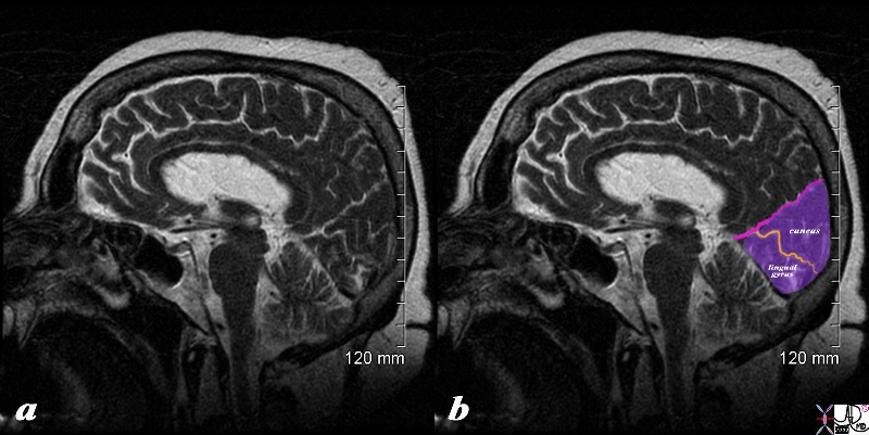
Occip[itoparietal Fissure Calcarine Fissure and Occipital Gyri |
|
The sagittal view of the brain using a T2 weighted sequence shows the parieto-occipital fissure (pink) (aka sulcus), that separates the parietal lobe anteriorly and the occipital lobe (purple) posteriorly. The calcarine fissure (orange) separates the cuneus above from the lingual gyrus inferiorly.
Courtesy Ashley Davidoff copyright 2010 all rights reserved 71430cd01b02.8s
|
DOMElement Object
(
    [schemaTypeInfo] =>
    [tagName] => table
    [firstElementChild] => (object value omitted)
    [lastElementChild] => (object value omitted)
    [childElementCount] => 1
    [previousElementSibling] => (object value omitted)
    [nextElementSibling] =>
    [nodeName] => table
    [nodeValue] =>


Occip[itoparietal Fissure Calcarine Fissure and Occipital Gyri


The sagittal view of the brain using a T2 weighted sequence shows the parieto-occipital fissure (pink) (aka sulcus), that separates the parietal lobe anteriorly and the occipital lobe (purple) posteriorly. The calcarine fissure (orange) separates the cuneus above from the lingual gyrus inferiorly.
Courtesy Ashley Davidoff copyright 2010 all rights reserved 71430cd01b02.8s


    [nodeType] => 1
    [parentNode] => (object value omitted)
    [childNodes] => (object value omitted)
    [firstChild] => (object value omitted)
    [lastChild] => (object value omitted)
    [previousSibling] => (object value omitted)
    [nextSibling] => (object value omitted)
    [attributes] => (object value omitted)
    [ownerDocument] => (object value omitted)
    [namespaceURI] =>
    [prefix] =>
    [localName] => table
    [baseURI] =>
    [textContent] =>


Occip[itoparietal Fissure Calcarine Fissure and Occipital Gyri


The sagittal view of the brain using a T2 weighted sequence shows the parieto-occipital fissure (pink) (aka sulcus), that separates the parietal lobe anteriorly and the occipital lobe (purple) posteriorly. The calcarine fissure (orange) separates the cuneus above from the lingual gyrus inferiorly.
Courtesy Ashley Davidoff copyright 2010 all rights reserved 71430cd01b02.8s


)
DOMElement Object
(
    [schemaTypeInfo] =>
    [tagName] => td
    [firstElementChild] => (object value omitted)
    [lastElementChild] => (object value omitted)
    [childElementCount] => 2
    [previousElementSibling] =>
    [nextElementSibling] =>
    [nodeName] => td
    [nodeValue] =>
The sagittal view of the brain using a T2 weighted sequence shows the parieto-occipital fissure (pink) (aka sulcus), that separates the parietal lobe anteriorly and the occipital lobe (purple) posteriorly. The calcarine fissure (orange) separates the cuneus above from the lingual gyrus inferiorly.
Courtesy Ashley Davidoff copyright 2010 all rights reserved 71430cd01b02.8s

    [nodeType] => 1
    [parentNode] => (object value omitted)
    [childNodes] => (object value omitted)
    [firstChild] => (object value omitted)
    [lastChild] => (object value omitted)
    [previousSibling] => (object value omitted)
    [nextSibling] => (object value omitted)
    [attributes] => (object value omitted)
    [ownerDocument] => (object value omitted)
    [namespaceURI] =>
    [prefix] =>
    [localName] => td
    [baseURI] =>
    [textContent] =>
The sagittal view of the brain using a T2 weighted sequence shows the parieto-occipital fissure (pink) (aka sulcus), that separates the parietal lobe anteriorly and the occipital lobe (purple) posteriorly. The calcarine fissure (orange) separates the cuneus above from the lingual gyrus inferiorly.
Courtesy Ashley Davidoff copyright 2010 all rights reserved 71430cd01b02.8s

)
DOMElement Object
(
    [schemaTypeInfo] =>
    [tagName] => td
    [firstElementChild] => (object value omitted)
    [lastElementChild] => (object value omitted)
    [childElementCount] => 2
    [previousElementSibling] =>
    [nextElementSibling] =>
    [nodeName] => td
    [nodeValue] =>

Occip[itoparietal Fissure Calcarine Fissure and Occipital Gyri
    [nodeType] => 1
    [parentNode] => (object value omitted)
    [childNodes] => (object value omitted)
    [firstChild] => (object value omitted)
    [lastChild] => (object value omitted)
    [previousSibling] => (object value omitted)
    [nextSibling] => (object value omitted)
    [attributes] => (object value omitted)
    [ownerDocument] => (object value omitted)
    [namespaceURI] =>
    [prefix] =>
    [localName] => td
    [baseURI] =>
    [textContent] =>

Occip[itoparietal Fissure Calcarine Fissure and Occipital Gyri
)

