Midbrain – Transverse View – Concepts
Ashley Davidoff MD
The Common Vein Copyright 2010
Introduction
On cross sectional imaging the midbrain is relatively short and has similar shape to other structures in the region. In general it is almost butterfly shaped.
|
Baseline Shape of the Midbrain Sting Like a Butterfly |
|
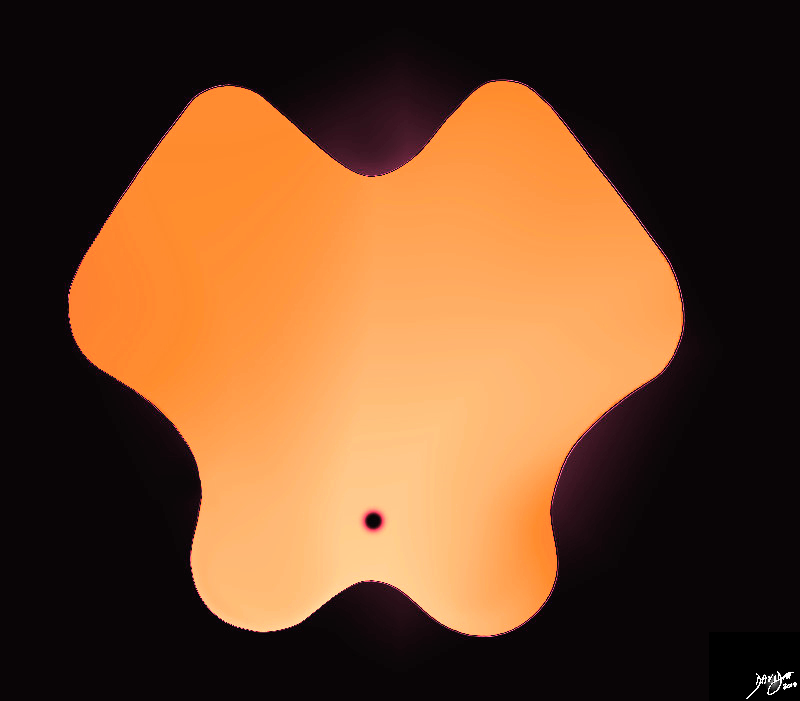
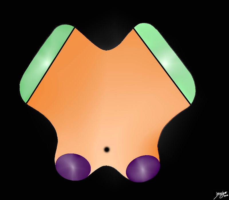
The midbrain is almost butterfly shaped with rounded posterior borders (colliculi) and rectangular shaped wings – the cerebral peduncles. It is very similar in shape to the pons and medulla on axial imaging but a a few characteristics distinguish it from these structures, most specific of which is the aqueduct of Sylvius that run right through its substance posteriorly shown as a black circle Davidoff art Courtesy Ashley Davidoff MD copyright 2010 all rights reserved 94074b08a.82s |
DOMElement Object
(
    [schemaTypeInfo] =>
    [tagName] => table
    [firstElementChild] => (object value omitted)
    [lastElementChild] => (object value omitted)
    [childElementCount] => 1
    [previousElementSibling] => (object value omitted)
    [nextElementSibling] =>
    [nodeName] => table
    [nodeValue] =>


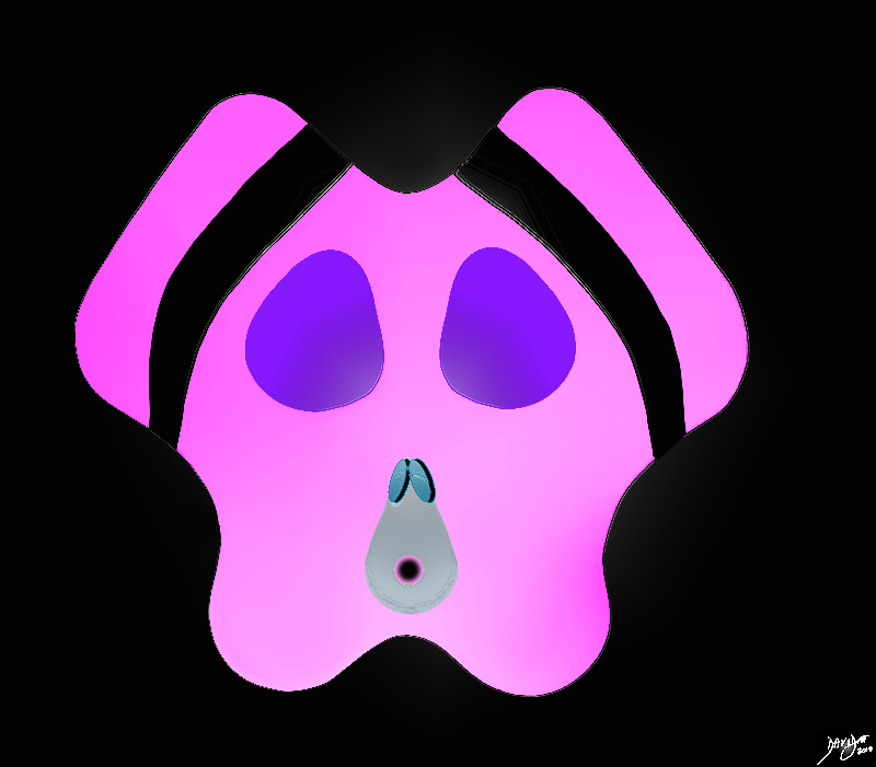
The Face of a Baby Pig


As we start to delve into the complexity of the nuclii of the midbrain the shape takes the form of the head of a baby pig
Davidoff art Courtesy Ashley Davidoff MD copyright 2010 all rights reserved 94074b08a06b1b.8s


    [nodeType] => 1
    [parentNode] => (object value omitted)
    [childNodes] => (object value omitted)
    [firstChild] => (object value omitted)
    [lastChild] => (object value omitted)
    [previousSibling] => (object value omitted)
    [nextSibling] => (object value omitted)
    [attributes] => (object value omitted)
    [ownerDocument] => (object value omitted)
    [namespaceURI] =>
    [prefix] =>
    [localName] => table
    [baseURI] =>
    [textContent] =>


The Face of a Baby Pig


As we start to delve into the complexity of the nuclii of the midbrain the shape takes the form of the head of a baby pig
Davidoff art Courtesy Ashley Davidoff MD copyright 2010 all rights reserved 94074b08a06b1b.8s


)
DOMElement Object
(
    [schemaTypeInfo] =>
    [tagName] => td
    [firstElementChild] => (object value omitted)
    [lastElementChild] => (object value omitted)
    [childElementCount] => 2
    [previousElementSibling] =>
    [nextElementSibling] =>
    [nodeName] => td
    [nodeValue] =>
As we start to delve into the complexity of the nuclii of the midbrain the shape takes the form of the head of a baby pig
Davidoff art Courtesy Ashley Davidoff MD copyright 2010 all rights reserved 94074b08a06b1b.8s

    [nodeType] => 1
    [parentNode] => (object value omitted)
    [childNodes] => (object value omitted)
    [firstChild] => (object value omitted)
    [lastChild] => (object value omitted)
    [previousSibling] => (object value omitted)
    [nextSibling] => (object value omitted)
    [attributes] => (object value omitted)
    [ownerDocument] => (object value omitted)
    [namespaceURI] =>
    [prefix] =>
    [localName] => td
    [baseURI] =>
    [textContent] =>
As we start to delve into the complexity of the nuclii of the midbrain the shape takes the form of the head of a baby pig
Davidoff art Courtesy Ashley Davidoff MD copyright 2010 all rights reserved 94074b08a06b1b.8s

)
DOMElement Object
(
    [schemaTypeInfo] =>
    [tagName] => td
    [firstElementChild] => (object value omitted)
    [lastElementChild] => (object value omitted)
    [childElementCount] => 2
    [previousElementSibling] =>
    [nextElementSibling] =>
    [nodeName] => td
    [nodeValue] =>
The Face of a Baby Pig

    [nodeType] => 1
    [parentNode] => (object value omitted)
    [childNodes] => (object value omitted)
    [firstChild] => (object value omitted)
    [lastChild] => (object value omitted)
    [previousSibling] => (object value omitted)
    [nextSibling] => (object value omitted)
    [attributes] => (object value omitted)
    [ownerDocument] => (object value omitted)
    [namespaceURI] =>
    [prefix] =>
    [localName] => td
    [baseURI] =>
    [textContent] =>
The Face of a Baby Pig

)
DOMElement Object
(
    [schemaTypeInfo] =>
    [tagName] => table
    [firstElementChild] => (object value omitted)
    [lastElementChild] => (object value omitted)
    [childElementCount] => 1
    [previousElementSibling] => (object value omitted)
    [nextElementSibling] => (object value omitted)
    [nodeName] => table
    [nodeValue] =>


The Cerebral arteries Characteristically Surround the Midbrain


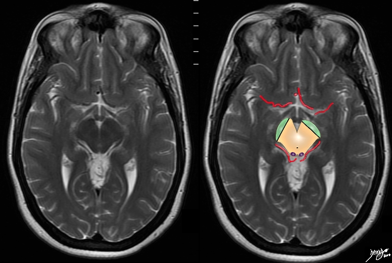
The MRI T2 weighted series now focuses on another very characteristic feature of the midbrain in that it is surrounded by the anterior and posterior cerebral circulations, and in this case portions of the arteries are seen with parts of the middle cerebral seen anteriorly and parts of the posterior cerebral seen posteriorly
Davidoff art Courteys Ashley Davidoff MD copyright 2010 all rights reserved 94081.4kc02.8s


    [nodeType] => 1
    [parentNode] => (object value omitted)
    [childNodes] => (object value omitted)
    [firstChild] => (object value omitted)
    [lastChild] => (object value omitted)
    [previousSibling] => (object value omitted)
    [nextSibling] => (object value omitted)
    [attributes] => (object value omitted)
    [ownerDocument] => (object value omitted)
    [namespaceURI] =>
    [prefix] =>
    [localName] => table
    [baseURI] =>
    [textContent] =>


The Cerebral arteries Characteristically Surround the Midbrain


The MRI T2 weighted series now focuses on another very characteristic feature of the midbrain in that it is surrounded by the anterior and posterior cerebral circulations, and in this case portions of the arteries are seen with parts of the middle cerebral seen anteriorly and parts of the posterior cerebral seen posteriorly
Davidoff art Courteys Ashley Davidoff MD copyright 2010 all rights reserved 94081.4kc02.8s


)
DOMElement Object
(
    [schemaTypeInfo] =>
    [tagName] => td
    [firstElementChild] => (object value omitted)
    [lastElementChild] => (object value omitted)
    [childElementCount] => 2
    [previousElementSibling] =>
    [nextElementSibling] =>
    [nodeName] => td
    [nodeValue] =>
The MRI T2 weighted series now focuses on another very characteristic feature of the midbrain in that it is surrounded by the anterior and posterior cerebral circulations, and in this case portions of the arteries are seen with parts of the middle cerebral seen anteriorly and parts of the posterior cerebral seen posteriorly
Davidoff art Courteys Ashley Davidoff MD copyright 2010 all rights reserved 94081.4kc02.8s

    [nodeType] => 1
    [parentNode] => (object value omitted)
    [childNodes] => (object value omitted)
    [firstChild] => (object value omitted)
    [lastChild] => (object value omitted)
    [previousSibling] => (object value omitted)
    [nextSibling] => (object value omitted)
    [attributes] => (object value omitted)
    [ownerDocument] => (object value omitted)
    [namespaceURI] =>
    [prefix] =>
    [localName] => td
    [baseURI] =>
    [textContent] =>
The MRI T2 weighted series now focuses on another very characteristic feature of the midbrain in that it is surrounded by the anterior and posterior cerebral circulations, and in this case portions of the arteries are seen with parts of the middle cerebral seen anteriorly and parts of the posterior cerebral seen posteriorly
Davidoff art Courteys Ashley Davidoff MD copyright 2010 all rights reserved 94081.4kc02.8s

)
DOMElement Object
(
    [schemaTypeInfo] =>
    [tagName] => td
    [firstElementChild] => (object value omitted)
    [lastElementChild] => (object value omitted)
    [childElementCount] => 2
    [previousElementSibling] =>
    [nextElementSibling] =>
    [nodeName] => td
    [nodeValue] =>
The Cerebral arteries Characteristically Surround the Midbrain

    [nodeType] => 1
    [parentNode] => (object value omitted)
    [childNodes] => (object value omitted)
    [firstChild] => (object value omitted)
    [lastChild] => (object value omitted)
    [previousSibling] => (object value omitted)
    [nextSibling] => (object value omitted)
    [attributes] => (object value omitted)
    [ownerDocument] => (object value omitted)
    [namespaceURI] =>
    [prefix] =>
    [localName] => td
    [baseURI] =>
    [textContent] =>
The Cerebral arteries Characteristically Surround the Midbrain

)
DOMElement Object
(
    [schemaTypeInfo] =>
    [tagName] => table
    [firstElementChild] => (object value omitted)
    [lastElementChild] => (object value omitted)
    [childElementCount] => 1
    [previousElementSibling] => (object value omitted)
    [nextElementSibling] => (object value omitted)
    [nodeName] => table
    [nodeValue] =>


The Cerebral Crura and the Colliculi
The Tegmentum and the Tectum


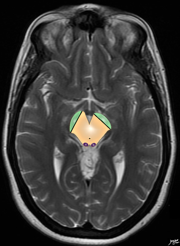
 The T2 weighted MRI series focuses on the midbrain with characteristic shape and the small aqueduct of Sylvius posteriorly. The cerebral crura are now shown anteriorly (green) in the cerebral peduncles, and the colliculi are shown posteriorly in the tectum or roof posteriorly
Davidoff art Courtesy Ashley Davidoff MD copyright 2010 all rights reserved 94081.4kb07.8s


    [nodeType] => 1
    [parentNode] => (object value omitted)
    [childNodes] => (object value omitted)
    [firstChild] => (object value omitted)
    [lastChild] => (object value omitted)
    [previousSibling] => (object value omitted)
    [nextSibling] => (object value omitted)
    [attributes] => (object value omitted)
    [ownerDocument] => (object value omitted)
    [namespaceURI] =>
    [prefix] =>
    [localName] => table
    [baseURI] =>
    [textContent] =>


The Cerebral Crura and the Colliculi
The Tegmentum and the Tectum


 The T2 weighted MRI series focuses on the midbrain with characteristic shape and the small aqueduct of Sylvius posteriorly. The cerebral crura are now shown anteriorly (green) in the cerebral peduncles, and the colliculi are shown posteriorly in the tectum or roof posteriorly
Davidoff art Courtesy Ashley Davidoff MD copyright 2010 all rights reserved 94081.4kb07.8s


)
DOMElement Object
(
    [schemaTypeInfo] =>
    [tagName] => td
    [firstElementChild] => (object value omitted)
    [lastElementChild] => (object value omitted)
    [childElementCount] => 2
    [previousElementSibling] =>
    [nextElementSibling] =>
    [nodeName] => td
    [nodeValue] =>
 The T2 weighted MRI series focuses on the midbrain with characteristic shape and the small aqueduct of Sylvius posteriorly. The cerebral crura are now shown anteriorly (green) in the cerebral peduncles, and the colliculi are shown posteriorly in the tectum or roof posteriorly
Davidoff art Courtesy Ashley Davidoff MD copyright 2010 all rights reserved 94081.4kb07.8s

    [nodeType] => 1
    [parentNode] => (object value omitted)
    [childNodes] => (object value omitted)
    [firstChild] => (object value omitted)
    [lastChild] => (object value omitted)
    [previousSibling] => (object value omitted)
    [nextSibling] => (object value omitted)
    [attributes] => (object value omitted)
    [ownerDocument] => (object value omitted)
    [namespaceURI] =>
    [prefix] =>
    [localName] => td
    [baseURI] =>
    [textContent] =>
 The T2 weighted MRI series focuses on the midbrain with characteristic shape and the small aqueduct of Sylvius posteriorly. The cerebral crura are now shown anteriorly (green) in the cerebral peduncles, and the colliculi are shown posteriorly in the tectum or roof posteriorly
Davidoff art Courtesy Ashley Davidoff MD copyright 2010 all rights reserved 94081.4kb07.8s

)
DOMElement Object
(
    [schemaTypeInfo] =>
    [tagName] => td
    [firstElementChild] => (object value omitted)
    [lastElementChild] => (object value omitted)
    [childElementCount] => 3
    [previousElementSibling] =>
    [nextElementSibling] =>
    [nodeName] => td
    [nodeValue] =>
The Cerebral Crura and the Colliculi
The Tegmentum and the Tectum

    [nodeType] => 1
    [parentNode] => (object value omitted)
    [childNodes] => (object value omitted)
    [firstChild] => (object value omitted)
    [lastChild] => (object value omitted)
    [previousSibling] => (object value omitted)
    [nextSibling] => (object value omitted)
    [attributes] => (object value omitted)
    [ownerDocument] => (object value omitted)
    [namespaceURI] =>
    [prefix] =>
    [localName] => td
    [baseURI] =>
    [textContent] =>
The Cerebral Crura and the Colliculi
The Tegmentum and the Tectum

)
DOMElement Object
(
    [schemaTypeInfo] =>
    [tagName] => table
    [firstElementChild] => (object value omitted)
    [lastElementChild] => (object value omitted)
    [childElementCount] => 1
    [previousElementSibling] => (object value omitted)
    [nextElementSibling] => (object value omitted)
    [nodeName] => table
    [nodeValue] =>


Peduncles Substantia Nigra Tegmentum Colliculi and Tectum


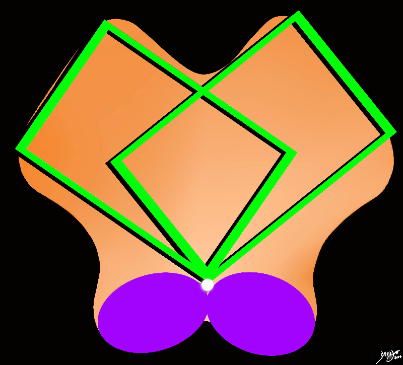
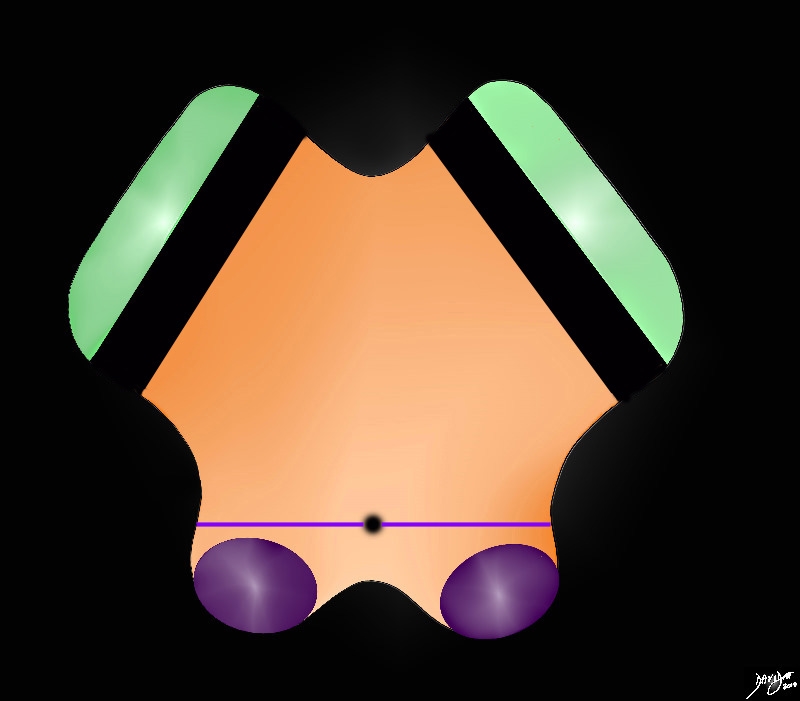
The anterior border iof the midbrain incorporates the cerbral peduncles(green), and the substantia nigra (black – just posterior to the peduncles. Between the substantia nigra and the aqueduct is an area of the midbrain called the tegmentum (floor of the midbrain) The posterior end of the midbrain is bordered by the colliculi (purple) in the tectum (roof) of the nidbrain.
Davidoff art Courtesy Ashley Davidoff MD copyright 2010 all rights reserved 94074b09b05b.83s


    [nodeType] => 1
    [parentNode] => (object value omitted)
    [childNodes] => (object value omitted)
    [firstChild] => (object value omitted)
    [lastChild] => (object value omitted)
    [previousSibling] => (object value omitted)
    [nextSibling] => (object value omitted)
    [attributes] => (object value omitted)
    [ownerDocument] => (object value omitted)
    [namespaceURI] =>
    [prefix] =>
    [localName] => table
    [baseURI] =>
    [textContent] =>


Peduncles Substantia Nigra Tegmentum Colliculi and Tectum


The anterior border iof the midbrain incorporates the cerbral peduncles(green), and the substantia nigra (black – just posterior to the peduncles. Between the substantia nigra and the aqueduct is an area of the midbrain called the tegmentum (floor of the midbrain) The posterior end of the midbrain is bordered by the colliculi (purple) in the tectum (roof) of the nidbrain.
Davidoff art Courtesy Ashley Davidoff MD copyright 2010 all rights reserved 94074b09b05b.83s


)
DOMElement Object
(
    [schemaTypeInfo] =>
    [tagName] => td
    [firstElementChild] => (object value omitted)
    [lastElementChild] => (object value omitted)
    [childElementCount] => 2
    [previousElementSibling] =>
    [nextElementSibling] =>
    [nodeName] => td
    [nodeValue] =>
The anterior border iof the midbrain incorporates the cerbral peduncles(green), and the substantia nigra (black – just posterior to the peduncles. Between the substantia nigra and the aqueduct is an area of the midbrain called the tegmentum (floor of the midbrain) The posterior end of the midbrain is bordered by the colliculi (purple) in the tectum (roof) of the nidbrain.
Davidoff art Courtesy Ashley Davidoff MD copyright 2010 all rights reserved 94074b09b05b.83s

    [nodeType] => 1
    [parentNode] => (object value omitted)
    [childNodes] => (object value omitted)
    [firstChild] => (object value omitted)
    [lastChild] => (object value omitted)
    [previousSibling] => (object value omitted)
    [nextSibling] => (object value omitted)
    [attributes] => (object value omitted)
    [ownerDocument] => (object value omitted)
    [namespaceURI] =>
    [prefix] =>
    [localName] => td
    [baseURI] =>
    [textContent] =>
The anterior border iof the midbrain incorporates the cerbral peduncles(green), and the substantia nigra (black – just posterior to the peduncles. Between the substantia nigra and the aqueduct is an area of the midbrain called the tegmentum (floor of the midbrain) The posterior end of the midbrain is bordered by the colliculi (purple) in the tectum (roof) of the nidbrain.
Davidoff art Courtesy Ashley Davidoff MD copyright 2010 all rights reserved 94074b09b05b.83s

)
DOMElement Object
(
    [schemaTypeInfo] =>
    [tagName] => td
    [firstElementChild] => (object value omitted)
    [lastElementChild] => (object value omitted)
    [childElementCount] => 2
    [previousElementSibling] =>
    [nextElementSibling] =>
    [nodeName] => td
    [nodeValue] =>
Peduncles Substantia Nigra Tegmentum Colliculi and Tectum

    [nodeType] => 1
    [parentNode] => (object value omitted)
    [childNodes] => (object value omitted)
    [firstChild] => (object value omitted)
    [lastChild] => (object value omitted)
    [previousSibling] => (object value omitted)
    [nextSibling] => (object value omitted)
    [attributes] => (object value omitted)
    [ownerDocument] => (object value omitted)
    [namespaceURI] =>
    [prefix] =>
    [localName] => td
    [baseURI] =>
    [textContent] =>
Peduncles Substantia Nigra Tegmentum Colliculi and Tectum

)
DOMElement Object
(
    [schemaTypeInfo] =>
    [tagName] => table
    [firstElementChild] => (object value omitted)
    [lastElementChild] => (object value omitted)
    [childElementCount] => 1
    [previousElementSibling] => (object value omitted)
    [nextElementSibling] => (object value omitted)
    [nodeName] => table
    [nodeValue] =>


Cerebral Crura and Colliculi
Tectum and Tegmentum


The posterior end of the midbrain is bordered by the colliculi (purple) in the tectum (roof) of the nidbrain. The anterior border is bordered by the crura cerebri (green) in the cerebral peduncles and tegmentum (floor of the midbrain)
Davidoff Drawing Davidoff art Courtesy Ashley Davidoff MD copyright 2010 all rights reserved 94074b09b05b.8s


    [nodeType] => 1
    [parentNode] => (object value omitted)
    [childNodes] => (object value omitted)
    [firstChild] => (object value omitted)
    [lastChild] => (object value omitted)
    [previousSibling] => (object value omitted)
    [nextSibling] => (object value omitted)
    [attributes] => (object value omitted)
    [ownerDocument] => (object value omitted)
    [namespaceURI] =>
    [prefix] =>
    [localName] => table
    [baseURI] =>
    [textContent] =>


Cerebral Crura and Colliculi
Tectum and Tegmentum


The posterior end of the midbrain is bordered by the colliculi (purple) in the tectum (roof) of the nidbrain. The anterior border is bordered by the crura cerebri (green) in the cerebral peduncles and tegmentum (floor of the midbrain)
Davidoff Drawing Davidoff art Courtesy Ashley Davidoff MD copyright 2010 all rights reserved 94074b09b05b.8s


)
DOMElement Object
(
    [schemaTypeInfo] =>
    [tagName] => td
    [firstElementChild] => (object value omitted)
    [lastElementChild] => (object value omitted)
    [childElementCount] => 2
    [previousElementSibling] =>
    [nextElementSibling] =>
    [nodeName] => td
    [nodeValue] =>
The posterior end of the midbrain is bordered by the colliculi (purple) in the tectum (roof) of the nidbrain. The anterior border is bordered by the crura cerebri (green) in the cerebral peduncles and tegmentum (floor of the midbrain)
Davidoff Drawing Davidoff art Courtesy Ashley Davidoff MD copyright 2010 all rights reserved 94074b09b05b.8s

    [nodeType] => 1
    [parentNode] => (object value omitted)
    [childNodes] => (object value omitted)
    [firstChild] => (object value omitted)
    [lastChild] => (object value omitted)
    [previousSibling] => (object value omitted)
    [nextSibling] => (object value omitted)
    [attributes] => (object value omitted)
    [ownerDocument] => (object value omitted)
    [namespaceURI] =>
    [prefix] =>
    [localName] => td
    [baseURI] =>
    [textContent] =>
The posterior end of the midbrain is bordered by the colliculi (purple) in the tectum (roof) of the nidbrain. The anterior border is bordered by the crura cerebri (green) in the cerebral peduncles and tegmentum (floor of the midbrain)
Davidoff Drawing Davidoff art Courtesy Ashley Davidoff MD copyright 2010 all rights reserved 94074b09b05b.8s

)
DOMElement Object
(
    [schemaTypeInfo] =>
    [tagName] => td
    [firstElementChild] => (object value omitted)
    [lastElementChild] => (object value omitted)
    [childElementCount] => 3
    [previousElementSibling] =>
    [nextElementSibling] =>
    [nodeName] => td
    [nodeValue] =>
Cerebral Crura and Colliculi
Tectum and Tegmentum

    [nodeType] => 1
    [parentNode] => (object value omitted)
    [childNodes] => (object value omitted)
    [firstChild] => (object value omitted)
    [lastChild] => (object value omitted)
    [previousSibling] => (object value omitted)
    [nextSibling] => (object value omitted)
    [attributes] => (object value omitted)
    [ownerDocument] => (object value omitted)
    [namespaceURI] =>
    [prefix] =>
    [localName] => td
    [baseURI] =>
    [textContent] =>
Cerebral Crura and Colliculi
Tectum and Tegmentum

)
DOMElement Object
(
    [schemaTypeInfo] =>
    [tagName] => table
    [firstElementChild] => (object value omitted)
    [lastElementChild] => (object value omitted)
    [childElementCount] => 1
    [previousElementSibling] => (object value omitted)
    [nextElementSibling] => (object value omitted)
    [nodeName] => table
    [nodeValue] =>


The Midbrain within the Context of the Whole Brain
T2 Weighted MRI


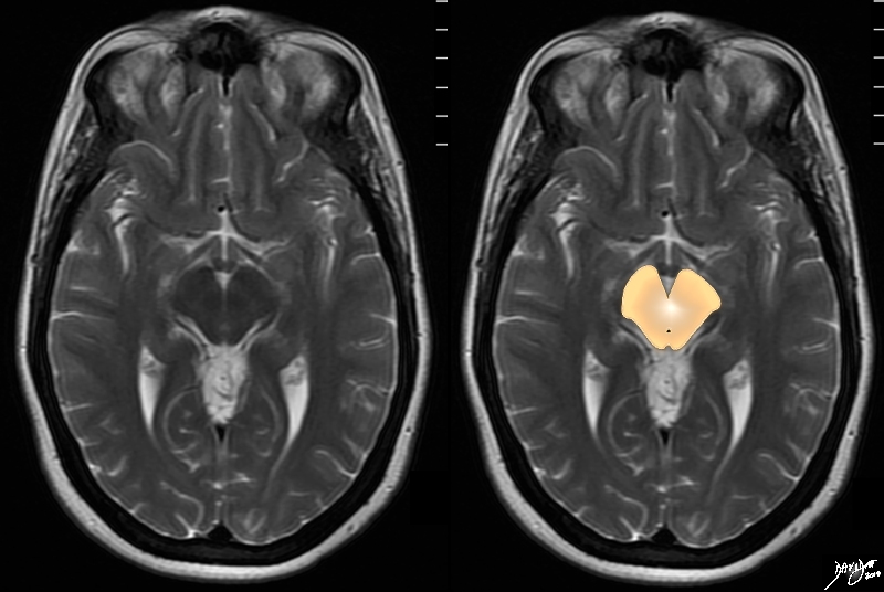
The T2 weighted MRI series focuses on the midbrain with characteristic shape and the small aqueduct of Sylvius posteriorly.  It is surrounded by portions of the frontal lobe anteriorly, temporal lobes laterally and perhaps small portions of the occipital lobes posteriorly.
Davidoff art Courtesy Ashley Davidoff MD copyright 2010 all rights reserved 94081.4kc01.8s


    [nodeType] => 1
    [parentNode] => (object value omitted)
    [childNodes] => (object value omitted)
    [firstChild] => (object value omitted)
    [lastChild] => (object value omitted)
    [previousSibling] => (object value omitted)
    [nextSibling] => (object value omitted)
    [attributes] => (object value omitted)
    [ownerDocument] => (object value omitted)
    [namespaceURI] =>
    [prefix] =>
    [localName] => table
    [baseURI] =>
    [textContent] =>


The Midbrain within the Context of the Whole Brain
T2 Weighted MRI


The T2 weighted MRI series focuses on the midbrain with characteristic shape and the small aqueduct of Sylvius posteriorly.  It is surrounded by portions of the frontal lobe anteriorly, temporal lobes laterally and perhaps small portions of the occipital lobes posteriorly.
Davidoff art Courtesy Ashley Davidoff MD copyright 2010 all rights reserved 94081.4kc01.8s


)
DOMElement Object
(
    [schemaTypeInfo] =>
    [tagName] => td
    [firstElementChild] => (object value omitted)
    [lastElementChild] => (object value omitted)
    [childElementCount] => 2
    [previousElementSibling] =>
    [nextElementSibling] =>
    [nodeName] => td
    [nodeValue] =>
The T2 weighted MRI series focuses on the midbrain with characteristic shape and the small aqueduct of Sylvius posteriorly.  It is surrounded by portions of the frontal lobe anteriorly, temporal lobes laterally and perhaps small portions of the occipital lobes posteriorly.
Davidoff art Courtesy Ashley Davidoff MD copyright 2010 all rights reserved 94081.4kc01.8s

    [nodeType] => 1
    [parentNode] => (object value omitted)
    [childNodes] => (object value omitted)
    [firstChild] => (object value omitted)
    [lastChild] => (object value omitted)
    [previousSibling] => (object value omitted)
    [nextSibling] => (object value omitted)
    [attributes] => (object value omitted)
    [ownerDocument] => (object value omitted)
    [namespaceURI] =>
    [prefix] =>
    [localName] => td
    [baseURI] =>
    [textContent] =>
The T2 weighted MRI series focuses on the midbrain with characteristic shape and the small aqueduct of Sylvius posteriorly.  It is surrounded by portions of the frontal lobe anteriorly, temporal lobes laterally and perhaps small portions of the occipital lobes posteriorly.
Davidoff art Courtesy Ashley Davidoff MD copyright 2010 all rights reserved 94081.4kc01.8s

)
DOMElement Object
(
    [schemaTypeInfo] =>
    [tagName] => td
    [firstElementChild] => (object value omitted)
    [lastElementChild] => (object value omitted)
    [childElementCount] => 3
    [previousElementSibling] =>
    [nextElementSibling] =>
    [nodeName] => td
    [nodeValue] =>
The Midbrain within the Context of the Whole Brain
T2 Weighted MRI

    [nodeType] => 1
    [parentNode] => (object value omitted)
    [childNodes] => (object value omitted)
    [firstChild] => (object value omitted)
    [lastChild] => (object value omitted)
    [previousSibling] => (object value omitted)
    [nextSibling] => (object value omitted)
    [attributes] => (object value omitted)
    [ownerDocument] => (object value omitted)
    [namespaceURI] =>
    [prefix] =>
    [localName] => td
    [baseURI] =>
    [textContent] =>
The Midbrain within the Context of the Whole Brain
T2 Weighted MRI

)
DOMElement Object
(
    [schemaTypeInfo] =>
    [tagName] => table
    [firstElementChild] => (object value omitted)
    [lastElementChild] => (object value omitted)
    [childElementCount] => 1
    [previousElementSibling] => (object value omitted)
    [nextElementSibling] => (object value omitted)
    [nodeName] => table
    [nodeValue] =>


94074b09b01a08.8s


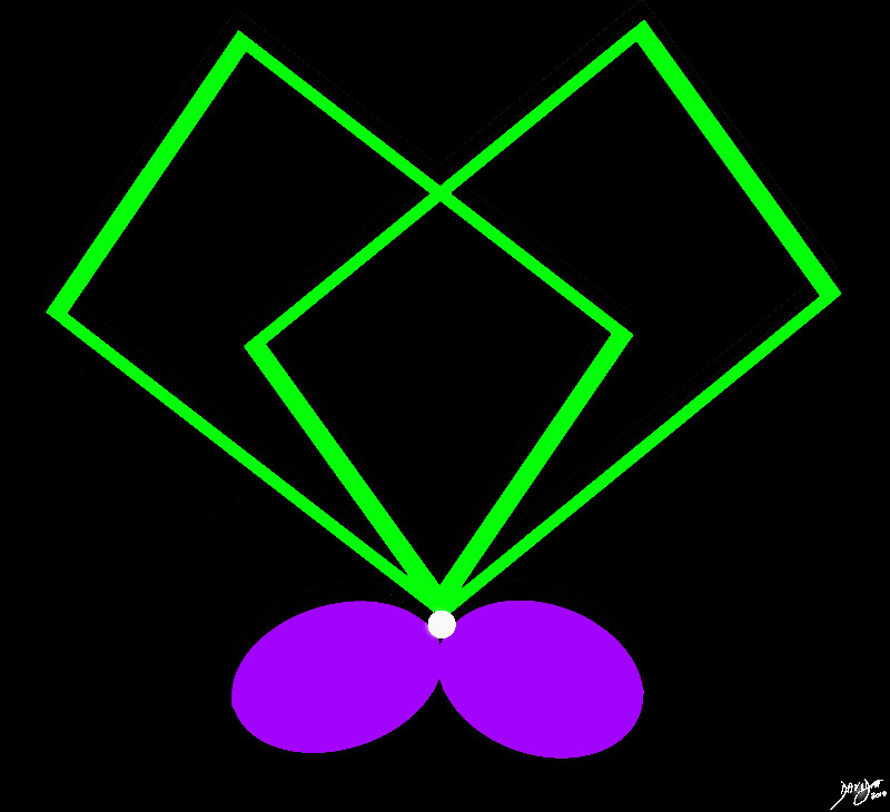
94074b09b01a08.8s The midbrain consists conceptually of two anteroleaterally positioned rectancles, and two posterocentrally placed ovals (purple), surrounding the small aqueduct of Sylvius (white).
Courtesy Ashley Davidoff MD copyright 2010 all rights reserved


    [nodeType] => 1
    [parentNode] => (object value omitted)
    [childNodes] => (object value omitted)
    [firstChild] => (object value omitted)
    [lastChild] => (object value omitted)
    [previousSibling] => (object value omitted)
    [nextSibling] => (object value omitted)
    [attributes] => (object value omitted)
    [ownerDocument] => (object value omitted)
    [namespaceURI] =>
    [prefix] =>
    [localName] => table
    [baseURI] =>
    [textContent] =>


94074b09b01a08.8s


94074b09b01a08.8s The midbrain consists conceptually of two anteroleaterally positioned rectancles, and two posterocentrally placed ovals (purple), surrounding the small aqueduct of Sylvius (white).
Courtesy Ashley Davidoff MD copyright 2010 all rights reserved


)
DOMElement Object
(
    [schemaTypeInfo] =>
    [tagName] => td
    [firstElementChild] => (object value omitted)
    [lastElementChild] => (object value omitted)
    [childElementCount] => 1
    [previousElementSibling] =>
    [nextElementSibling] =>
    [nodeName] => td
    [nodeValue] => 94074b09b01a08.8s The midbrain consists conceptually of two anteroleaterally positioned rectancles, and two posterocentrally placed ovals (purple), surrounding the small aqueduct of Sylvius (white).
Courtesy Ashley Davidoff MD copyright 2010 all rights reserved

    [nodeType] => 1
    [parentNode] => (object value omitted)
    [childNodes] => (object value omitted)
    [firstChild] => (object value omitted)
    [lastChild] => (object value omitted)
    [previousSibling] => (object value omitted)
    [nextSibling] => (object value omitted)
    [attributes] => (object value omitted)
    [ownerDocument] => (object value omitted)
    [namespaceURI] =>
    [prefix] =>
    [localName] => td
    [baseURI] =>
    [textContent] => 94074b09b01a08.8s The midbrain consists conceptually of two anteroleaterally positioned rectancles, and two posterocentrally placed ovals (purple), surrounding the small aqueduct of Sylvius (white).
Courtesy Ashley Davidoff MD copyright 2010 all rights reserved

)
DOMElement Object
(
    [schemaTypeInfo] =>
    [tagName] => td
    [firstElementChild] => (object value omitted)
    [lastElementChild] => (object value omitted)
    [childElementCount] => 2
    [previousElementSibling] =>
    [nextElementSibling] =>
    [nodeName] => td
    [nodeValue] =>
94074b09b01a08.8s

    [nodeType] => 1
    [parentNode] => (object value omitted)
    [childNodes] => (object value omitted)
    [firstChild] => (object value omitted)
    [lastChild] => (object value omitted)
    [previousSibling] => (object value omitted)
    [nextSibling] => (object value omitted)
    [attributes] => (object value omitted)
    [ownerDocument] => (object value omitted)
    [namespaceURI] =>
    [prefix] =>
    [localName] => td
    [baseURI] =>
    [textContent] =>
94074b09b01a08.8s

)
DOMElement Object
(
    [schemaTypeInfo] =>
    [tagName] => table
    [firstElementChild] => (object value omitted)
    [lastElementChild] => (object value omitted)
    [childElementCount] => 1
    [previousElementSibling] => (object value omitted)
    [nextElementSibling] => (object value omitted)
    [nodeName] => table
    [nodeValue] =>


Baseline Shape of the Midbrain
Sting Like a Butterfly


The midbrain is almost butterfly shaped with rounded posterior borders (colliculi) and rectangular shaped wings – the cerebral peduncles. It is very similar in shape to the pons and medulla on axial imaging but a a few characteristics distinguish it from these structures, most specific of which is the aqueduct of Sylvius that run right through its substance posteriorly shown as a black circle
Davidoff art Courtesy Ashley Davidoff MD copyright 2010 all rights reserved 94074b08a.82s


    [nodeType] => 1
    [parentNode] => (object value omitted)
    [childNodes] => (object value omitted)
    [firstChild] => (object value omitted)
    [lastChild] => (object value omitted)
    [previousSibling] => (object value omitted)
    [nextSibling] => (object value omitted)
    [attributes] => (object value omitted)
    [ownerDocument] => (object value omitted)
    [namespaceURI] =>
    [prefix] =>
    [localName] => table
    [baseURI] =>
    [textContent] =>


Baseline Shape of the Midbrain
Sting Like a Butterfly


The midbrain is almost butterfly shaped with rounded posterior borders (colliculi) and rectangular shaped wings – the cerebral peduncles. It is very similar in shape to the pons and medulla on axial imaging but a a few characteristics distinguish it from these structures, most specific of which is the aqueduct of Sylvius that run right through its substance posteriorly shown as a black circle
Davidoff art Courtesy Ashley Davidoff MD copyright 2010 all rights reserved 94074b08a.82s


)
DOMElement Object
(
    [schemaTypeInfo] =>
    [tagName] => td
    [firstElementChild] => (object value omitted)
    [lastElementChild] => (object value omitted)
    [childElementCount] => 2
    [previousElementSibling] =>
    [nextElementSibling] =>
    [nodeName] => td
    [nodeValue] =>
The midbrain is almost butterfly shaped with rounded posterior borders (colliculi) and rectangular shaped wings – the cerebral peduncles. It is very similar in shape to the pons and medulla on axial imaging but a a few characteristics distinguish it from these structures, most specific of which is the aqueduct of Sylvius that run right through its substance posteriorly shown as a black circle
Davidoff art Courtesy Ashley Davidoff MD copyright 2010 all rights reserved 94074b08a.82s

    [nodeType] => 1
    [parentNode] => (object value omitted)
    [childNodes] => (object value omitted)
    [firstChild] => (object value omitted)
    [lastChild] => (object value omitted)
    [previousSibling] => (object value omitted)
    [nextSibling] => (object value omitted)
    [attributes] => (object value omitted)
    [ownerDocument] => (object value omitted)
    [namespaceURI] =>
    [prefix] =>
    [localName] => td
    [baseURI] =>
    [textContent] =>
The midbrain is almost butterfly shaped with rounded posterior borders (colliculi) and rectangular shaped wings – the cerebral peduncles. It is very similar in shape to the pons and medulla on axial imaging but a a few characteristics distinguish it from these structures, most specific of which is the aqueduct of Sylvius that run right through its substance posteriorly shown as a black circle
Davidoff art Courtesy Ashley Davidoff MD copyright 2010 all rights reserved 94074b08a.82s

)
DOMElement Object
(
    [schemaTypeInfo] =>
    [tagName] => td
    [firstElementChild] => (object value omitted)
    [lastElementChild] => (object value omitted)
    [childElementCount] => 3
    [previousElementSibling] =>
    [nextElementSibling] =>
    [nodeName] => td
    [nodeValue] =>

Baseline Shape of the Midbrain
Sting Like a Butterfly
    [nodeType] => 1
    [parentNode] => (object value omitted)
    [childNodes] => (object value omitted)
    [firstChild] => (object value omitted)
    [lastChild] => (object value omitted)
    [previousSibling] => (object value omitted)
    [nextSibling] => (object value omitted)
    [attributes] => (object value omitted)
    [ownerDocument] => (object value omitted)
    [namespaceURI] =>
    [prefix] =>
    [localName] => td
    [baseURI] =>
    [textContent] =>

Baseline Shape of the Midbrain
Sting Like a Butterfly
)
DOMElement Object
(
    [schemaTypeInfo] =>
    [tagName] => table
    [firstElementChild] => (object value omitted)
    [lastElementChild] => (object value omitted)
    [childElementCount] => 1
    [previousElementSibling] => (object value omitted)
    [nextElementSibling] => (object value omitted)
    [nodeName] => table
    [nodeValue] =>


Two Rectangles
Two Ovals and a Tiny Sphere


The midbrain consists conceptually of twoanteroleaterally positioned rectancles, and two posterocentrally placed ovals (purple), surrounding the small aqueduct of Sylvius (white).
Courtesy Ashley Davidoff MD copyright 2010 all rights reserved  94074b09b01a0910.8s


    [nodeType] => 1
    [parentNode] => (object value omitted)
    [childNodes] => (object value omitted)
    [firstChild] => (object value omitted)
    [lastChild] => (object value omitted)
    [previousSibling] => (object value omitted)
    [nextSibling] => (object value omitted)
    [attributes] => (object value omitted)
    [ownerDocument] => (object value omitted)
    [namespaceURI] =>
    [prefix] =>
    [localName] => table
    [baseURI] =>
    [textContent] =>


Two Rectangles
Two Ovals and a Tiny Sphere


The midbrain consists conceptually of twoanteroleaterally positioned rectancles, and two posterocentrally placed ovals (purple), surrounding the small aqueduct of Sylvius (white).
Courtesy Ashley Davidoff MD copyright 2010 all rights reserved  94074b09b01a0910.8s


)
DOMElement Object
(
    [schemaTypeInfo] =>
    [tagName] => td
    [firstElementChild] => (object value omitted)
    [lastElementChild] => (object value omitted)
    [childElementCount] => 2
    [previousElementSibling] =>
    [nextElementSibling] =>
    [nodeName] => td
    [nodeValue] =>
The midbrain consists conceptually of twoanteroleaterally positioned rectancles, and two posterocentrally placed ovals (purple), surrounding the small aqueduct of Sylvius (white).
Courtesy Ashley Davidoff MD copyright 2010 all rights reserved  94074b09b01a0910.8s

    [nodeType] => 1
    [parentNode] => (object value omitted)
    [childNodes] => (object value omitted)
    [firstChild] => (object value omitted)
    [lastChild] => (object value omitted)
    [previousSibling] => (object value omitted)
    [nextSibling] => (object value omitted)
    [attributes] => (object value omitted)
    [ownerDocument] => (object value omitted)
    [namespaceURI] =>
    [prefix] =>
    [localName] => td
    [baseURI] =>
    [textContent] =>
The midbrain consists conceptually of twoanteroleaterally positioned rectancles, and two posterocentrally placed ovals (purple), surrounding the small aqueduct of Sylvius (white).
Courtesy Ashley Davidoff MD copyright 2010 all rights reserved  94074b09b01a0910.8s

)
DOMElement Object
(
    [schemaTypeInfo] =>
    [tagName] => td
    [firstElementChild] => (object value omitted)
    [lastElementChild] => (object value omitted)
    [childElementCount] => 3
    [previousElementSibling] =>
    [nextElementSibling] =>
    [nodeName] => td
    [nodeValue] =>
Two Rectangles
Two Ovals and a Tiny Sphere

    [nodeType] => 1
    [parentNode] => (object value omitted)
    [childNodes] => (object value omitted)
    [firstChild] => (object value omitted)
    [lastChild] => (object value omitted)
    [previousSibling] => (object value omitted)
    [nextSibling] => (object value omitted)
    [attributes] => (object value omitted)
    [ownerDocument] => (object value omitted)
    [namespaceURI] =>
    [prefix] =>
    [localName] => td
    [baseURI] =>
    [textContent] =>
Two Rectangles
Two Ovals and a Tiny Sphere

)

