Middle Frontal Gyrus
Sumit Karia MD
The Common Vein opyright 2010
Definition
Structure
The middle frontal gyrus is a convolution located in the frontal lobe between between the superior and inferior frontal sulci. It has a superior and an inferior segment. The superior segment, located in the superior lateral portion of the frontal lobe is limited inferiorly by the inferior frontal sulcus. The superior portion is contiguous with the orbital segment of the superior frontal gyrus.
The inferior segment, called the orbitary segment is very large and occupies ¾ of the inferior face of the frontal lobe being medially limited by the olfactory sulcus and laterally by a parallel sulcus called lateral orbitary sulcus.
Function
It participates, along with the primary motor cortex, in the control and initiation of voluntary movements. Likewise, it also aids the superior frontal gyrus in the higher cognitive functions related to personality and insight. In essence, it shares many of the functions of the superior frontal gyrus, since it is also part of the supplementary motor area. However, it also constitutes the prefrontal association cortex, which is involved in memory recognition, working memory, categorization, drawing and planning/ creation of strategies.
Disease
The middle frontal gyrus is supplied by the anterior-medial division of the anterior cerebral artery. A stroke causing a lesion in this area may result in tonic deviation of the eyes towards the side of the injury. In regards to its responsibility for higher functions, the stroke can cause apathy, personality changes, abulia, lack of ability to plan tasks.

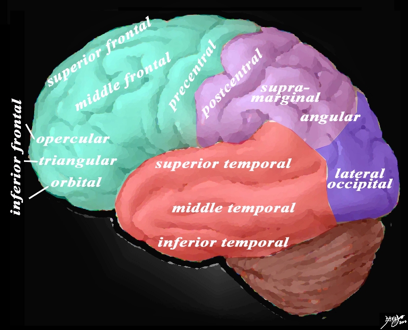
General View of the Gyri – Lateral View of the Brain |
|
The lateral view of the brain shows the frontal lobe (green) parietal lobe (light mauve), the occipital lobe (purple) and the temporal lobe (red) In this view the frontal lobe gyri that are visible are; superior frontal, middle frontal, inferior frontal and precentral gyri. The inferior frontal gyri include the opercular, triangular and orbital. The parietal gyri include the post central, supramarginal, and angular gyri. The occipital gyrus that is visible is the lateral occipital. The temporal lobe gyri include the superior, middle and inferior temporal gyri.
Courtesy Ashley Davidoff MD Copyright 2010 83029d05g.8
|
DOMElement Object
(
    [schemaTypeInfo] =>
    [tagName] => table
    [firstElementChild] => (object value omitted)
    [lastElementChild] => (object value omitted)
    [childElementCount] => 1
    [previousElementSibling] => (object value omitted)
    [nextElementSibling] =>
    [nodeName] => table
    [nodeValue] =>


General View of the Gyri – Lateral View of the Brain


The lateral view of the brain shows the frontal lobe (green) parietal lobe (light mauve), the occipital lobe (purple) and the temporal lobe (red) In this view the frontal lobe gyri that are visible are; superior frontal, middle frontal, inferior frontal and precentral gyri. The inferior frontal gyri include the opercular, triangular and orbital. The parietal gyri include the post central, supramarginal, and angular gyri. The occipital gyrus that is visible is the lateral occipital. The temporal lobe gyri include the superior, middle and inferior temporal gyri.
Courtesy Ashley Davidoff MD Copyright 2010 83029d05g.8


    [nodeType] => 1
    [parentNode] => (object value omitted)
    [childNodes] => (object value omitted)
    [firstChild] => (object value omitted)
    [lastChild] => (object value omitted)
    [previousSibling] => (object value omitted)
    [nextSibling] => (object value omitted)
    [attributes] => (object value omitted)
    [ownerDocument] => (object value omitted)
    [namespaceURI] =>
    [prefix] =>
    [localName] => table
    [baseURI] =>
    [textContent] =>


General View of the Gyri – Lateral View of the Brain


The lateral view of the brain shows the frontal lobe (green) parietal lobe (light mauve), the occipital lobe (purple) and the temporal lobe (red) In this view the frontal lobe gyri that are visible are; superior frontal, middle frontal, inferior frontal and precentral gyri. The inferior frontal gyri include the opercular, triangular and orbital. The parietal gyri include the post central, supramarginal, and angular gyri. The occipital gyrus that is visible is the lateral occipital. The temporal lobe gyri include the superior, middle and inferior temporal gyri.
Courtesy Ashley Davidoff MD Copyright 2010 83029d05g.8


)
DOMElement Object
(
    [schemaTypeInfo] =>
    [tagName] => td
    [firstElementChild] => (object value omitted)
    [lastElementChild] => (object value omitted)
    [childElementCount] => 2
    [previousElementSibling] =>
    [nextElementSibling] =>
    [nodeName] => td
    [nodeValue] =>
The lateral view of the brain shows the frontal lobe (green) parietal lobe (light mauve), the occipital lobe (purple) and the temporal lobe (red) In this view the frontal lobe gyri that are visible are; superior frontal, middle frontal, inferior frontal and precentral gyri. The inferior frontal gyri include the opercular, triangular and orbital. The parietal gyri include the post central, supramarginal, and angular gyri. The occipital gyrus that is visible is the lateral occipital. The temporal lobe gyri include the superior, middle and inferior temporal gyri.
Courtesy Ashley Davidoff MD Copyright 2010 83029d05g.8

    [nodeType] => 1
    [parentNode] => (object value omitted)
    [childNodes] => (object value omitted)
    [firstChild] => (object value omitted)
    [lastChild] => (object value omitted)
    [previousSibling] => (object value omitted)
    [nextSibling] => (object value omitted)
    [attributes] => (object value omitted)
    [ownerDocument] => (object value omitted)
    [namespaceURI] =>
    [prefix] =>
    [localName] => td
    [baseURI] =>
    [textContent] =>
The lateral view of the brain shows the frontal lobe (green) parietal lobe (light mauve), the occipital lobe (purple) and the temporal lobe (red) In this view the frontal lobe gyri that are visible are; superior frontal, middle frontal, inferior frontal and precentral gyri. The inferior frontal gyri include the opercular, triangular and orbital. The parietal gyri include the post central, supramarginal, and angular gyri. The occipital gyrus that is visible is the lateral occipital. The temporal lobe gyri include the superior, middle and inferior temporal gyri.
Courtesy Ashley Davidoff MD Copyright 2010 83029d05g.8

)
DOMElement Object
(
    [schemaTypeInfo] =>
    [tagName] => td
    [firstElementChild] => (object value omitted)
    [lastElementChild] => (object value omitted)
    [childElementCount] => 2
    [previousElementSibling] =>
    [nextElementSibling] =>
    [nodeName] => td
    [nodeValue] =>
General View of the Gyri – Lateral View of the Brain
    [nodeType] => 1
    [parentNode] => (object value omitted)
    [childNodes] => (object value omitted)
    [firstChild] => (object value omitted)
    [lastChild] => (object value omitted)
    [previousSibling] => (object value omitted)
    [nextSibling] => (object value omitted)
    [attributes] => (object value omitted)
    [ownerDocument] => (object value omitted)
    [namespaceURI] =>
    [prefix] =>
    [localName] => td
    [baseURI] =>
    [textContent] =>
General View of the Gyri – Lateral View of the Brain
)

