Veins
Ashley Davidoff MD
The Common Vein Copyright 2010
Introduction
|

Miodsagittal View – The Veins of the Frontal Parietal and Occipital Lobes |
|
The midsagittal section view of brain reveals the dark vascular structures that lie in the sulci of the frontal parietal and occipital lobes. The arteries are empty of blood best exemplified as two whiter structures anterior to the genu of the corpus callosum (cc)
The distinction between the character of the cerebral cortex which has a creamy color and the white matter exemplified by the corpus callosum (cc white), and the midbrain (mb), pons (p) and medulla (m) which are off white in color as opposed to the color of the cerebellum (c) which is light salmon pink in color.
The relative sizes of the forebrain, midbrain and hindbrain are also well appreciated in this section.
Image Courtesy of Thomas W.Smith, MD; Department of Pathology, University of Massachusetts Medical School. 97805b01
|
Forebrain
The veins that drain into the sinuses are divided into to two systems ; superficial and deep
Superficial System
The superficial system consists of two divisions; superior and inferior
Superior System
There are 10-15 superior cerebral veins that drains syuperior surface of hemispheres that includes the frontal and parietal lobes. These veins drain into the superior sagittal sinus. The veins then run in the subarachnoid space, and for a short distance in the subdural space before enetering superior sagittal sinus. When they are within the subdural space they are vulnerable to tearing.
Inferior System
The inferior cerebral veins drain drain the lower inferior surface of hemispheres including the temporal and occipital lobes. hey drain into the transverse sinus and the superior petrosal sinus.
The most consistent vessel of the inferior cerebral veins is the middle cerebral vein which runs along the Sylvian fissure and drains in to the cavernous sinus. It is through this vessel that the superior and inferior systems connect.
The superior anastomotic vein of Trolard connects the middle cerebral vein (and hence the inferior cerebral vein) with a superior cerebral vein.
The inferior anastomotic vein of Labbe connects the superficial middle vein with the transverse sinus
Deep System
The single vessel responsible fror draining the deep system ids the vein of Galen.
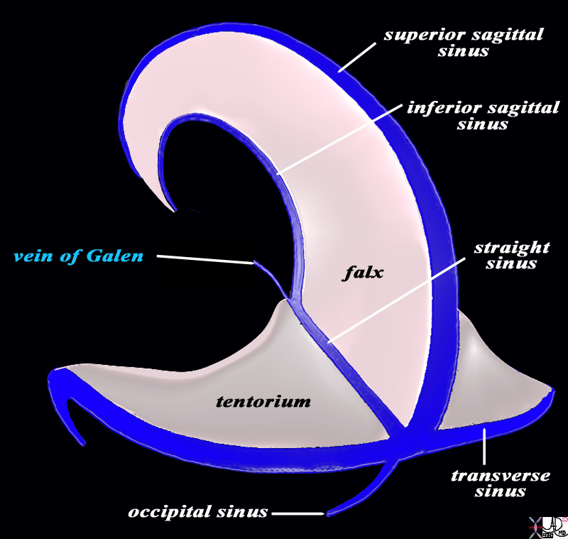
The Final Common Pathway into the Sinuses – Vein of Galen |
|
In this diagram the vein of Galen (blue text) is the common pathway of the deep veins of the brain, and it enters the straight sinus which lies on the superior edge of the tentorium. In addition, the diagram in the parasagittal plane, shows the basic plan of the dural sinuses and relationship to the tentorium and falx. The superior sagittal sinus runs on the superior aspect of the falx and abuts the vertex of the skull. It usually empties into the right transverse sinus. The inferior sagittal sinus runs on the inferior aspect of the falx and empties into the straight sinus which in turn tends to empty into the left transverse sinus The transverse sinuses in turn empty into the sigmoid sinuses which exit the skull to enter the internal jugular veins.
Courtesy Ashley Davidoff MD Copyright 2010 All rights reserved 98057b05.81Ls
|
The vein of Galen receives two major branches; internal cerebral vcein and the vein of Rosenthal.. The basal vein of Rosenthal receives its branches
The vein of Galen drains into the straight sinus.
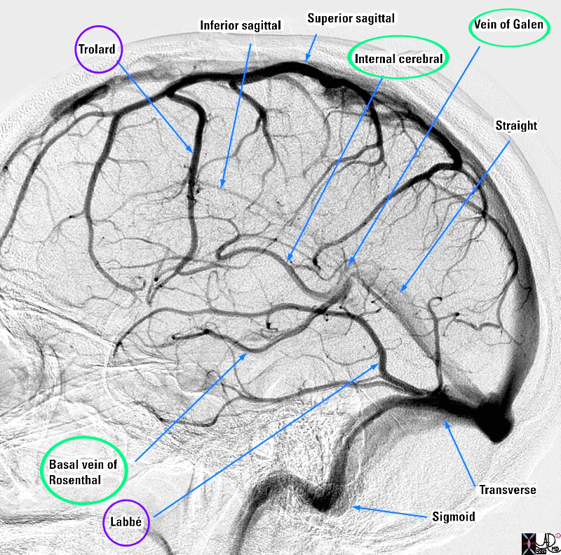
Veins of the Brain |
|
The diagram shows a venogram of the dural sinuses and highlighting the veins of the brain in lateral projection. The superficial veins (ringed in purple) consist of superior and inferior cerebral veins (not labeled). A branch of the inferior cerebral vein called the middle cerebral vein (not shown) provides collateral (vein of Trollard (purple ring)) that connects the inferior cerebral vein with the superior system by utilizing one of the superior cerebral veins. The vein of Labbe connects the middle cerebral vein with the transverse sinus . The common vein for the deep system (ringed in green) is the vein of Galen which empties into the straight sinus. It receives vessels from the internal cerebral vein and the vein of Rosenthal (both ringed in green) Image Courtesy Elisa Flower MD and Alex Norbash MD Copyright 2010 All rights reserved 97627b01.81s
|
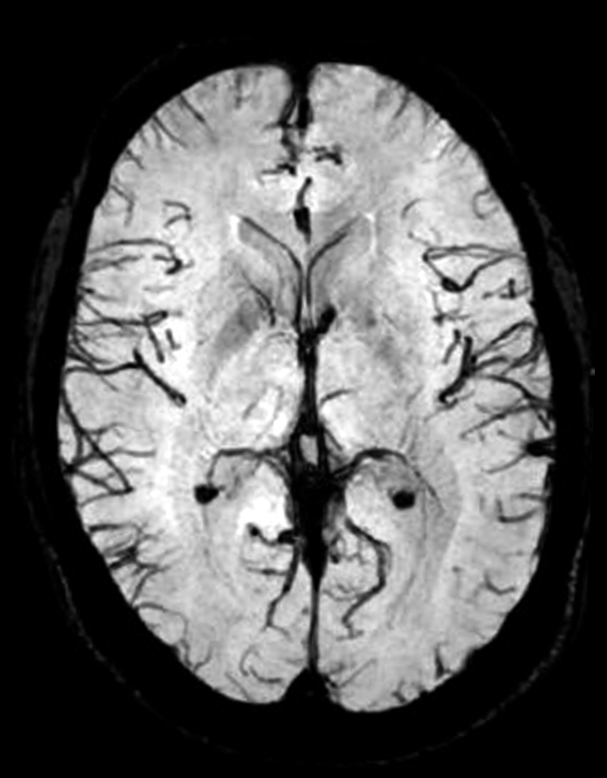
MRI of the Cortical Veins
|
|
This axial MRI using ?venous BOLD” sequence enables the visualization of the normal cortical veins.
Courtesy Philips Medical Systems 92394.8
|
Midbrain and Hindbrain veins
There are superior and inferior cerebellar venous systems and pontine and medullary venous systems.
Applied Biology
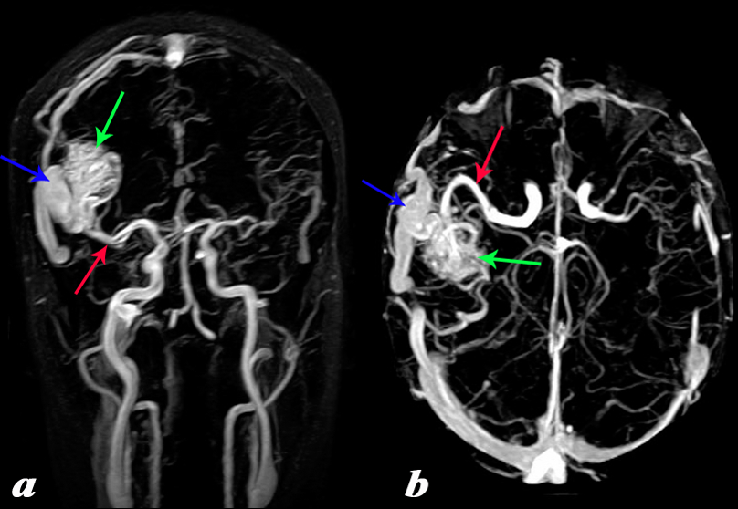
Temperoparietal AVM Large Feeding Artery and Large Draining Vein |
|
The MRA with 2d TOF in the coronal projection (a) and in the axial projection(b). It shows an arteriovenous malformation (AVM) in the right temperoparietal region fed by an enlarged middle cerebral artery (red arrow) and draining into a large draining vein (blue arrow) resulting in an enlarged transverse sinus. Note the difference in size between the right and left transverse sinuses.
Courtesy Philips Medical Systems 92465c01L.8
|
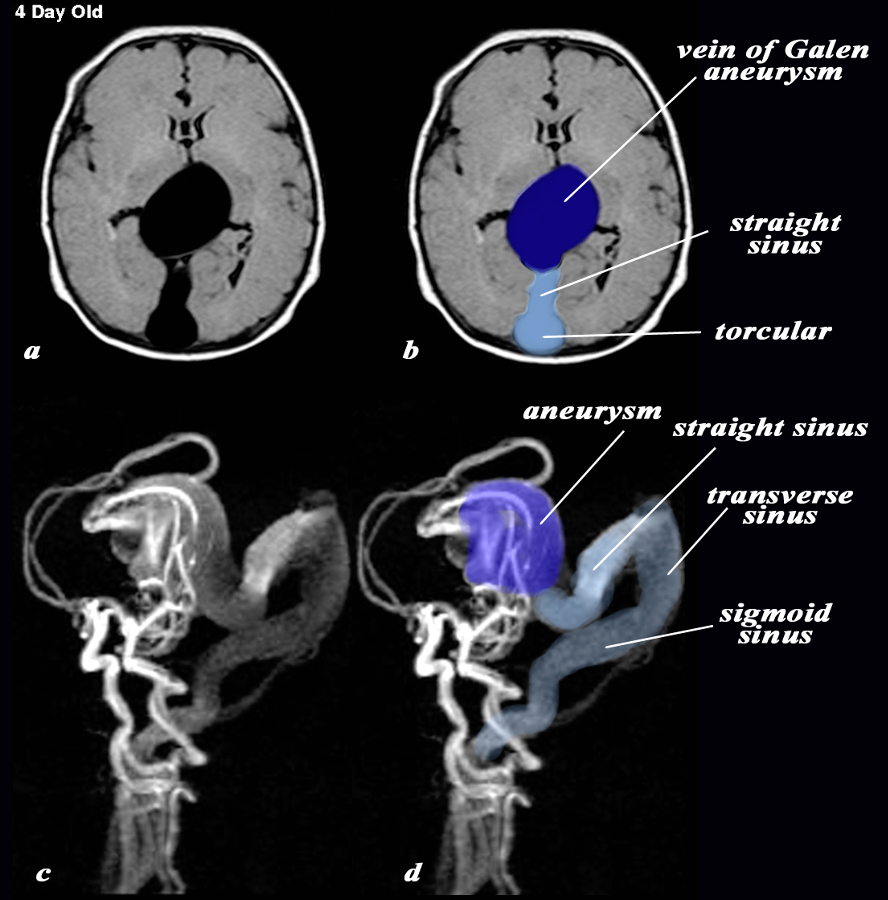
Vein of Galen Aneurysm – 4day Old with AVM |
|
The MRI with a T1 weighted axial sequence (a,b) and with 3d TOF (c,d) shows a large aneurysm of the vein of Galen (dark blue (b,d) an enlarged straight sinus (light blue b) and enlarged draining transverse sinus and sigmoid sinus (c,d) The feeding vessels of the arteriovenous malformation (AVM) which is the cause of the aneurysm, seem to arise predominantly from the vertebro-basilar system and likely from the posterior cerebral artery, but the middle cerebral artery also appears to contribute to the AVM.
Courtesy Philips Medical Systems 92432cL.9
|
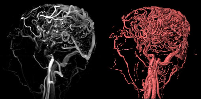
Extensive Cerebral Varicocities – AVM
Caput Medusa |
|
Exetnsive varicocities are seen in the parieto-occipital regionin the 2D TOF and volume rendered image. The draining transverse sinus and sigmoid sinus are also enlarged.
Courtesy Philips Medical Systems 92455c.8
|
DOMElement Object
(
    [schemaTypeInfo] =>
    [tagName] => table
    [firstElementChild] => (object value omitted)
    [lastElementChild] => (object value omitted)
    [childElementCount] => 1
    [previousElementSibling] => (object value omitted)
    [nextElementSibling] =>
    [nodeName] => table
    [nodeValue] =>


Extensive Cerebral Varicocities – AVM
Caput Medusa


Exetnsive varicocities are seen in the parieto-occipital regionin the 2D TOF and volume rendered image. The draining transverse sinus and sigmoid sinus are also enlarged.
Courtesy Philips Medical Systems 92455c.8


    [nodeType] => 1
    [parentNode] => (object value omitted)
    [childNodes] => (object value omitted)
    [firstChild] => (object value omitted)
    [lastChild] => (object value omitted)
    [previousSibling] => (object value omitted)
    [nextSibling] => (object value omitted)
    [attributes] => (object value omitted)
    [ownerDocument] => (object value omitted)
    [namespaceURI] =>
    [prefix] =>
    [localName] => table
    [baseURI] =>
    [textContent] =>


Extensive Cerebral Varicocities – AVM
Caput Medusa


Exetnsive varicocities are seen in the parieto-occipital regionin the 2D TOF and volume rendered image. The draining transverse sinus and sigmoid sinus are also enlarged.
Courtesy Philips Medical Systems 92455c.8


)
DOMElement Object
(
    [schemaTypeInfo] =>
    [tagName] => td
    [firstElementChild] => (object value omitted)
    [lastElementChild] => (object value omitted)
    [childElementCount] => 2
    [previousElementSibling] =>
    [nextElementSibling] =>
    [nodeName] => td
    [nodeValue] =>
Exetnsive varicocities are seen in the parieto-occipital regionin the 2D TOF and volume rendered image. The draining transverse sinus and sigmoid sinus are also enlarged.
Courtesy Philips Medical Systems 92455c.8

    [nodeType] => 1
    [parentNode] => (object value omitted)
    [childNodes] => (object value omitted)
    [firstChild] => (object value omitted)
    [lastChild] => (object value omitted)
    [previousSibling] => (object value omitted)
    [nextSibling] => (object value omitted)
    [attributes] => (object value omitted)
    [ownerDocument] => (object value omitted)
    [namespaceURI] =>
    [prefix] =>
    [localName] => td
    [baseURI] =>
    [textContent] =>
Exetnsive varicocities are seen in the parieto-occipital regionin the 2D TOF and volume rendered image. The draining transverse sinus and sigmoid sinus are also enlarged.
Courtesy Philips Medical Systems 92455c.8

)
DOMElement Object
(
    [schemaTypeInfo] =>
    [tagName] => td
    [firstElementChild] => (object value omitted)
    [lastElementChild] => (object value omitted)
    [childElementCount] => 3
    [previousElementSibling] =>
    [nextElementSibling] =>
    [nodeName] => td
    [nodeValue] =>
Extensive Cerebral Varicocities – AVM
Caput Medusa
    [nodeType] => 1
    [parentNode] => (object value omitted)
    [childNodes] => (object value omitted)
    [firstChild] => (object value omitted)
    [lastChild] => (object value omitted)
    [previousSibling] => (object value omitted)
    [nextSibling] => (object value omitted)
    [attributes] => (object value omitted)
    [ownerDocument] => (object value omitted)
    [namespaceURI] =>
    [prefix] =>
    [localName] => td
    [baseURI] =>
    [textContent] =>
Extensive Cerebral Varicocities – AVM
Caput Medusa
)
DOMElement Object
(
    [schemaTypeInfo] =>
    [tagName] => table
    [firstElementChild] => (object value omitted)
    [lastElementChild] => (object value omitted)
    [childElementCount] => 1
    [previousElementSibling] => (object value omitted)
    [nextElementSibling] => (object value omitted)
    [nodeName] => table
    [nodeValue] =>


Vein of Galen Aneurysm – 4day Old with AVM


The MRI with a T1 weighted axial sequence (a,b) and with 3d TOF (c,d) shows a large aneurysm of the vein of Galen (dark blue (b,d) an enlarged straight sinus (light blue b) and enlarged draining transverse sinus and sigmoid sinus (c,d) The feeding vessels of the arteriovenous malformation (AVM) which is the cause of the aneurysm, seem to arise predominantly from the vertebro-basilar system and likely from the posterior cerebral artery, but the middle cerebral artery also appears to contribute to the AVM.
Courtesy Philips Medical Systems 92432cL.9


    [nodeType] => 1
    [parentNode] => (object value omitted)
    [childNodes] => (object value omitted)
    [firstChild] => (object value omitted)
    [lastChild] => (object value omitted)
    [previousSibling] => (object value omitted)
    [nextSibling] => (object value omitted)
    [attributes] => (object value omitted)
    [ownerDocument] => (object value omitted)
    [namespaceURI] =>
    [prefix] =>
    [localName] => table
    [baseURI] =>
    [textContent] =>


Vein of Galen Aneurysm – 4day Old with AVM


The MRI with a T1 weighted axial sequence (a,b) and with 3d TOF (c,d) shows a large aneurysm of the vein of Galen (dark blue (b,d) an enlarged straight sinus (light blue b) and enlarged draining transverse sinus and sigmoid sinus (c,d) The feeding vessels of the arteriovenous malformation (AVM) which is the cause of the aneurysm, seem to arise predominantly from the vertebro-basilar system and likely from the posterior cerebral artery, but the middle cerebral artery also appears to contribute to the AVM.
Courtesy Philips Medical Systems 92432cL.9


)
DOMElement Object
(
    [schemaTypeInfo] =>
    [tagName] => td
    [firstElementChild] => (object value omitted)
    [lastElementChild] => (object value omitted)
    [childElementCount] => 2
    [previousElementSibling] =>
    [nextElementSibling] =>
    [nodeName] => td
    [nodeValue] =>
The MRI with a T1 weighted axial sequence (a,b) and with 3d TOF (c,d) shows a large aneurysm of the vein of Galen (dark blue (b,d) an enlarged straight sinus (light blue b) and enlarged draining transverse sinus and sigmoid sinus (c,d) The feeding vessels of the arteriovenous malformation (AVM) which is the cause of the aneurysm, seem to arise predominantly from the vertebro-basilar system and likely from the posterior cerebral artery, but the middle cerebral artery also appears to contribute to the AVM.
Courtesy Philips Medical Systems 92432cL.9

    [nodeType] => 1
    [parentNode] => (object value omitted)
    [childNodes] => (object value omitted)
    [firstChild] => (object value omitted)
    [lastChild] => (object value omitted)
    [previousSibling] => (object value omitted)
    [nextSibling] => (object value omitted)
    [attributes] => (object value omitted)
    [ownerDocument] => (object value omitted)
    [namespaceURI] =>
    [prefix] =>
    [localName] => td
    [baseURI] =>
    [textContent] =>
The MRI with a T1 weighted axial sequence (a,b) and with 3d TOF (c,d) shows a large aneurysm of the vein of Galen (dark blue (b,d) an enlarged straight sinus (light blue b) and enlarged draining transverse sinus and sigmoid sinus (c,d) The feeding vessels of the arteriovenous malformation (AVM) which is the cause of the aneurysm, seem to arise predominantly from the vertebro-basilar system and likely from the posterior cerebral artery, but the middle cerebral artery also appears to contribute to the AVM.
Courtesy Philips Medical Systems 92432cL.9

)
DOMElement Object
(
    [schemaTypeInfo] =>
    [tagName] => td
    [firstElementChild] => (object value omitted)
    [lastElementChild] => (object value omitted)
    [childElementCount] => 2
    [previousElementSibling] =>
    [nextElementSibling] =>
    [nodeName] => td
    [nodeValue] =>
Vein of Galen Aneurysm – 4day Old with AVM
    [nodeType] => 1
    [parentNode] => (object value omitted)
    [childNodes] => (object value omitted)
    [firstChild] => (object value omitted)
    [lastChild] => (object value omitted)
    [previousSibling] => (object value omitted)
    [nextSibling] => (object value omitted)
    [attributes] => (object value omitted)
    [ownerDocument] => (object value omitted)
    [namespaceURI] =>
    [prefix] =>
    [localName] => td
    [baseURI] =>
    [textContent] =>
Vein of Galen Aneurysm – 4day Old with AVM
)
DOMElement Object
(
    [schemaTypeInfo] =>
    [tagName] => table
    [firstElementChild] => (object value omitted)
    [lastElementChild] => (object value omitted)
    [childElementCount] => 1
    [previousElementSibling] => (object value omitted)
    [nextElementSibling] => (object value omitted)
    [nodeName] => table
    [nodeValue] =>


Temperoparietal AVM Large Feeding Artery and Large Draining Vein


The MRA with 2d TOF in the coronal projection (a) and in the axial projection(b). It shows an arteriovenous malformation (AVM) in the right temperoparietal region fed by an enlarged middle cerebral artery (red arrow) and draining into a large draining vein (blue arrow) resulting in an enlarged transverse sinus. Note the difference in size between the right and left transverse sinuses.
Courtesy Philips Medical Systems  92465c01L.8


    [nodeType] => 1
    [parentNode] => (object value omitted)
    [childNodes] => (object value omitted)
    [firstChild] => (object value omitted)
    [lastChild] => (object value omitted)
    [previousSibling] => (object value omitted)
    [nextSibling] => (object value omitted)
    [attributes] => (object value omitted)
    [ownerDocument] => (object value omitted)
    [namespaceURI] =>
    [prefix] =>
    [localName] => table
    [baseURI] =>
    [textContent] =>


Temperoparietal AVM Large Feeding Artery and Large Draining Vein


The MRA with 2d TOF in the coronal projection (a) and in the axial projection(b). It shows an arteriovenous malformation (AVM) in the right temperoparietal region fed by an enlarged middle cerebral artery (red arrow) and draining into a large draining vein (blue arrow) resulting in an enlarged transverse sinus. Note the difference in size between the right and left transverse sinuses.
Courtesy Philips Medical Systems  92465c01L.8


)
DOMElement Object
(
    [schemaTypeInfo] =>
    [tagName] => td
    [firstElementChild] => (object value omitted)
    [lastElementChild] => (object value omitted)
    [childElementCount] => 2
    [previousElementSibling] =>
    [nextElementSibling] =>
    [nodeName] => td
    [nodeValue] =>
The MRA with 2d TOF in the coronal projection (a) and in the axial projection(b). It shows an arteriovenous malformation (AVM) in the right temperoparietal region fed by an enlarged middle cerebral artery (red arrow) and draining into a large draining vein (blue arrow) resulting in an enlarged transverse sinus. Note the difference in size between the right and left transverse sinuses.
Courtesy Philips Medical Systems  92465c01L.8

    [nodeType] => 1
    [parentNode] => (object value omitted)
    [childNodes] => (object value omitted)
    [firstChild] => (object value omitted)
    [lastChild] => (object value omitted)
    [previousSibling] => (object value omitted)
    [nextSibling] => (object value omitted)
    [attributes] => (object value omitted)
    [ownerDocument] => (object value omitted)
    [namespaceURI] =>
    [prefix] =>
    [localName] => td
    [baseURI] =>
    [textContent] =>
The MRA with 2d TOF in the coronal projection (a) and in the axial projection(b). It shows an arteriovenous malformation (AVM) in the right temperoparietal region fed by an enlarged middle cerebral artery (red arrow) and draining into a large draining vein (blue arrow) resulting in an enlarged transverse sinus. Note the difference in size between the right and left transverse sinuses.
Courtesy Philips Medical Systems  92465c01L.8

)
DOMElement Object
(
    [schemaTypeInfo] =>
    [tagName] => td
    [firstElementChild] => (object value omitted)
    [lastElementChild] => (object value omitted)
    [childElementCount] => 2
    [previousElementSibling] =>
    [nextElementSibling] =>
    [nodeName] => td
    [nodeValue] =>
Temperoparietal AVM Large Feeding Artery and Large Draining Vein
    [nodeType] => 1
    [parentNode] => (object value omitted)
    [childNodes] => (object value omitted)
    [firstChild] => (object value omitted)
    [lastChild] => (object value omitted)
    [previousSibling] => (object value omitted)
    [nextSibling] => (object value omitted)
    [attributes] => (object value omitted)
    [ownerDocument] => (object value omitted)
    [namespaceURI] =>
    [prefix] =>
    [localName] => td
    [baseURI] =>
    [textContent] =>
Temperoparietal AVM Large Feeding Artery and Large Draining Vein
)
DOMElement Object
(
    [schemaTypeInfo] =>
    [tagName] => table
    [firstElementChild] => (object value omitted)
    [lastElementChild] => (object value omitted)
    [childElementCount] => 1
    [previousElementSibling] => (object value omitted)
    [nextElementSibling] => (object value omitted)
    [nodeName] => table
    [nodeValue] =>


MRI of the Cortical Veins


This axial MRI using ?venous BOLD” sequence enables the visualization of the normal cortical veins.
Courtesy Philips Medical Systems 92394.8


    [nodeType] => 1
    [parentNode] => (object value omitted)
    [childNodes] => (object value omitted)
    [firstChild] => (object value omitted)
    [lastChild] => (object value omitted)
    [previousSibling] => (object value omitted)
    [nextSibling] => (object value omitted)
    [attributes] => (object value omitted)
    [ownerDocument] => (object value omitted)
    [namespaceURI] =>
    [prefix] =>
    [localName] => table
    [baseURI] =>
    [textContent] =>


MRI of the Cortical Veins


This axial MRI using ?venous BOLD” sequence enables the visualization of the normal cortical veins.
Courtesy Philips Medical Systems 92394.8


)
DOMElement Object
(
    [schemaTypeInfo] =>
    [tagName] => td
    [firstElementChild] => (object value omitted)
    [lastElementChild] => (object value omitted)
    [childElementCount] => 2
    [previousElementSibling] =>
    [nextElementSibling] =>
    [nodeName] => td
    [nodeValue] =>
This axial MRI using ?venous BOLD” sequence enables the visualization of the normal cortical veins.
Courtesy Philips Medical Systems 92394.8

    [nodeType] => 1
    [parentNode] => (object value omitted)
    [childNodes] => (object value omitted)
    [firstChild] => (object value omitted)
    [lastChild] => (object value omitted)
    [previousSibling] => (object value omitted)
    [nextSibling] => (object value omitted)
    [attributes] => (object value omitted)
    [ownerDocument] => (object value omitted)
    [namespaceURI] =>
    [prefix] =>
    [localName] => td
    [baseURI] =>
    [textContent] =>
This axial MRI using ?venous BOLD” sequence enables the visualization of the normal cortical veins.
Courtesy Philips Medical Systems 92394.8

)
DOMElement Object
(
    [schemaTypeInfo] =>
    [tagName] => td
    [firstElementChild] => (object value omitted)
    [lastElementChild] => (object value omitted)
    [childElementCount] => 2
    [previousElementSibling] =>
    [nextElementSibling] =>
    [nodeName] => td
    [nodeValue] =>
MRI of the Cortical Veins

    [nodeType] => 1
    [parentNode] => (object value omitted)
    [childNodes] => (object value omitted)
    [firstChild] => (object value omitted)
    [lastChild] => (object value omitted)
    [previousSibling] => (object value omitted)
    [nextSibling] => (object value omitted)
    [attributes] => (object value omitted)
    [ownerDocument] => (object value omitted)
    [namespaceURI] =>
    [prefix] =>
    [localName] => td
    [baseURI] =>
    [textContent] =>
MRI of the Cortical Veins

)
DOMElement Object
(
    [schemaTypeInfo] =>
    [tagName] => table
    [firstElementChild] => (object value omitted)
    [lastElementChild] => (object value omitted)
    [childElementCount] => 1
    [previousElementSibling] => (object value omitted)
    [nextElementSibling] => (object value omitted)
    [nodeName] => table
    [nodeValue] =>


Veins of the Brain


The diagram shows a venogram of the dural sinuses and highlighting the veins of the brain in lateral projection. The superficial veins (ringed in purple) consist of superior and inferior cerebral veins (not labeled). A branch of the inferior cerebral vein called the middle cerebral vein (not shown) provides collateral (vein of Trollard (purple ring)) that connects the inferior cerebral vein with the superior system by utilizing one of the superior cerebral veins. The vein of Labbe connects the middle cerebral vein with the transverse sinus . The common vein for the deep system (ringed in green) is the vein of Galen which empties into the straight sinus. It receives vessels from the internal cerebral vein and the vein of Rosenthal (both ringed in green) Image Courtesy Elisa Flower MD and Alex Norbash MD Copyright 2010 All rights reserved 97627b01.81s


    [nodeType] => 1
    [parentNode] => (object value omitted)
    [childNodes] => (object value omitted)
    [firstChild] => (object value omitted)
    [lastChild] => (object value omitted)
    [previousSibling] => (object value omitted)
    [nextSibling] => (object value omitted)
    [attributes] => (object value omitted)
    [ownerDocument] => (object value omitted)
    [namespaceURI] =>
    [prefix] =>
    [localName] => table
    [baseURI] =>
    [textContent] =>


Veins of the Brain


The diagram shows a venogram of the dural sinuses and highlighting the veins of the brain in lateral projection. The superficial veins (ringed in purple) consist of superior and inferior cerebral veins (not labeled). A branch of the inferior cerebral vein called the middle cerebral vein (not shown) provides collateral (vein of Trollard (purple ring)) that connects the inferior cerebral vein with the superior system by utilizing one of the superior cerebral veins. The vein of Labbe connects the middle cerebral vein with the transverse sinus . The common vein for the deep system (ringed in green) is the vein of Galen which empties into the straight sinus. It receives vessels from the internal cerebral vein and the vein of Rosenthal (both ringed in green) Image Courtesy Elisa Flower MD and Alex Norbash MD Copyright 2010 All rights reserved 97627b01.81s


)
DOMElement Object
(
    [schemaTypeInfo] =>
    [tagName] => td
    [firstElementChild] => (object value omitted)
    [lastElementChild] => (object value omitted)
    [childElementCount] => 1
    [previousElementSibling] =>
    [nextElementSibling] =>
    [nodeName] => td
    [nodeValue] =>
The diagram shows a venogram of the dural sinuses and highlighting the veins of the brain in lateral projection. The superficial veins (ringed in purple) consist of superior and inferior cerebral veins (not labeled). A branch of the inferior cerebral vein called the middle cerebral vein (not shown) provides collateral (vein of Trollard (purple ring)) that connects the inferior cerebral vein with the superior system by utilizing one of the superior cerebral veins. The vein of Labbe connects the middle cerebral vein with the transverse sinus . The common vein for the deep system (ringed in green) is the vein of Galen which empties into the straight sinus. It receives vessels from the internal cerebral vein and the vein of Rosenthal (both ringed in green) Image Courtesy Elisa Flower MD and Alex Norbash MD Copyright 2010 All rights reserved 97627b01.81s

    [nodeType] => 1
    [parentNode] => (object value omitted)
    [childNodes] => (object value omitted)
    [firstChild] => (object value omitted)
    [lastChild] => (object value omitted)
    [previousSibling] => (object value omitted)
    [nextSibling] => (object value omitted)
    [attributes] => (object value omitted)
    [ownerDocument] => (object value omitted)
    [namespaceURI] =>
    [prefix] =>
    [localName] => td
    [baseURI] =>
    [textContent] =>
The diagram shows a venogram of the dural sinuses and highlighting the veins of the brain in lateral projection. The superficial veins (ringed in purple) consist of superior and inferior cerebral veins (not labeled). A branch of the inferior cerebral vein called the middle cerebral vein (not shown) provides collateral (vein of Trollard (purple ring)) that connects the inferior cerebral vein with the superior system by utilizing one of the superior cerebral veins. The vein of Labbe connects the middle cerebral vein with the transverse sinus . The common vein for the deep system (ringed in green) is the vein of Galen which empties into the straight sinus. It receives vessels from the internal cerebral vein and the vein of Rosenthal (both ringed in green) Image Courtesy Elisa Flower MD and Alex Norbash MD Copyright 2010 All rights reserved 97627b01.81s

)
DOMElement Object
(
    [schemaTypeInfo] =>
    [tagName] => td
    [firstElementChild] => (object value omitted)
    [lastElementChild] => (object value omitted)
    [childElementCount] => 2
    [previousElementSibling] =>
    [nextElementSibling] =>
    [nodeName] => td
    [nodeValue] =>
Veins of the Brain
    [nodeType] => 1
    [parentNode] => (object value omitted)
    [childNodes] => (object value omitted)
    [firstChild] => (object value omitted)
    [lastChild] => (object value omitted)
    [previousSibling] => (object value omitted)
    [nextSibling] => (object value omitted)
    [attributes] => (object value omitted)
    [ownerDocument] => (object value omitted)
    [namespaceURI] =>
    [prefix] =>
    [localName] => td
    [baseURI] =>
    [textContent] =>
Veins of the Brain
)
DOMElement Object
(
    [schemaTypeInfo] =>
    [tagName] => table
    [firstElementChild] => (object value omitted)
    [lastElementChild] => (object value omitted)
    [childElementCount] => 1
    [previousElementSibling] => (object value omitted)
    [nextElementSibling] => (object value omitted)
    [nodeName] => table
    [nodeValue] =>


The Final Common Pathway into the Sinuses – Vein of Galen


In this diagram the vein of Galen (blue text) is the common pathway of the deep veins of the brain, and it enters the straight sinus which lies on the superior edge of the tentorium. In addition, the diagram in the parasagittal plane, shows the basic plan of the dural sinuses and relationship to the tentorium and falx. The superior sagittal sinus runs on the superior aspect of the falx and abuts the vertex of the skull. It usually empties into the right transverse sinus. The inferior sagittal sinus runs on the inferior aspect of the falx and empties into the straight sinus which in turn tends to empty into the left transverse sinus The transverse sinuses in turn empty into the sigmoid sinuses which exit the skull to enter the internal jugular veins.
Courtesy Ashley Davidoff MD Copyright 2010 All rights reserved 98057b05.81Ls


    [nodeType] => 1
    [parentNode] => (object value omitted)
    [childNodes] => (object value omitted)
    [firstChild] => (object value omitted)
    [lastChild] => (object value omitted)
    [previousSibling] => (object value omitted)
    [nextSibling] => (object value omitted)
    [attributes] => (object value omitted)
    [ownerDocument] => (object value omitted)
    [namespaceURI] =>
    [prefix] =>
    [localName] => table
    [baseURI] =>
    [textContent] =>


The Final Common Pathway into the Sinuses – Vein of Galen


In this diagram the vein of Galen (blue text) is the common pathway of the deep veins of the brain, and it enters the straight sinus which lies on the superior edge of the tentorium. In addition, the diagram in the parasagittal plane, shows the basic plan of the dural sinuses and relationship to the tentorium and falx. The superior sagittal sinus runs on the superior aspect of the falx and abuts the vertex of the skull. It usually empties into the right transverse sinus. The inferior sagittal sinus runs on the inferior aspect of the falx and empties into the straight sinus which in turn tends to empty into the left transverse sinus The transverse sinuses in turn empty into the sigmoid sinuses which exit the skull to enter the internal jugular veins.
Courtesy Ashley Davidoff MD Copyright 2010 All rights reserved 98057b05.81Ls


)
DOMElement Object
(
    [schemaTypeInfo] =>
    [tagName] => td
    [firstElementChild] => (object value omitted)
    [lastElementChild] => (object value omitted)
    [childElementCount] => 2
    [previousElementSibling] =>
    [nextElementSibling] =>
    [nodeName] => td
    [nodeValue] =>
In this diagram the vein of Galen (blue text) is the common pathway of the deep veins of the brain, and it enters the straight sinus which lies on the superior edge of the tentorium. In addition, the diagram in the parasagittal plane, shows the basic plan of the dural sinuses and relationship to the tentorium and falx. The superior sagittal sinus runs on the superior aspect of the falx and abuts the vertex of the skull. It usually empties into the right transverse sinus. The inferior sagittal sinus runs on the inferior aspect of the falx and empties into the straight sinus which in turn tends to empty into the left transverse sinus The transverse sinuses in turn empty into the sigmoid sinuses which exit the skull to enter the internal jugular veins.
Courtesy Ashley Davidoff MD Copyright 2010 All rights reserved 98057b05.81Ls

    [nodeType] => 1
    [parentNode] => (object value omitted)
    [childNodes] => (object value omitted)
    [firstChild] => (object value omitted)
    [lastChild] => (object value omitted)
    [previousSibling] => (object value omitted)
    [nextSibling] => (object value omitted)
    [attributes] => (object value omitted)
    [ownerDocument] => (object value omitted)
    [namespaceURI] =>
    [prefix] =>
    [localName] => td
    [baseURI] =>
    [textContent] =>
In this diagram the vein of Galen (blue text) is the common pathway of the deep veins of the brain, and it enters the straight sinus which lies on the superior edge of the tentorium. In addition, the diagram in the parasagittal plane, shows the basic plan of the dural sinuses and relationship to the tentorium and falx. The superior sagittal sinus runs on the superior aspect of the falx and abuts the vertex of the skull. It usually empties into the right transverse sinus. The inferior sagittal sinus runs on the inferior aspect of the falx and empties into the straight sinus which in turn tends to empty into the left transverse sinus The transverse sinuses in turn empty into the sigmoid sinuses which exit the skull to enter the internal jugular veins.
Courtesy Ashley Davidoff MD Copyright 2010 All rights reserved 98057b05.81Ls

)
DOMElement Object
(
    [schemaTypeInfo] =>
    [tagName] => td
    [firstElementChild] => (object value omitted)
    [lastElementChild] => (object value omitted)
    [childElementCount] => 2
    [previousElementSibling] =>
    [nextElementSibling] =>
    [nodeName] => td
    [nodeValue] =>
The Final Common Pathway into the Sinuses – Vein of Galen
    [nodeType] => 1
    [parentNode] => (object value omitted)
    [childNodes] => (object value omitted)
    [firstChild] => (object value omitted)
    [lastChild] => (object value omitted)
    [previousSibling] => (object value omitted)
    [nextSibling] => (object value omitted)
    [attributes] => (object value omitted)
    [ownerDocument] => (object value omitted)
    [namespaceURI] =>
    [prefix] =>
    [localName] => td
    [baseURI] =>
    [textContent] =>
The Final Common Pathway into the Sinuses – Vein of Galen
)
DOMElement Object
(
    [schemaTypeInfo] =>
    [tagName] => table
    [firstElementChild] => (object value omitted)
    [lastElementChild] => (object value omitted)
    [childElementCount] => 1
    [previousElementSibling] => (object value omitted)
    [nextElementSibling] => (object value omitted)
    [nodeName] => table
    [nodeValue] =>


Miodsagittal View – The Veins of the Frontal Parietal and Occipital Lobes


The midsagittal section view of brain reveals the dark vascular structures that lie in the sulci of the frontal parietal and occipital lobes.  The arteries are empty of blood best exemplified as two whiter structures anterior to the genu of the corpus callosum (cc)
The distinction between the character of the cerebral cortex which has a creamy color and the white matter exemplified by the corpus callosum (cc white), and the midbrain (mb), pons (p) and medulla (m) which are off white in color  as opposed to the color of the cerebellum  (c) which is light salmon pink in color.
The relative sizes of the forebrain, midbrain and hindbrain are also well appreciated in this section.
Image Courtesy of Thomas W.Smith, MD; Department of Pathology, University of Massachusetts Medical School. 97805b01


    [nodeType] => 1
    [parentNode] => (object value omitted)
    [childNodes] => (object value omitted)
    [firstChild] => (object value omitted)
    [lastChild] => (object value omitted)
    [previousSibling] => (object value omitted)
    [nextSibling] => (object value omitted)
    [attributes] => (object value omitted)
    [ownerDocument] => (object value omitted)
    [namespaceURI] =>
    [prefix] =>
    [localName] => table
    [baseURI] =>
    [textContent] =>


Miodsagittal View – The Veins of the Frontal Parietal and Occipital Lobes


The midsagittal section view of brain reveals the dark vascular structures that lie in the sulci of the frontal parietal and occipital lobes.  The arteries are empty of blood best exemplified as two whiter structures anterior to the genu of the corpus callosum (cc)
The distinction between the character of the cerebral cortex which has a creamy color and the white matter exemplified by the corpus callosum (cc white), and the midbrain (mb), pons (p) and medulla (m) which are off white in color  as opposed to the color of the cerebellum  (c) which is light salmon pink in color.
The relative sizes of the forebrain, midbrain and hindbrain are also well appreciated in this section.
Image Courtesy of Thomas W.Smith, MD; Department of Pathology, University of Massachusetts Medical School. 97805b01


)
DOMElement Object
(
    [schemaTypeInfo] =>
    [tagName] => td
    [firstElementChild] => (object value omitted)
    [lastElementChild] => (object value omitted)
    [childElementCount] => 4
    [previousElementSibling] =>
    [nextElementSibling] =>
    [nodeName] => td
    [nodeValue] =>
The midsagittal section view of brain reveals the dark vascular structures that lie in the sulci of the frontal parietal and occipital lobes.  The arteries are empty of blood best exemplified as two whiter structures anterior to the genu of the corpus callosum (cc)
The distinction between the character of the cerebral cortex which has a creamy color and the white matter exemplified by the corpus callosum (cc white), and the midbrain (mb), pons (p) and medulla (m) which are off white in color  as opposed to the color of the cerebellum  (c) which is light salmon pink in color.
The relative sizes of the forebrain, midbrain and hindbrain are also well appreciated in this section.
Image Courtesy of Thomas W.Smith, MD; Department of Pathology, University of Massachusetts Medical School. 97805b01

    [nodeType] => 1
    [parentNode] => (object value omitted)
    [childNodes] => (object value omitted)
    [firstChild] => (object value omitted)
    [lastChild] => (object value omitted)
    [previousSibling] => (object value omitted)
    [nextSibling] => (object value omitted)
    [attributes] => (object value omitted)
    [ownerDocument] => (object value omitted)
    [namespaceURI] =>
    [prefix] =>
    [localName] => td
    [baseURI] =>
    [textContent] =>
The midsagittal section view of brain reveals the dark vascular structures that lie in the sulci of the frontal parietal and occipital lobes.  The arteries are empty of blood best exemplified as two whiter structures anterior to the genu of the corpus callosum (cc)
The distinction between the character of the cerebral cortex which has a creamy color and the white matter exemplified by the corpus callosum (cc white), and the midbrain (mb), pons (p) and medulla (m) which are off white in color  as opposed to the color of the cerebellum  (c) which is light salmon pink in color.
The relative sizes of the forebrain, midbrain and hindbrain are also well appreciated in this section.
Image Courtesy of Thomas W.Smith, MD; Department of Pathology, University of Massachusetts Medical School. 97805b01

)
DOMElement Object
(
    [schemaTypeInfo] =>
    [tagName] => td
    [firstElementChild] => (object value omitted)
    [lastElementChild] => (object value omitted)
    [childElementCount] => 2
    [previousElementSibling] =>
    [nextElementSibling] =>
    [nodeName] => td
    [nodeValue] =>

Miodsagittal View – The Veins of the Frontal Parietal and Occipital Lobes
    [nodeType] => 1
    [parentNode] => (object value omitted)
    [childNodes] => (object value omitted)
    [firstChild] => (object value omitted)
    [lastChild] => (object value omitted)
    [previousSibling] => (object value omitted)
    [nextSibling] => (object value omitted)
    [attributes] => (object value omitted)
    [ownerDocument] => (object value omitted)
    [namespaceURI] =>
    [prefix] =>
    [localName] => td
    [baseURI] =>
    [textContent] =>

Miodsagittal View – The Veins of the Frontal Parietal and Occipital Lobes
)


