Somatosensory Cortex
Ashley Davidoff MD
Copyright 2010
Somatosensory Cortex in the Parietal Lobe
The somatosensory cortex is part of the somatosensory system and is characterized by its parietal location in the brain and by its ability to perceive and localize the pain.
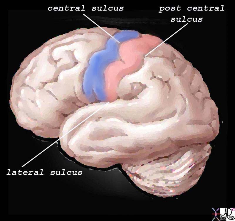
Structurally, the cortex lies as the anterior most structure of the parietal lobe, positioned between between the motor cortex of the frontal lobe and the central sulcus anteriorly, the post central sulcus posteriorly, and the lateral sulcus inferiorly.

The Somatosensory Cortex – Post Central Gyrus
|
| The somatosensory cortex is overlaid in light rose pink in the diagram above and represents the most anterior structure of the parietal lobe. It lies posterior to the motor cortex (blue) which is part of the frontal lobe, behind the central sulcus and in front of the post central sulcus. It serves to perceive, localize and evaluate intensity of the pain, as well as initiate the response to the pain.
83029b01.b1.81s pink = somatosensory cortex in post central gyrus blue = motor cortex The Common vein Davidoff art copyright 2008 |
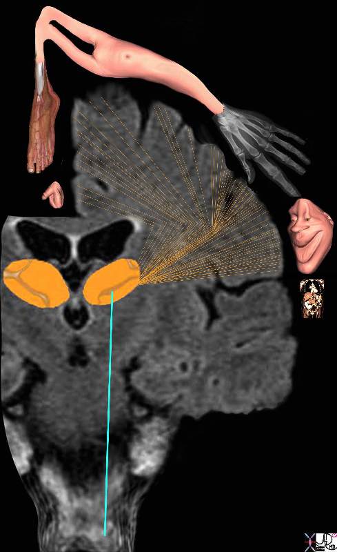
The regions of the body have a specific location in the somatosensory cortex and, depending on the number of nociceptors, will have a a correlatively sized distribution. Thus for example, the lips, mouth, hands, feet, and genitalia will have a much larger representation in the somatosensory cortex than the limbs, trunk, and viscera. Subsequently, the various structures have a descending order of reperesentation and consequently a descending order of sensitivity. The homunculus figure represents this concept and is diagrammed below.
If the somatosensory cortex is viewed in the coronal plane, the homunculus (literally “little man”) is draped over the sensory cortex with genitalia, and legs draped medially, thighs and trunk superiorly, and hands, head, mouth, lips, pharynx, tongue and viscera draped laterally. The size of organ representation is not only specific to pain fibres but related to the sensory and motor system as a whole.
 
Somatosensory Cortex in the Parietal Lobe
Localization and the Homunculus Man
|
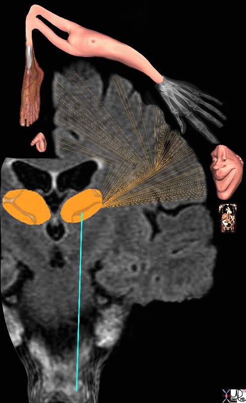
| The diagram reflects the relative functional sensory space each body part occupies in the somatosensory cortex. Those structures with high density of sensory receptors are represented by a larger size, while those with a lesser concentration of sensory apparatus shown as being “smaller” in size. Hence the mouth lips, hands feet and genitalia have a relatively large representation. The homunculus man (literally the “little man”) is the distorted figure drawn to reflect the concept of size of organ paralleling the size of the sensory innervation.
somatosensory cortex (sensory homunculus) spinothalamic tract spinal cord thalamus sensory cortex homunculus man penis clitoris genitals genitalia foot body thigh abdomen chest and face mouth eyes lips viscera somatosensory Davidoff art Copyright 2008 38610b09.46k.8s |
The function of the somatosensory cortex is that of a higher processing centre for touch, temperature, pain, and proprioception serving to amplify awareness of the sensations enabled by the thalamus. Sensation from the left side of the body are processed in the right somatosensory cortex and similarly those from the right side are processed on the left. The higher function of the somatosensory cortex allows us to localise the pain to a specific site, perceive the character and intensity of the stimulus, and sometimes helps identify the shape of the originating object.
The somatosensory cortex is not the final level of the somatosensory system since it also relays impulses to other cerebral areas of perception and reaction. Thus it sends signals via the white matter to other centres in the cortex to enable integration with visual and auditory input, and with other higher cortical functions such as emotion and memory for example. The full experience is then “seen” by the brain enabling the consequent reaction to be as discriminating and prudent as the nature and experience of the person allows. The difference between the reaction of an infant, child and an adult to the “shot at the doctors” speaks volumes about this latter function.
DOMElement Object
(
    [schemaTypeInfo] =>
    [tagName] => table
    [firstElementChild] => (object value omitted)
    [lastElementChild] => (object value omitted)
    [childElementCount] => 1
    [previousElementSibling] => (object value omitted)
    [nextElementSibling] => (object value omitted)
    [nodeName] => table
    [nodeValue] =>


Somatosensory Cortex in the Parietal Lobe
Localization and the Homunculus Man


The diagram reflects the relative functional sensory space each body part occupies in the somatosensory cortex.  Those structures with  high density of sensory receptors are represented by a larger size, while those with a lesser concentration of sensory apparatus shown as being “smaller” in size. Hence the mouth lips, hands feet and genitalia have a relatively large representation. The homunculus man (literally the “little man”) is the distorted figure drawn to reflect the concept of size of organ paralleling the size of the sensory innervation.
somatosensory cortex (sensory homunculus) spinothalamic tract spinal cord thalamus sensory cortex homunculus man penis clitoris genitals genitalia foot body thigh abdomen chest and face mouth eyes lips viscera somatosensory Davidoff art Copyright 2008 38610b09.46k.8s


    [nodeType] => 1
    [parentNode] => (object value omitted)
    [childNodes] => (object value omitted)
    [firstChild] => (object value omitted)
    [lastChild] => (object value omitted)
    [previousSibling] => (object value omitted)
    [nextSibling] => (object value omitted)
    [attributes] => (object value omitted)
    [ownerDocument] => (object value omitted)
    [namespaceURI] =>
    [prefix] =>
    [localName] => table
    [baseURI] =>
    [textContent] =>


Somatosensory Cortex in the Parietal Lobe
Localization and the Homunculus Man


The diagram reflects the relative functional sensory space each body part occupies in the somatosensory cortex.  Those structures with  high density of sensory receptors are represented by a larger size, while those with a lesser concentration of sensory apparatus shown as being “smaller” in size. Hence the mouth lips, hands feet and genitalia have a relatively large representation. The homunculus man (literally the “little man”) is the distorted figure drawn to reflect the concept of size of organ paralleling the size of the sensory innervation.
somatosensory cortex (sensory homunculus) spinothalamic tract spinal cord thalamus sensory cortex homunculus man penis clitoris genitals genitalia foot body thigh abdomen chest and face mouth eyes lips viscera somatosensory Davidoff art Copyright 2008 38610b09.46k.8s


)
DOMElement Object
(
    [schemaTypeInfo] =>
    [tagName] => td
    [firstElementChild] => (object value omitted)
    [lastElementChild] => (object value omitted)
    [childElementCount] => 2
    [previousElementSibling] =>
    [nextElementSibling] =>
    [nodeName] => td
    [nodeValue] => The diagram reflects the relative functional sensory space each body part occupies in the somatosensory cortex.  Those structures with  high density of sensory receptors are represented by a larger size, while those with a lesser concentration of sensory apparatus shown as being “smaller” in size. Hence the mouth lips, hands feet and genitalia have a relatively large representation. The homunculus man (literally the “little man”) is the distorted figure drawn to reflect the concept of size of organ paralleling the size of the sensory innervation.
somatosensory cortex (sensory homunculus) spinothalamic tract spinal cord thalamus sensory cortex homunculus man penis clitoris genitals genitalia foot body thigh abdomen chest and face mouth eyes lips viscera somatosensory Davidoff art Copyright 2008 38610b09.46k.8s
    [nodeType] => 1
    [parentNode] => (object value omitted)
    [childNodes] => (object value omitted)
    [firstChild] => (object value omitted)
    [lastChild] => (object value omitted)
    [previousSibling] => (object value omitted)
    [nextSibling] => (object value omitted)
    [attributes] => (object value omitted)
    [ownerDocument] => (object value omitted)
    [namespaceURI] =>
    [prefix] =>
    [localName] => td
    [baseURI] =>
    [textContent] => The diagram reflects the relative functional sensory space each body part occupies in the somatosensory cortex.  Those structures with  high density of sensory receptors are represented by a larger size, while those with a lesser concentration of sensory apparatus shown as being “smaller” in size. Hence the mouth lips, hands feet and genitalia have a relatively large representation. The homunculus man (literally the “little man”) is the distorted figure drawn to reflect the concept of size of organ paralleling the size of the sensory innervation.
somatosensory cortex (sensory homunculus) spinothalamic tract spinal cord thalamus sensory cortex homunculus man penis clitoris genitals genitalia foot body thigh abdomen chest and face mouth eyes lips viscera somatosensory Davidoff art Copyright 2008 38610b09.46k.8s
)
DOMElement Object
(
    [schemaTypeInfo] =>
    [tagName] => td
    [firstElementChild] => (object value omitted)
    [lastElementChild] => (object value omitted)
    [childElementCount] => 3
    [previousElementSibling] =>
    [nextElementSibling] =>
    [nodeName] => td
    [nodeValue] =>
Somatosensory Cortex in the Parietal Lobe
Localization and the Homunculus Man

    [nodeType] => 1
    [parentNode] => (object value omitted)
    [childNodes] => (object value omitted)
    [firstChild] => (object value omitted)
    [lastChild] => (object value omitted)
    [previousSibling] => (object value omitted)
    [nextSibling] => (object value omitted)
    [attributes] => (object value omitted)
    [ownerDocument] => (object value omitted)
    [namespaceURI] =>
    [prefix] =>
    [localName] => td
    [baseURI] =>
    [textContent] =>
Somatosensory Cortex in the Parietal Lobe
Localization and the Homunculus Man

)
https://beta.thecommonvein.net/wp-content/uploads/2023/09/38610b09.46k.8s.jpg
http://thecommonvein.net/media/38610b09.46k.8s_1.jpg
DOMElement Object
(
    [schemaTypeInfo] =>
    [tagName] => table
    [firstElementChild] => (object value omitted)
    [lastElementChild] => (object value omitted)
    [childElementCount] => 1
    [previousElementSibling] => (object value omitted)
    [nextElementSibling] => (object value omitted)
    [nodeName] => table
    [nodeValue] =>


The Somatosensory Cortex – Post Central Gyrus


The somatosensory cortex is overlaid in light rose pink in the diagram above and represents the most anterior structure of the parietal lobe.  It lies posterior to the motor cortex (blue) which is part of the frontal lobe, behind the central sulcus and in front of the post central sulcus.  It serves to perceive, localize and evaluate intensity of the pain, as well as initiate the response to the pain.
 83029b01.b1.81s pink = somatosensory cortex in post central gyrus blue = motor cortex The Common vein Davidoff art copyright 2008


    [nodeType] => 1
    [parentNode] => (object value omitted)
    [childNodes] => (object value omitted)
    [firstChild] => (object value omitted)
    [lastChild] => (object value omitted)
    [previousSibling] => (object value omitted)
    [nextSibling] => (object value omitted)
    [attributes] => (object value omitted)
    [ownerDocument] => (object value omitted)
    [namespaceURI] =>
    [prefix] =>
    [localName] => table
    [baseURI] =>
    [textContent] =>


The Somatosensory Cortex – Post Central Gyrus


The somatosensory cortex is overlaid in light rose pink in the diagram above and represents the most anterior structure of the parietal lobe.  It lies posterior to the motor cortex (blue) which is part of the frontal lobe, behind the central sulcus and in front of the post central sulcus.  It serves to perceive, localize and evaluate intensity of the pain, as well as initiate the response to the pain.
 83029b01.b1.81s pink = somatosensory cortex in post central gyrus blue = motor cortex The Common vein Davidoff art copyright 2008


)
DOMElement Object
(
    [schemaTypeInfo] =>
    [tagName] => td
    [firstElementChild] => (object value omitted)
    [lastElementChild] => (object value omitted)
    [childElementCount] => 2
    [previousElementSibling] =>
    [nextElementSibling] =>
    [nodeName] => td
    [nodeValue] => The somatosensory cortex is overlaid in light rose pink in the diagram above and represents the most anterior structure of the parietal lobe.  It lies posterior to the motor cortex (blue) which is part of the frontal lobe, behind the central sulcus and in front of the post central sulcus.  It serves to perceive, localize and evaluate intensity of the pain, as well as initiate the response to the pain.
 83029b01.b1.81s pink = somatosensory cortex in post central gyrus blue = motor cortex The Common vein Davidoff art copyright 2008
    [nodeType] => 1
    [parentNode] => (object value omitted)
    [childNodes] => (object value omitted)
    [firstChild] => (object value omitted)
    [lastChild] => (object value omitted)
    [previousSibling] => (object value omitted)
    [nextSibling] => (object value omitted)
    [attributes] => (object value omitted)
    [ownerDocument] => (object value omitted)
    [namespaceURI] =>
    [prefix] =>
    [localName] => td
    [baseURI] =>
    [textContent] => The somatosensory cortex is overlaid in light rose pink in the diagram above and represents the most anterior structure of the parietal lobe.  It lies posterior to the motor cortex (blue) which is part of the frontal lobe, behind the central sulcus and in front of the post central sulcus.  It serves to perceive, localize and evaluate intensity of the pain, as well as initiate the response to the pain.
 83029b01.b1.81s pink = somatosensory cortex in post central gyrus blue = motor cortex The Common vein Davidoff art copyright 2008
)
DOMElement Object
(
    [schemaTypeInfo] =>
    [tagName] => td
    [firstElementChild] => (object value omitted)
    [lastElementChild] => (object value omitted)
    [childElementCount] => 2
    [previousElementSibling] =>
    [nextElementSibling] =>
    [nodeName] => td
    [nodeValue] =>
The Somatosensory Cortex – Post Central Gyrus

    [nodeType] => 1
    [parentNode] => (object value omitted)
    [childNodes] => (object value omitted)
    [firstChild] => (object value omitted)
    [lastChild] => (object value omitted)
    [previousSibling] => (object value omitted)
    [nextSibling] => (object value omitted)
    [attributes] => (object value omitted)
    [ownerDocument] => (object value omitted)
    [namespaceURI] =>
    [prefix] =>
    [localName] => td
    [baseURI] =>
    [textContent] =>
The Somatosensory Cortex – Post Central Gyrus

)


