
Ashley Davidoff MD
The Common Vein Copyright 2010
Introduction
Shape of the Endometrium
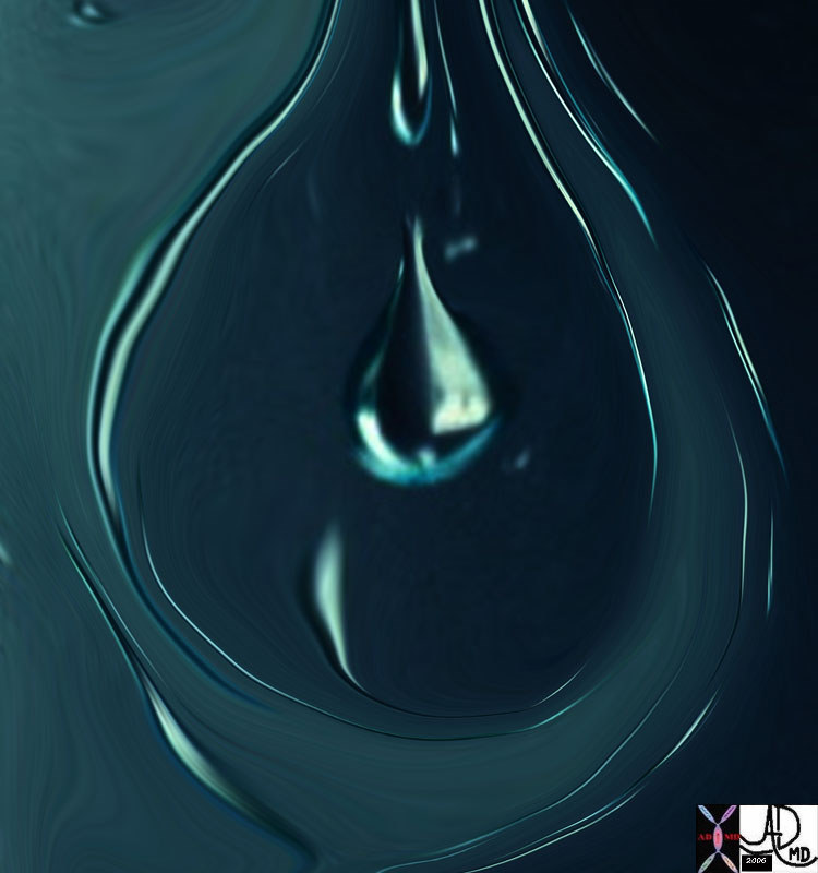
Tear Drop |
| 02504pb03 water droplets window glass teardrop shape Davidoff photography water |

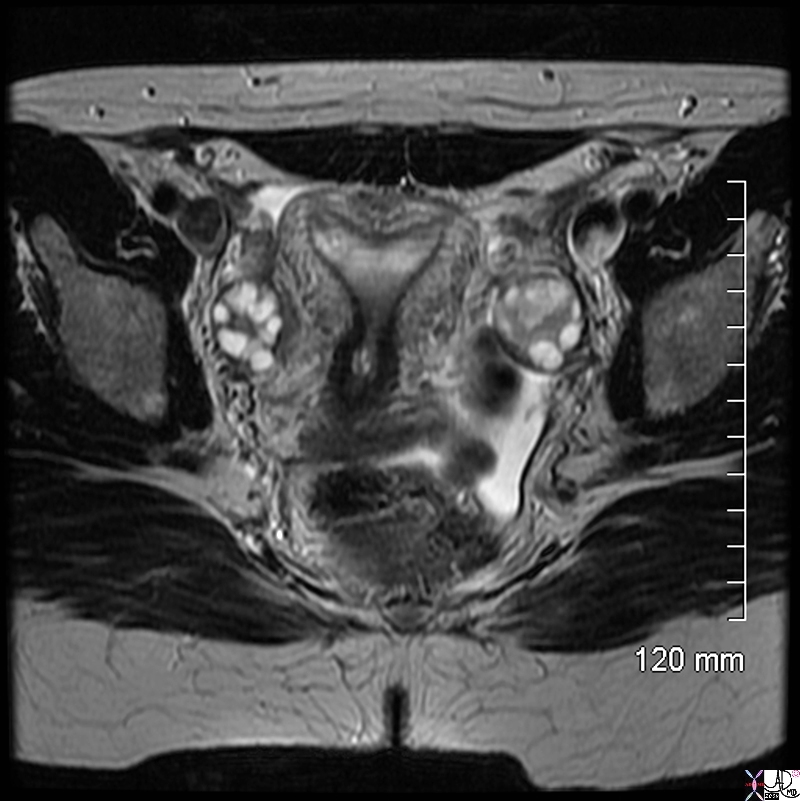
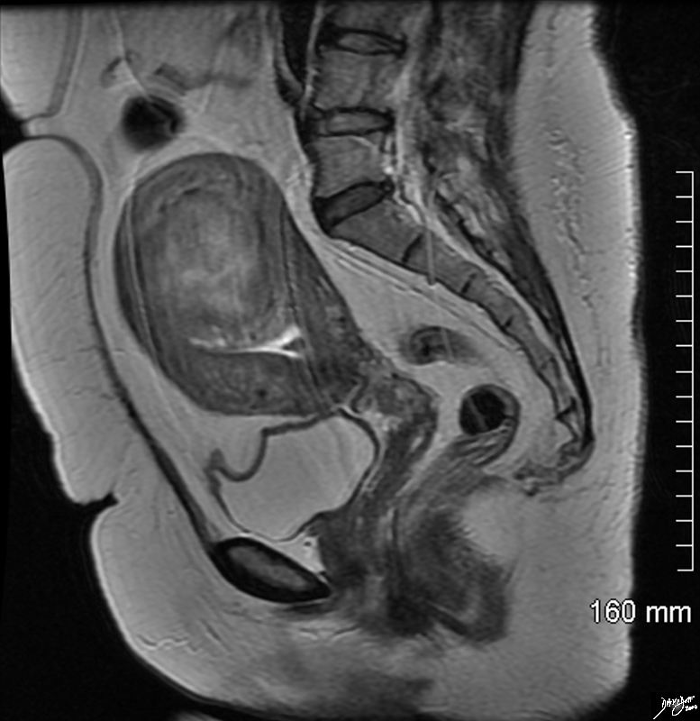
Normal Pear Shaped Uterys |
| hx young female presentingwith vaginal bleeding for 2 weeks, hematocrit of 25, left lower quadrant pain, and LLQ mass HCG 7000 fx uterus endometrial stripe ovary fx no intrauterine pregnancy
46749 Davidoff MD |
|
Premenstrual |
| 46317 uterus endometrium shape size normal anatomy USscan Davidoff MD |

Arcuate Uterus – T2 Weighted MRI |
|
The T2 weighted MRI in axial projection of a 31 year old female shows the endometrial cavity in coronal view with an arcuate shape. This is a normal variant and has no clinical significance. The follicles in the ovaries are also well demonstrated . CODE uterus endometrial cavity ovary ovaries follicles fx arcuate shape dx normal variant Image Courtesy Ashley Davidoff MD Copyright 2010 99420.8s |
Introduction
|
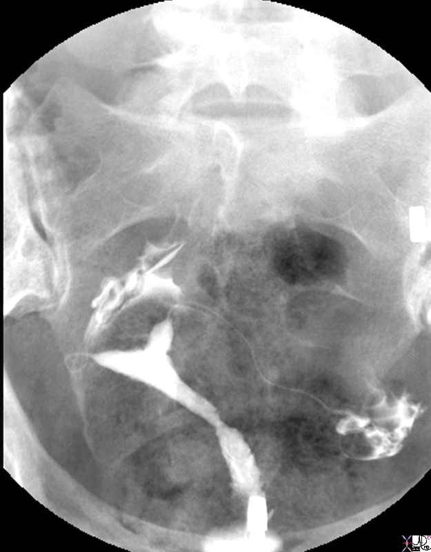
Position and Shape Relates to the Position of the Uterus |
| 40 year old female with normal endometrial cavity and cervical canal and fallopian tubes normal anatomy cervix uterus HSG hysterosalpingogram
Courtesy Ashley Davidoff MD Copyright 2009 all rights reserved 83700.8s |
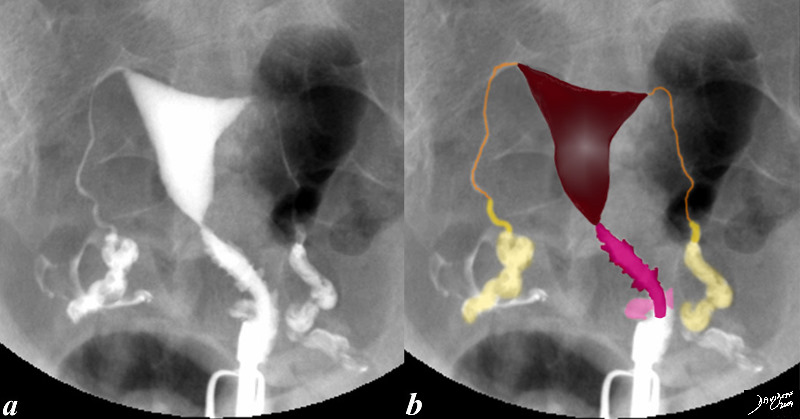
83690c02.8s |
|
This hysterosalpingogram is from a 26 year old female with normaL endometrial cavity and cervical canal. Note the irregular shape to the cervical canal while the endometrial lining is smooth. Note also the free spillage of contrast from the tubes into the peritoneal cavity (white contrast in d, indicating patent tubes. codefallopian tubes and fimbrae normal anatomy cervix uterus HSG hysterosalpingogram Courtesy Ashley Davidoff MD Copyright 2009 all rights reserved 83690c02.8s |
|
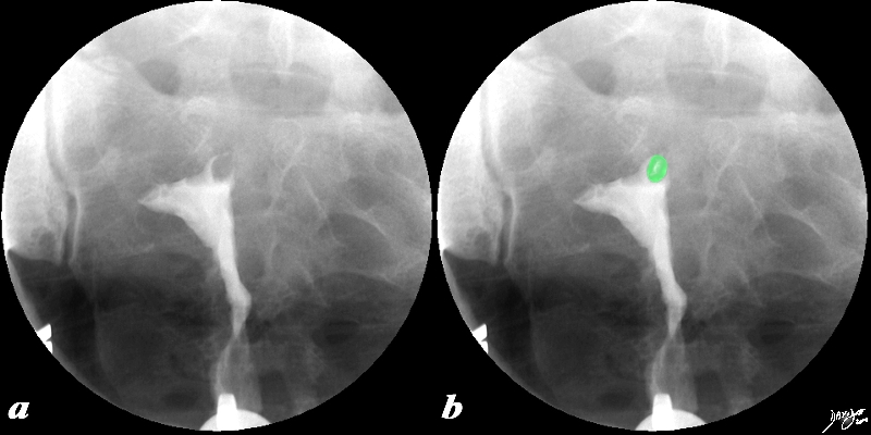
Filling Defect at the Left Cornu |
|
This 38 year old female presents with a history of infertility. The hysterosalpingogram shows non filling of both Fallopian tubes and a filling defect (green) in the region of the patient?s left cornu that likely is the cause of the obstructed left tube and in part the cause of the infertility. The lesion is most likely a submucosal fibroid. Courtesy Ashley Davidoff MD Copyright 2010 All rights reserved 96897c02.8s |
|
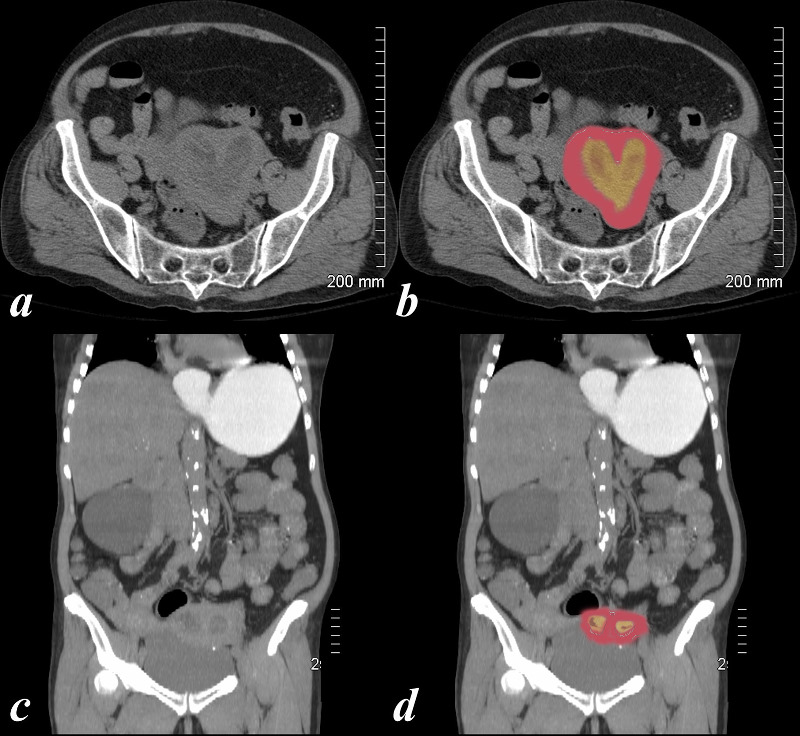
Heart Shaped Carcinoma of the Cervix in a Bicornuate Uterus Complicated by Obstruction |
| 74 year old female with bicornuate uterus and dilated endometrial cavities Diagnosis is carcinoma of the cervix with obstruction . The myometrium is overlaid in dark pink, and the endometrial cavity is a heterogeneous orange consisting of both fluid and soft tissue elements.
Courtesy Ashley Davidoff MD copyright 2009 all rights reserved 83438c01.8s |
|
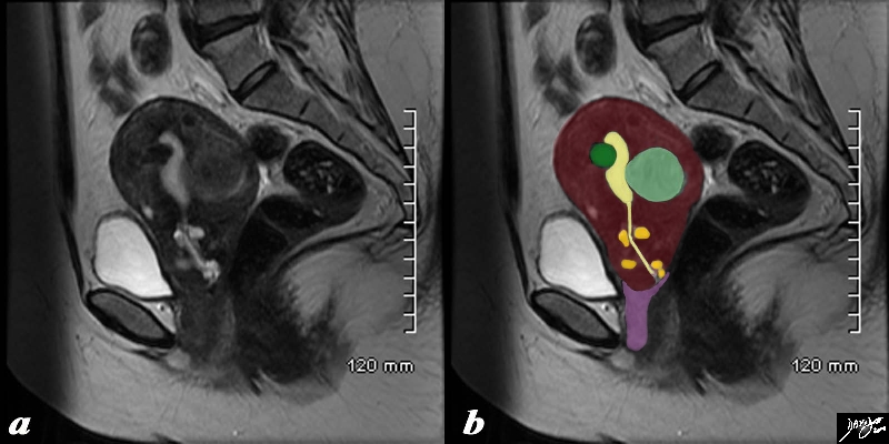
Fibroid Impingement on the Endometrium |
|
In this sagittal view of a T2 weighted MRI of the uterus of a 42 year old female with multiple fibroids two of which impinge on the endometrial cavity (the larger (light green) from posterior and the smaller (dark green) from anterior resulting in a sigmoid shaped cavity (yellow). In addition incidentally noted Nabothian cysts (orange) are seen within the cervix. Courtesy Ashley Davidoff MD Copyright 2010 All rights reserved 96531c02.8s |
|
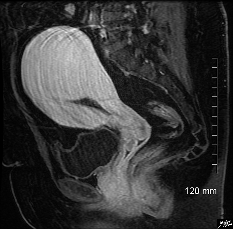
Deformity of the Endometrial Cavity Caused by an Intramural Fibroid |
|
In this sagittal view of the T1 weighted contrast enhanced study of the the uterus of a 46 year old female. The uterus roughly retains its pear shaped structure but the shape of the endometrial cavity is distinctly abnormal as if there is something pushing on it from above. This patient has a diffusely enhancing isointense leiomyoma that impinges on the endometrial cavity. Courtesy Ashley Davidoff MD Copyright 2010 All rights reserved 96577.8s |
|
Large Intramural Fibroid Deforming the Stripe T2 Weighted Image |
|
In this sagittal view of the T2 weighted MRI of the uterus of a 46 year old female an almost 6cms slightly hyperintense fibroid is seen pushing on the endometrial cavity from above. This patient has a large fibroid aka leiomyoma that impinges on the endometrial cavity. Courtesy Ashley Davidoff MD Copyright 2010 All rights reserved 96570.8s |
|
Filling Defect at the Left Cornu |
|
This 38 year old female presents with a history of infertility. The hysterosalpingogram shows non filling of both Fallopian tubes and a filling defect (green) in the region of the patient?s left cornu that likely is the cause of the obstructed left tube and in part the cause of the infertility. The lesion is most likely a submucosal fibroid. Courtesy Ashley Davidoff MD Copyright 2010 All rights reserved 96897c02.8s |
|
Arcuate Uterus – T2 Weighted MRI |
|
The T2 weighted MRI in axial projection of a 31 year old female shows the endometrial cavity in coronal view with an arcuate shape. This is a normal variant and has no clinical significance. The follicles in the ovaries are also well demonstrated . CODE uterus endometrial cavity ovary ovaries follicles fx arcuate shape dx normal variant Image Courtesy Ashley Davidoff MD Copyright 2010 99420.8s |
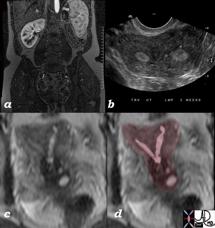
Bicornuate Uterus T2 Weighted Image |
|
The series of images from a young adult female with Bicornuate uterus with agenesis of the left kidney. Image a is a coronal T1 weighted contrast enhanced image that shows absence of the left kidney. Image b is an ultrasound in transverse dimension that shows two endometrial cavities. Image c is a T2 weighted image that shows 2 separate horns that fuse above a single cervix and overlaid in pink in image d These findings are consistent with bicornuate uterus with agenesis of the left kidney. Combined congenital anomalies of the uterus and renal system are a well known association. 46527c01 Image Courtesy Ashley DAvidoff MD Copyright |
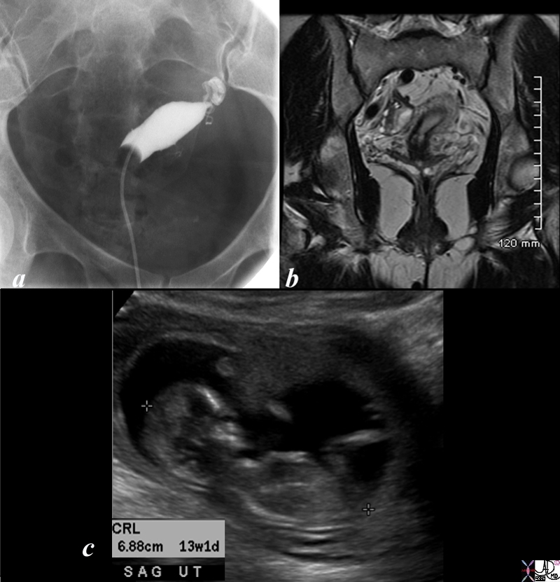
Unicornuate Uterus with Subsequent Pregnancy |
|
34 year old female who presented with infertility had a hysterosalpingogram which showed a unicornuate uterus (a). An MRI confirms the diagnosis using a T2 weighted sequence(b). The patient subsequently fell pregnant and was scanned with a normal 13 week pregnancy(c). Courtesy Ashley Davidoff MD copyright 2010 all rights reserved 85711cL.8s |
DOMElement Object
(
    [schemaTypeInfo] =>
    [tagName] => table
    [firstElementChild] => (object value omitted)
    [lastElementChild] => (object value omitted)
    [childElementCount] => 1
    [previousElementSibling] => (object value omitted)
    [nextElementSibling] =>
    [nodeName] => table
    [nodeValue] =>


Unicornuate Uterus with Subsequent Pregnancy


34 year old female who presented with infertility had a hysterosalpingogram which showed a unicornuate uterus (a). An MRI confirms the diagnosis using a T2 weighted sequence(b). The patient subsequently fell pregnant and was scanned with a normal 13 week pregnancy(c).
Courtesy Ashley Davidoff MD copyright 2010 all rights reserved 85711cL.8s


    [nodeType] => 1
    [parentNode] => (object value omitted)
    [childNodes] => (object value omitted)
    [firstChild] => (object value omitted)
    [lastChild] => (object value omitted)
    [previousSibling] => (object value omitted)
    [nextSibling] => (object value omitted)
    [attributes] => (object value omitted)
    [ownerDocument] => (object value omitted)
    [namespaceURI] =>
    [prefix] =>
    [localName] => table
    [baseURI] =>
    [textContent] =>


Unicornuate Uterus with Subsequent Pregnancy


34 year old female who presented with infertility had a hysterosalpingogram which showed a unicornuate uterus (a). An MRI confirms the diagnosis using a T2 weighted sequence(b). The patient subsequently fell pregnant and was scanned with a normal 13 week pregnancy(c).
Courtesy Ashley Davidoff MD copyright 2010 all rights reserved 85711cL.8s


)
DOMElement Object
(
    [schemaTypeInfo] =>
    [tagName] => td
    [firstElementChild] => (object value omitted)
    [lastElementChild] => (object value omitted)
    [childElementCount] => 2
    [previousElementSibling] =>
    [nextElementSibling] =>
    [nodeName] => td
    [nodeValue] =>
34 year old female who presented with infertility had a hysterosalpingogram which showed a unicornuate uterus (a). An MRI confirms the diagnosis using a T2 weighted sequence(b). The patient subsequently fell pregnant and was scanned with a normal 13 week pregnancy(c).
Courtesy Ashley Davidoff MD copyright 2010 all rights reserved 85711cL.8s

    [nodeType] => 1
    [parentNode] => (object value omitted)
    [childNodes] => (object value omitted)
    [firstChild] => (object value omitted)
    [lastChild] => (object value omitted)
    [previousSibling] => (object value omitted)
    [nextSibling] => (object value omitted)
    [attributes] => (object value omitted)
    [ownerDocument] => (object value omitted)
    [namespaceURI] =>
    [prefix] =>
    [localName] => td
    [baseURI] =>
    [textContent] =>
34 year old female who presented with infertility had a hysterosalpingogram which showed a unicornuate uterus (a). An MRI confirms the diagnosis using a T2 weighted sequence(b). The patient subsequently fell pregnant and was scanned with a normal 13 week pregnancy(c).
Courtesy Ashley Davidoff MD copyright 2010 all rights reserved 85711cL.8s

)
DOMElement Object
(
    [schemaTypeInfo] =>
    [tagName] => td
    [firstElementChild] => (object value omitted)
    [lastElementChild] => (object value omitted)
    [childElementCount] => 2
    [previousElementSibling] =>
    [nextElementSibling] =>
    [nodeName] => td
    [nodeValue] =>
Unicornuate Uterus with Subsequent Pregnancy
    [nodeType] => 1
    [parentNode] => (object value omitted)
    [childNodes] => (object value omitted)
    [firstChild] => (object value omitted)
    [lastChild] => (object value omitted)
    [previousSibling] => (object value omitted)
    [nextSibling] => (object value omitted)
    [attributes] => (object value omitted)
    [ownerDocument] => (object value omitted)
    [namespaceURI] =>
    [prefix] =>
    [localName] => td
    [baseURI] =>
    [textContent] =>
Unicornuate Uterus with Subsequent Pregnancy
)
DOMElement Object
(
    [schemaTypeInfo] =>
    [tagName] => table
    [firstElementChild] => (object value omitted)
    [lastElementChild] => (object value omitted)
    [childElementCount] => 1
    [previousElementSibling] => (object value omitted)
    [nextElementSibling] => (object value omitted)
    [nodeName] => table
    [nodeValue] =>


Bicornuate Uterus T2 Weighted Image


The series of images from a young adult female with Bicornuate uterus with agenesis of the left kidney.
Image a is a coronal T1 weighted contrast enhanced image that shows absence of the left kidney.
Image b is an ultrasound in transverse dimension that shows two endometrial cavities.
Image c is a T2 weighted image that shows 2 separate horns that fuse above a single cervix and overlaid in pink in image d
These findings are consistent with bicornuate uterus with agenesis of the left kidney.  Combined congenital anomalies of the uterus and renal system are a well known association.
46527c01 Image Courtesy Ashley DAvidoff MD Copyright


    [nodeType] => 1
    [parentNode] => (object value omitted)
    [childNodes] => (object value omitted)
    [firstChild] => (object value omitted)
    [lastChild] => (object value omitted)
    [previousSibling] => (object value omitted)
    [nextSibling] => (object value omitted)
    [attributes] => (object value omitted)
    [ownerDocument] => (object value omitted)
    [namespaceURI] =>
    [prefix] =>
    [localName] => table
    [baseURI] =>
    [textContent] =>


Bicornuate Uterus T2 Weighted Image


The series of images from a young adult female with Bicornuate uterus with agenesis of the left kidney.
Image a is a coronal T1 weighted contrast enhanced image that shows absence of the left kidney.
Image b is an ultrasound in transverse dimension that shows two endometrial cavities.
Image c is a T2 weighted image that shows 2 separate horns that fuse above a single cervix and overlaid in pink in image d
These findings are consistent with bicornuate uterus with agenesis of the left kidney.  Combined congenital anomalies of the uterus and renal system are a well known association.
46527c01 Image Courtesy Ashley DAvidoff MD Copyright


)
DOMElement Object
(
    [schemaTypeInfo] =>
    [tagName] => td
    [firstElementChild] => (object value omitted)
    [lastElementChild] => (object value omitted)
    [childElementCount] => 6
    [previousElementSibling] =>
    [nextElementSibling] =>
    [nodeName] => td
    [nodeValue] =>
The series of images from a young adult female with Bicornuate uterus with agenesis of the left kidney.
Image a is a coronal T1 weighted contrast enhanced image that shows absence of the left kidney.
Image b is an ultrasound in transverse dimension that shows two endometrial cavities.
Image c is a T2 weighted image that shows 2 separate horns that fuse above a single cervix and overlaid in pink in image d
These findings are consistent with bicornuate uterus with agenesis of the left kidney.  Combined congenital anomalies of the uterus and renal system are a well known association.
46527c01 Image Courtesy Ashley DAvidoff MD Copyright

    [nodeType] => 1
    [parentNode] => (object value omitted)
    [childNodes] => (object value omitted)
    [firstChild] => (object value omitted)
    [lastChild] => (object value omitted)
    [previousSibling] => (object value omitted)
    [nextSibling] => (object value omitted)
    [attributes] => (object value omitted)
    [ownerDocument] => (object value omitted)
    [namespaceURI] =>
    [prefix] =>
    [localName] => td
    [baseURI] =>
    [textContent] =>
The series of images from a young adult female with Bicornuate uterus with agenesis of the left kidney.
Image a is a coronal T1 weighted contrast enhanced image that shows absence of the left kidney.
Image b is an ultrasound in transverse dimension that shows two endometrial cavities.
Image c is a T2 weighted image that shows 2 separate horns that fuse above a single cervix and overlaid in pink in image d
These findings are consistent with bicornuate uterus with agenesis of the left kidney.  Combined congenital anomalies of the uterus and renal system are a well known association.
46527c01 Image Courtesy Ashley DAvidoff MD Copyright

)
DOMElement Object
(
    [schemaTypeInfo] =>
    [tagName] => td
    [firstElementChild] => (object value omitted)
    [lastElementChild] => (object value omitted)
    [childElementCount] => 2
    [previousElementSibling] =>
    [nextElementSibling] =>
    [nodeName] => td
    [nodeValue] =>
Bicornuate Uterus T2 Weighted Image
    [nodeType] => 1
    [parentNode] => (object value omitted)
    [childNodes] => (object value omitted)
    [firstChild] => (object value omitted)
    [lastChild] => (object value omitted)
    [previousSibling] => (object value omitted)
    [nextSibling] => (object value omitted)
    [attributes] => (object value omitted)
    [ownerDocument] => (object value omitted)
    [namespaceURI] =>
    [prefix] =>
    [localName] => td
    [baseURI] =>
    [textContent] =>
Bicornuate Uterus T2 Weighted Image
)
DOMElement Object
(
    [schemaTypeInfo] =>
    [tagName] => table
    [firstElementChild] => (object value omitted)
    [lastElementChild] => (object value omitted)
    [childElementCount] => 1
    [previousElementSibling] => (object value omitted)
    [nextElementSibling] => (object value omitted)
    [nodeName] => table
    [nodeValue] =>


Arcuate Uterus –  T2 Weighted MRI


The T2 weighted MRI in axial projection of a 31 year old female shows the endometrial cavity in coronal view with an arcuate shape. This is a normal variant and has no clinical significance. The follicles in the ovaries are also well demonstrated . CODE uterus endometrial cavity ovary ovaries follicles fx arcuate shape dx normal variant
Image Courtesy Ashley Davidoff MD Copyright 2010 99420.8s


    [nodeType] => 1
    [parentNode] => (object value omitted)
    [childNodes] => (object value omitted)
    [firstChild] => (object value omitted)
    [lastChild] => (object value omitted)
    [previousSibling] => (object value omitted)
    [nextSibling] => (object value omitted)
    [attributes] => (object value omitted)
    [ownerDocument] => (object value omitted)
    [namespaceURI] =>
    [prefix] =>
    [localName] => table
    [baseURI] =>
    [textContent] =>


Arcuate Uterus –  T2 Weighted MRI


The T2 weighted MRI in axial projection of a 31 year old female shows the endometrial cavity in coronal view with an arcuate shape. This is a normal variant and has no clinical significance. The follicles in the ovaries are also well demonstrated . CODE uterus endometrial cavity ovary ovaries follicles fx arcuate shape dx normal variant
Image Courtesy Ashley Davidoff MD Copyright 2010 99420.8s


)
DOMElement Object
(
    [schemaTypeInfo] =>
    [tagName] => td
    [firstElementChild] => (object value omitted)
    [lastElementChild] => (object value omitted)
    [childElementCount] => 2
    [previousElementSibling] =>
    [nextElementSibling] =>
    [nodeName] => td
    [nodeValue] =>
The T2 weighted MRI in axial projection of a 31 year old female shows the endometrial cavity in coronal view with an arcuate shape. This is a normal variant and has no clinical significance. The follicles in the ovaries are also well demonstrated . CODE uterus endometrial cavity ovary ovaries follicles fx arcuate shape dx normal variant
Image Courtesy Ashley Davidoff MD Copyright 2010 99420.8s

    [nodeType] => 1
    [parentNode] => (object value omitted)
    [childNodes] => (object value omitted)
    [firstChild] => (object value omitted)
    [lastChild] => (object value omitted)
    [previousSibling] => (object value omitted)
    [nextSibling] => (object value omitted)
    [attributes] => (object value omitted)
    [ownerDocument] => (object value omitted)
    [namespaceURI] =>
    [prefix] =>
    [localName] => td
    [baseURI] =>
    [textContent] =>
The T2 weighted MRI in axial projection of a 31 year old female shows the endometrial cavity in coronal view with an arcuate shape. This is a normal variant and has no clinical significance. The follicles in the ovaries are also well demonstrated . CODE uterus endometrial cavity ovary ovaries follicles fx arcuate shape dx normal variant
Image Courtesy Ashley Davidoff MD Copyright 2010 99420.8s

)
DOMElement Object
(
    [schemaTypeInfo] =>
    [tagName] => td
    [firstElementChild] => (object value omitted)
    [lastElementChild] => (object value omitted)
    [childElementCount] => 2
    [previousElementSibling] =>
    [nextElementSibling] =>
    [nodeName] => td
    [nodeValue] =>
Arcuate Uterus –  T2 Weighted MRI
    [nodeType] => 1
    [parentNode] => (object value omitted)
    [childNodes] => (object value omitted)
    [firstChild] => (object value omitted)
    [lastChild] => (object value omitted)
    [previousSibling] => (object value omitted)
    [nextSibling] => (object value omitted)
    [attributes] => (object value omitted)
    [ownerDocument] => (object value omitted)
    [namespaceURI] =>
    [prefix] =>
    [localName] => td
    [baseURI] =>
    [textContent] =>
Arcuate Uterus –  T2 Weighted MRI
)
DOMElement Object
(
    [schemaTypeInfo] =>
    [tagName] => table
    [firstElementChild] => (object value omitted)
    [lastElementChild] => (object value omitted)
    [childElementCount] => 1
    [previousElementSibling] => (object value omitted)
    [nextElementSibling] => (object value omitted)
    [nodeName] => table
    [nodeValue] =>


Filling Defect at the Left Cornu


This 38 year old female presents with a history of infertility. The hysterosalpingogram shows non filling of both Fallopian tubes and a filling defect (green) in the region of the patient?s left cornu that likely is the cause of the obstructed left tube and in part the cause of the infertility. The lesion is most likely a submucosal fibroid.
Courtesy Ashley Davidoff MD Copyright 2010 All rights reserved 96897c02.8s


    [nodeType] => 1
    [parentNode] => (object value omitted)
    [childNodes] => (object value omitted)
    [firstChild] => (object value omitted)
    [lastChild] => (object value omitted)
    [previousSibling] => (object value omitted)
    [nextSibling] => (object value omitted)
    [attributes] => (object value omitted)
    [ownerDocument] => (object value omitted)
    [namespaceURI] =>
    [prefix] =>
    [localName] => table
    [baseURI] =>
    [textContent] =>


Filling Defect at the Left Cornu


This 38 year old female presents with a history of infertility. The hysterosalpingogram shows non filling of both Fallopian tubes and a filling defect (green) in the region of the patient?s left cornu that likely is the cause of the obstructed left tube and in part the cause of the infertility. The lesion is most likely a submucosal fibroid.
Courtesy Ashley Davidoff MD Copyright 2010 All rights reserved 96897c02.8s


)
DOMElement Object
(
    [schemaTypeInfo] =>
    [tagName] => td
    [firstElementChild] => (object value omitted)
    [lastElementChild] => (object value omitted)
    [childElementCount] => 2
    [previousElementSibling] =>
    [nextElementSibling] =>
    [nodeName] => td
    [nodeValue] =>
This 38 year old female presents with a history of infertility. The hysterosalpingogram shows non filling of both Fallopian tubes and a filling defect (green) in the region of the patient?s left cornu that likely is the cause of the obstructed left tube and in part the cause of the infertility. The lesion is most likely a submucosal fibroid.
Courtesy Ashley Davidoff MD Copyright 2010 All rights reserved 96897c02.8s

    [nodeType] => 1
    [parentNode] => (object value omitted)
    [childNodes] => (object value omitted)
    [firstChild] => (object value omitted)
    [lastChild] => (object value omitted)
    [previousSibling] => (object value omitted)
    [nextSibling] => (object value omitted)
    [attributes] => (object value omitted)
    [ownerDocument] => (object value omitted)
    [namespaceURI] =>
    [prefix] =>
    [localName] => td
    [baseURI] =>
    [textContent] =>
This 38 year old female presents with a history of infertility. The hysterosalpingogram shows non filling of both Fallopian tubes and a filling defect (green) in the region of the patient?s left cornu that likely is the cause of the obstructed left tube and in part the cause of the infertility. The lesion is most likely a submucosal fibroid.
Courtesy Ashley Davidoff MD Copyright 2010 All rights reserved 96897c02.8s

)
DOMElement Object
(
    [schemaTypeInfo] =>
    [tagName] => td
    [firstElementChild] => (object value omitted)
    [lastElementChild] => (object value omitted)
    [childElementCount] => 2
    [previousElementSibling] =>
    [nextElementSibling] =>
    [nodeName] => td
    [nodeValue] =>

Filling Defect at the Left Cornu
    [nodeType] => 1
    [parentNode] => (object value omitted)
    [childNodes] => (object value omitted)
    [firstChild] => (object value omitted)
    [lastChild] => (object value omitted)
    [previousSibling] => (object value omitted)
    [nextSibling] => (object value omitted)
    [attributes] => (object value omitted)
    [ownerDocument] => (object value omitted)
    [namespaceURI] =>
    [prefix] =>
    [localName] => td
    [baseURI] =>
    [textContent] =>

Filling Defect at the Left Cornu
)
DOMElement Object
(
    [schemaTypeInfo] =>
    [tagName] => table
    [firstElementChild] => (object value omitted)
    [lastElementChild] => (object value omitted)
    [childElementCount] => 1
    [previousElementSibling] => (object value omitted)
    [nextElementSibling] => (object value omitted)
    [nodeName] => table
    [nodeValue] =>


Large Intramural Fibroid Deforming the Stripe
T2 Weighted Image


In this sagittal view of the T2 weighted MRI of the uterus of a 46 year old female an almost 6cms slightly hyperintense fibroid is seen pushing on the endometrial cavity from above. This patient has a large fibroid aka leiomyoma that impinges on the endometrial cavity.
Courtesy Ashley Davidoff MD Copyright 2010 All rights reserved 96570.8s


    [nodeType] => 1
    [parentNode] => (object value omitted)
    [childNodes] => (object value omitted)
    [firstChild] => (object value omitted)
    [lastChild] => (object value omitted)
    [previousSibling] => (object value omitted)
    [nextSibling] => (object value omitted)
    [attributes] => (object value omitted)
    [ownerDocument] => (object value omitted)
    [namespaceURI] =>
    [prefix] =>
    [localName] => table
    [baseURI] =>
    [textContent] =>


Large Intramural Fibroid Deforming the Stripe
T2 Weighted Image


In this sagittal view of the T2 weighted MRI of the uterus of a 46 year old female an almost 6cms slightly hyperintense fibroid is seen pushing on the endometrial cavity from above. This patient has a large fibroid aka leiomyoma that impinges on the endometrial cavity.
Courtesy Ashley Davidoff MD Copyright 2010 All rights reserved 96570.8s


)
DOMElement Object
(
    [schemaTypeInfo] =>
    [tagName] => td
    [firstElementChild] => (object value omitted)
    [lastElementChild] => (object value omitted)
    [childElementCount] => 2
    [previousElementSibling] =>
    [nextElementSibling] =>
    [nodeName] => td
    [nodeValue] =>
In this sagittal view of the T2 weighted MRI of the uterus of a 46 year old female an almost 6cms slightly hyperintense fibroid is seen pushing on the endometrial cavity from above. This patient has a large fibroid aka leiomyoma that impinges on the endometrial cavity.
Courtesy Ashley Davidoff MD Copyright 2010 All rights reserved 96570.8s

    [nodeType] => 1
    [parentNode] => (object value omitted)
    [childNodes] => (object value omitted)
    [firstChild] => (object value omitted)
    [lastChild] => (object value omitted)
    [previousSibling] => (object value omitted)
    [nextSibling] => (object value omitted)
    [attributes] => (object value omitted)
    [ownerDocument] => (object value omitted)
    [namespaceURI] =>
    [prefix] =>
    [localName] => td
    [baseURI] =>
    [textContent] =>
In this sagittal view of the T2 weighted MRI of the uterus of a 46 year old female an almost 6cms slightly hyperintense fibroid is seen pushing on the endometrial cavity from above. This patient has a large fibroid aka leiomyoma that impinges on the endometrial cavity.
Courtesy Ashley Davidoff MD Copyright 2010 All rights reserved 96570.8s

)
DOMElement Object
(
    [schemaTypeInfo] =>
    [tagName] => td
    [firstElementChild] => (object value omitted)
    [lastElementChild] => (object value omitted)
    [childElementCount] => 3
    [previousElementSibling] =>
    [nextElementSibling] =>
    [nodeName] => td
    [nodeValue] =>

Large Intramural Fibroid Deforming the Stripe
T2 Weighted Image
    [nodeType] => 1
    [parentNode] => (object value omitted)
    [childNodes] => (object value omitted)
    [firstChild] => (object value omitted)
    [lastChild] => (object value omitted)
    [previousSibling] => (object value omitted)
    [nextSibling] => (object value omitted)
    [attributes] => (object value omitted)
    [ownerDocument] => (object value omitted)
    [namespaceURI] =>
    [prefix] =>
    [localName] => td
    [baseURI] =>
    [textContent] =>

Large Intramural Fibroid Deforming the Stripe
T2 Weighted Image
)
DOMElement Object
(
    [schemaTypeInfo] =>
    [tagName] => table
    [firstElementChild] => (object value omitted)
    [lastElementChild] => (object value omitted)
    [childElementCount] => 1
    [previousElementSibling] => (object value omitted)
    [nextElementSibling] => (object value omitted)
    [nodeName] => table
    [nodeValue] =>


Deformity of the Endometrial Cavity Caused by an Intramural Fibroid


In this sagittal view of the T1 weighted contrast enhanced study of the the uterus of a 46 year old female. The uterus roughly retains its pear shaped structure but the shape of the endometrial cavity is distinctly abnormal as if there is something pushing on it from above. This patient has a diffusely enhancing isointense leiomyoma that impinges on the endometrial cavity.
Courtesy Ashley Davidoff MD Copyright 2010 All rights reserved 96577.8s


    [nodeType] => 1
    [parentNode] => (object value omitted)
    [childNodes] => (object value omitted)
    [firstChild] => (object value omitted)
    [lastChild] => (object value omitted)
    [previousSibling] => (object value omitted)
    [nextSibling] => (object value omitted)
    [attributes] => (object value omitted)
    [ownerDocument] => (object value omitted)
    [namespaceURI] =>
    [prefix] =>
    [localName] => table
    [baseURI] =>
    [textContent] =>


Deformity of the Endometrial Cavity Caused by an Intramural Fibroid


In this sagittal view of the T1 weighted contrast enhanced study of the the uterus of a 46 year old female. The uterus roughly retains its pear shaped structure but the shape of the endometrial cavity is distinctly abnormal as if there is something pushing on it from above. This patient has a diffusely enhancing isointense leiomyoma that impinges on the endometrial cavity.
Courtesy Ashley Davidoff MD Copyright 2010 All rights reserved 96577.8s


)
DOMElement Object
(
    [schemaTypeInfo] =>
    [tagName] => td
    [firstElementChild] => (object value omitted)
    [lastElementChild] => (object value omitted)
    [childElementCount] => 2
    [previousElementSibling] =>
    [nextElementSibling] =>
    [nodeName] => td
    [nodeValue] =>
In this sagittal view of the T1 weighted contrast enhanced study of the the uterus of a 46 year old female. The uterus roughly retains its pear shaped structure but the shape of the endometrial cavity is distinctly abnormal as if there is something pushing on it from above. This patient has a diffusely enhancing isointense leiomyoma that impinges on the endometrial cavity.
Courtesy Ashley Davidoff MD Copyright 2010 All rights reserved 96577.8s

    [nodeType] => 1
    [parentNode] => (object value omitted)
    [childNodes] => (object value omitted)
    [firstChild] => (object value omitted)
    [lastChild] => (object value omitted)
    [previousSibling] => (object value omitted)
    [nextSibling] => (object value omitted)
    [attributes] => (object value omitted)
    [ownerDocument] => (object value omitted)
    [namespaceURI] =>
    [prefix] =>
    [localName] => td
    [baseURI] =>
    [textContent] =>
In this sagittal view of the T1 weighted contrast enhanced study of the the uterus of a 46 year old female. The uterus roughly retains its pear shaped structure but the shape of the endometrial cavity is distinctly abnormal as if there is something pushing on it from above. This patient has a diffusely enhancing isointense leiomyoma that impinges on the endometrial cavity.
Courtesy Ashley Davidoff MD Copyright 2010 All rights reserved 96577.8s

)
DOMElement Object
(
    [schemaTypeInfo] =>
    [tagName] => td
    [firstElementChild] => (object value omitted)
    [lastElementChild] => (object value omitted)
    [childElementCount] => 2
    [previousElementSibling] =>
    [nextElementSibling] =>
    [nodeName] => td
    [nodeValue] =>

Deformity of the Endometrial Cavity Caused by an Intramural Fibroid
    [nodeType] => 1
    [parentNode] => (object value omitted)
    [childNodes] => (object value omitted)
    [firstChild] => (object value omitted)
    [lastChild] => (object value omitted)
    [previousSibling] => (object value omitted)
    [nextSibling] => (object value omitted)
    [attributes] => (object value omitted)
    [ownerDocument] => (object value omitted)
    [namespaceURI] =>
    [prefix] =>
    [localName] => td
    [baseURI] =>
    [textContent] =>

Deformity of the Endometrial Cavity Caused by an Intramural Fibroid
)
DOMElement Object
(
    [schemaTypeInfo] =>
    [tagName] => table
    [firstElementChild] => (object value omitted)
    [lastElementChild] => (object value omitted)
    [childElementCount] => 1
    [previousElementSibling] => (object value omitted)
    [nextElementSibling] => (object value omitted)
    [nodeName] => table
    [nodeValue] =>


Fibroid Impingement on the Endometrium


In this sagittal view of a T2 weighted MRI of the uterus of a 42 year old female with multiple fibroids two of which impinge on the endometrial cavity (the larger (light green) from posterior and the smaller (dark green) from anterior resulting in a sigmoid shaped cavity (yellow). In addition incidentally noted Nabothian cysts (orange) are seen within the cervix.
Courtesy Ashley Davidoff MD Copyright 2010 All rights reserved 96531c02.8s


    [nodeType] => 1
    [parentNode] => (object value omitted)
    [childNodes] => (object value omitted)
    [firstChild] => (object value omitted)
    [lastChild] => (object value omitted)
    [previousSibling] => (object value omitted)
    [nextSibling] => (object value omitted)
    [attributes] => (object value omitted)
    [ownerDocument] => (object value omitted)
    [namespaceURI] =>
    [prefix] =>
    [localName] => table
    [baseURI] =>
    [textContent] =>


Fibroid Impingement on the Endometrium


In this sagittal view of a T2 weighted MRI of the uterus of a 42 year old female with multiple fibroids two of which impinge on the endometrial cavity (the larger (light green) from posterior and the smaller (dark green) from anterior resulting in a sigmoid shaped cavity (yellow). In addition incidentally noted Nabothian cysts (orange) are seen within the cervix.
Courtesy Ashley Davidoff MD Copyright 2010 All rights reserved 96531c02.8s


)
DOMElement Object
(
    [schemaTypeInfo] =>
    [tagName] => td
    [firstElementChild] => (object value omitted)
    [lastElementChild] => (object value omitted)
    [childElementCount] => 2
    [previousElementSibling] =>
    [nextElementSibling] =>
    [nodeName] => td
    [nodeValue] =>
In this sagittal view of a T2 weighted MRI of the uterus of a 42 year old female with multiple fibroids two of which impinge on the endometrial cavity (the larger (light green) from posterior and the smaller (dark green) from anterior resulting in a sigmoid shaped cavity (yellow). In addition incidentally noted Nabothian cysts (orange) are seen within the cervix.
Courtesy Ashley Davidoff MD Copyright 2010 All rights reserved 96531c02.8s

    [nodeType] => 1
    [parentNode] => (object value omitted)
    [childNodes] => (object value omitted)
    [firstChild] => (object value omitted)
    [lastChild] => (object value omitted)
    [previousSibling] => (object value omitted)
    [nextSibling] => (object value omitted)
    [attributes] => (object value omitted)
    [ownerDocument] => (object value omitted)
    [namespaceURI] =>
    [prefix] =>
    [localName] => td
    [baseURI] =>
    [textContent] =>
In this sagittal view of a T2 weighted MRI of the uterus of a 42 year old female with multiple fibroids two of which impinge on the endometrial cavity (the larger (light green) from posterior and the smaller (dark green) from anterior resulting in a sigmoid shaped cavity (yellow). In addition incidentally noted Nabothian cysts (orange) are seen within the cervix.
Courtesy Ashley Davidoff MD Copyright 2010 All rights reserved 96531c02.8s

)
DOMElement Object
(
    [schemaTypeInfo] =>
    [tagName] => td
    [firstElementChild] => (object value omitted)
    [lastElementChild] => (object value omitted)
    [childElementCount] => 2
    [previousElementSibling] =>
    [nextElementSibling] =>
    [nodeName] => td
    [nodeValue] =>

Fibroid Impingement on the Endometrium
    [nodeType] => 1
    [parentNode] => (object value omitted)
    [childNodes] => (object value omitted)
    [firstChild] => (object value omitted)
    [lastChild] => (object value omitted)
    [previousSibling] => (object value omitted)
    [nextSibling] => (object value omitted)
    [attributes] => (object value omitted)
    [ownerDocument] => (object value omitted)
    [namespaceURI] =>
    [prefix] =>
    [localName] => td
    [baseURI] =>
    [textContent] =>

Fibroid Impingement on the Endometrium
)
DOMElement Object
(
    [schemaTypeInfo] =>
    [tagName] => table
    [firstElementChild] => (object value omitted)
    [lastElementChild] => (object value omitted)
    [childElementCount] => 1
    [previousElementSibling] => (object value omitted)
    [nextElementSibling] => (object value omitted)
    [nodeName] => table
    [nodeValue] =>


Heart Shaped
Carcinoma of the Cervix in a Bicornuate Uterus Complicated by Obstruction


 74 year old female with bicornuate uterus and dilated endometrial cavities Diagnosis is carcinoma of the cervix with obstruction . The myometrium is overlaid in dark pink, and the endometrial cavity is a heterogeneous orange consisting of both fluid and soft tissue elements.
 Courtesy Ashley Davidoff MD copyright 2009 all rights reserved 83438c01.8s


    [nodeType] => 1
    [parentNode] => (object value omitted)
    [childNodes] => (object value omitted)
    [firstChild] => (object value omitted)
    [lastChild] => (object value omitted)
    [previousSibling] => (object value omitted)
    [nextSibling] => (object value omitted)
    [attributes] => (object value omitted)
    [ownerDocument] => (object value omitted)
    [namespaceURI] =>
    [prefix] =>
    [localName] => table
    [baseURI] =>
    [textContent] =>


Heart Shaped
Carcinoma of the Cervix in a Bicornuate Uterus Complicated by Obstruction


 74 year old female with bicornuate uterus and dilated endometrial cavities Diagnosis is carcinoma of the cervix with obstruction . The myometrium is overlaid in dark pink, and the endometrial cavity is a heterogeneous orange consisting of both fluid and soft tissue elements.
 Courtesy Ashley Davidoff MD copyright 2009 all rights reserved 83438c01.8s


)
DOMElement Object
(
    [schemaTypeInfo] =>
    [tagName] => td
    [firstElementChild] => (object value omitted)
    [lastElementChild] => (object value omitted)
    [childElementCount] => 2
    [previousElementSibling] =>
    [nextElementSibling] =>
    [nodeName] => td
    [nodeValue] =>  74 year old female with bicornuate uterus and dilated endometrial cavities Diagnosis is carcinoma of the cervix with obstruction . The myometrium is overlaid in dark pink, and the endometrial cavity is a heterogeneous orange consisting of both fluid and soft tissue elements.
 Courtesy Ashley Davidoff MD copyright 2009 all rights reserved 83438c01.8s

    [nodeType] => 1
    [parentNode] => (object value omitted)
    [childNodes] => (object value omitted)
    [firstChild] => (object value omitted)
    [lastChild] => (object value omitted)
    [previousSibling] => (object value omitted)
    [nextSibling] => (object value omitted)
    [attributes] => (object value omitted)
    [ownerDocument] => (object value omitted)
    [namespaceURI] =>
    [prefix] =>
    [localName] => td
    [baseURI] =>
    [textContent] =>  74 year old female with bicornuate uterus and dilated endometrial cavities Diagnosis is carcinoma of the cervix with obstruction . The myometrium is overlaid in dark pink, and the endometrial cavity is a heterogeneous orange consisting of both fluid and soft tissue elements.
 Courtesy Ashley Davidoff MD copyright 2009 all rights reserved 83438c01.8s

)
DOMElement Object
(
    [schemaTypeInfo] =>
    [tagName] => td
    [firstElementChild] => (object value omitted)
    [lastElementChild] => (object value omitted)
    [childElementCount] => 3
    [previousElementSibling] =>
    [nextElementSibling] =>
    [nodeName] => td
    [nodeValue] =>

Heart Shaped
Carcinoma of the Cervix in a Bicornuate Uterus Complicated by Obstruction
    [nodeType] => 1
    [parentNode] => (object value omitted)
    [childNodes] => (object value omitted)
    [firstChild] => (object value omitted)
    [lastChild] => (object value omitted)
    [previousSibling] => (object value omitted)
    [nextSibling] => (object value omitted)
    [attributes] => (object value omitted)
    [ownerDocument] => (object value omitted)
    [namespaceURI] =>
    [prefix] =>
    [localName] => td
    [baseURI] =>
    [textContent] =>

Heart Shaped
Carcinoma of the Cervix in a Bicornuate Uterus Complicated by Obstruction
)
DOMElement Object
(
    [schemaTypeInfo] =>
    [tagName] => table
    [firstElementChild] => (object value omitted)
    [lastElementChild] => (object value omitted)
    [childElementCount] => 1
    [previousElementSibling] => (object value omitted)
    [nextElementSibling] => (object value omitted)
    [nodeName] => table
    [nodeValue] =>


Filling Defect at the Left Cornu


This 38 year old female presents with a history of infertility. The hysterosalpingogram shows non filling of both Fallopian tubes and a filling defect (green) in the region of the patient?s left cornu that likely is the cause of the obstructed left tube and in part the cause of the infertility. The lesion is most likely a submucosal fibroid.
Courtesy Ashley Davidoff MD Copyright 2010 All rights reserved 96897c02.8s


    [nodeType] => 1
    [parentNode] => (object value omitted)
    [childNodes] => (object value omitted)
    [firstChild] => (object value omitted)
    [lastChild] => (object value omitted)
    [previousSibling] => (object value omitted)
    [nextSibling] => (object value omitted)
    [attributes] => (object value omitted)
    [ownerDocument] => (object value omitted)
    [namespaceURI] =>
    [prefix] =>
    [localName] => table
    [baseURI] =>
    [textContent] =>


Filling Defect at the Left Cornu


This 38 year old female presents with a history of infertility. The hysterosalpingogram shows non filling of both Fallopian tubes and a filling defect (green) in the region of the patient?s left cornu that likely is the cause of the obstructed left tube and in part the cause of the infertility. The lesion is most likely a submucosal fibroid.
Courtesy Ashley Davidoff MD Copyright 2010 All rights reserved 96897c02.8s


)
DOMElement Object
(
    [schemaTypeInfo] =>
    [tagName] => td
    [firstElementChild] => (object value omitted)
    [lastElementChild] => (object value omitted)
    [childElementCount] => 2
    [previousElementSibling] =>
    [nextElementSibling] =>
    [nodeName] => td
    [nodeValue] =>
This 38 year old female presents with a history of infertility. The hysterosalpingogram shows non filling of both Fallopian tubes and a filling defect (green) in the region of the patient?s left cornu that likely is the cause of the obstructed left tube and in part the cause of the infertility. The lesion is most likely a submucosal fibroid.
Courtesy Ashley Davidoff MD Copyright 2010 All rights reserved 96897c02.8s

    [nodeType] => 1
    [parentNode] => (object value omitted)
    [childNodes] => (object value omitted)
    [firstChild] => (object value omitted)
    [lastChild] => (object value omitted)
    [previousSibling] => (object value omitted)
    [nextSibling] => (object value omitted)
    [attributes] => (object value omitted)
    [ownerDocument] => (object value omitted)
    [namespaceURI] =>
    [prefix] =>
    [localName] => td
    [baseURI] =>
    [textContent] =>
This 38 year old female presents with a history of infertility. The hysterosalpingogram shows non filling of both Fallopian tubes and a filling defect (green) in the region of the patient?s left cornu that likely is the cause of the obstructed left tube and in part the cause of the infertility. The lesion is most likely a submucosal fibroid.
Courtesy Ashley Davidoff MD Copyright 2010 All rights reserved 96897c02.8s

)
DOMElement Object
(
    [schemaTypeInfo] =>
    [tagName] => td
    [firstElementChild] => (object value omitted)
    [lastElementChild] => (object value omitted)
    [childElementCount] => 2
    [previousElementSibling] =>
    [nextElementSibling] =>
    [nodeName] => td
    [nodeValue] =>

Filling Defect at the Left Cornu
    [nodeType] => 1
    [parentNode] => (object value omitted)
    [childNodes] => (object value omitted)
    [firstChild] => (object value omitted)
    [lastChild] => (object value omitted)
    [previousSibling] => (object value omitted)
    [nextSibling] => (object value omitted)
    [attributes] => (object value omitted)
    [ownerDocument] => (object value omitted)
    [namespaceURI] =>
    [prefix] =>
    [localName] => td
    [baseURI] =>
    [textContent] =>

Filling Defect at the Left Cornu
)
DOMElement Object
(
    [schemaTypeInfo] =>
    [tagName] => table
    [firstElementChild] => (object value omitted)
    [lastElementChild] => (object value omitted)
    [childElementCount] => 1
    [previousElementSibling] => (object value omitted)
    [nextElementSibling] => (object value omitted)
    [nodeName] => table
    [nodeValue] =>


83690c02.8s


This hysterosalpingogram is from a 26 year old female with normaL endometrial cavity and cervical canal. Note the irregular shape to the cervical canal while the endometrial lining is smooth. Note also the free spillage of contrast from the tubes into the peritoneal cavity (white contrast in d, indicating patent tubes. codefallopian tubes and fimbrae normal anatomy cervix uterus HSG hysterosalpingogram
Courtesy Ashley Davidoff MD Copyright 2009 all rights reserved 83690c02.8s


    [nodeType] => 1
    [parentNode] => (object value omitted)
    [childNodes] => (object value omitted)
    [firstChild] => (object value omitted)
    [lastChild] => (object value omitted)
    [previousSibling] => (object value omitted)
    [nextSibling] => (object value omitted)
    [attributes] => (object value omitted)
    [ownerDocument] => (object value omitted)
    [namespaceURI] =>
    [prefix] =>
    [localName] => table
    [baseURI] =>
    [textContent] =>


83690c02.8s


This hysterosalpingogram is from a 26 year old female with normaL endometrial cavity and cervical canal. Note the irregular shape to the cervical canal while the endometrial lining is smooth. Note also the free spillage of contrast from the tubes into the peritoneal cavity (white contrast in d, indicating patent tubes. codefallopian tubes and fimbrae normal anatomy cervix uterus HSG hysterosalpingogram
Courtesy Ashley Davidoff MD Copyright 2009 all rights reserved 83690c02.8s


)
DOMElement Object
(
    [schemaTypeInfo] =>
    [tagName] => td
    [firstElementChild] => (object value omitted)
    [lastElementChild] => (object value omitted)
    [childElementCount] => 2
    [previousElementSibling] =>
    [nextElementSibling] =>
    [nodeName] => td
    [nodeValue] =>
This hysterosalpingogram is from a 26 year old female with normaL endometrial cavity and cervical canal. Note the irregular shape to the cervical canal while the endometrial lining is smooth. Note also the free spillage of contrast from the tubes into the peritoneal cavity (white contrast in d, indicating patent tubes. codefallopian tubes and fimbrae normal anatomy cervix uterus HSG hysterosalpingogram
Courtesy Ashley Davidoff MD Copyright 2009 all rights reserved 83690c02.8s

    [nodeType] => 1
    [parentNode] => (object value omitted)
    [childNodes] => (object value omitted)
    [firstChild] => (object value omitted)
    [lastChild] => (object value omitted)
    [previousSibling] => (object value omitted)
    [nextSibling] => (object value omitted)
    [attributes] => (object value omitted)
    [ownerDocument] => (object value omitted)
    [namespaceURI] =>
    [prefix] =>
    [localName] => td
    [baseURI] =>
    [textContent] =>
This hysterosalpingogram is from a 26 year old female with normaL endometrial cavity and cervical canal. Note the irregular shape to the cervical canal while the endometrial lining is smooth. Note also the free spillage of contrast from the tubes into the peritoneal cavity (white contrast in d, indicating patent tubes. codefallopian tubes and fimbrae normal anatomy cervix uterus HSG hysterosalpingogram
Courtesy Ashley Davidoff MD Copyright 2009 all rights reserved 83690c02.8s

)
DOMElement Object
(
    [schemaTypeInfo] =>
    [tagName] => td
    [firstElementChild] => (object value omitted)
    [lastElementChild] => (object value omitted)
    [childElementCount] => 2
    [previousElementSibling] =>
    [nextElementSibling] =>
    [nodeName] => td
    [nodeValue] =>
83690c02.8s
    [nodeType] => 1
    [parentNode] => (object value omitted)
    [childNodes] => (object value omitted)
    [firstChild] => (object value omitted)
    [lastChild] => (object value omitted)
    [previousSibling] => (object value omitted)
    [nextSibling] => (object value omitted)
    [attributes] => (object value omitted)
    [ownerDocument] => (object value omitted)
    [namespaceURI] =>
    [prefix] =>
    [localName] => td
    [baseURI] =>
    [textContent] =>
83690c02.8s
)
DOMElement Object
(
    [schemaTypeInfo] =>
    [tagName] => table
    [firstElementChild] => (object value omitted)
    [lastElementChild] => (object value omitted)
    [childElementCount] => 1
    [previousElementSibling] => (object value omitted)
    [nextElementSibling] => (object value omitted)
    [nodeName] => table
    [nodeValue] =>


Position and Shape Relates to the Position of the Uterus


40 year old female with normal endometrial cavity and cervical canal and fallopian tubes normal anatomy cervix uterus HSG hysterosalpingogram
Courtesy Ashley Davidoff MD Copyright 2009 all rights reserved 83700.8s


    [nodeType] => 1
    [parentNode] => (object value omitted)
    [childNodes] => (object value omitted)
    [firstChild] => (object value omitted)
    [lastChild] => (object value omitted)
    [previousSibling] => (object value omitted)
    [nextSibling] => (object value omitted)
    [attributes] => (object value omitted)
    [ownerDocument] => (object value omitted)
    [namespaceURI] =>
    [prefix] =>
    [localName] => table
    [baseURI] =>
    [textContent] =>


Position and Shape Relates to the Position of the Uterus


40 year old female with normal endometrial cavity and cervical canal and fallopian tubes normal anatomy cervix uterus HSG hysterosalpingogram
Courtesy Ashley Davidoff MD Copyright 2009 all rights reserved 83700.8s


)
DOMElement Object
(
    [schemaTypeInfo] =>
    [tagName] => td
    [firstElementChild] => (object value omitted)
    [lastElementChild] => (object value omitted)
    [childElementCount] => 2
    [previousElementSibling] =>
    [nextElementSibling] =>
    [nodeName] => td
    [nodeValue] => 40 year old female with normal endometrial cavity and cervical canal and fallopian tubes normal anatomy cervix uterus HSG hysterosalpingogram
Courtesy Ashley Davidoff MD Copyright 2009 all rights reserved 83700.8s
    [nodeType] => 1
    [parentNode] => (object value omitted)
    [childNodes] => (object value omitted)
    [firstChild] => (object value omitted)
    [lastChild] => (object value omitted)
    [previousSibling] => (object value omitted)
    [nextSibling] => (object value omitted)
    [attributes] => (object value omitted)
    [ownerDocument] => (object value omitted)
    [namespaceURI] =>
    [prefix] =>
    [localName] => td
    [baseURI] =>
    [textContent] => 40 year old female with normal endometrial cavity and cervical canal and fallopian tubes normal anatomy cervix uterus HSG hysterosalpingogram
Courtesy Ashley Davidoff MD Copyright 2009 all rights reserved 83700.8s
)
DOMElement Object
(
    [schemaTypeInfo] =>
    [tagName] => td
    [firstElementChild] => (object value omitted)
    [lastElementChild] => (object value omitted)
    [childElementCount] => 2
    [previousElementSibling] =>
    [nextElementSibling] =>
    [nodeName] => td
    [nodeValue] =>

Position and Shape Relates to the Position of the Uterus
    [nodeType] => 1
    [parentNode] => (object value omitted)
    [childNodes] => (object value omitted)
    [firstChild] => (object value omitted)
    [lastChild] => (object value omitted)
    [previousSibling] => (object value omitted)
    [nextSibling] => (object value omitted)
    [attributes] => (object value omitted)
    [ownerDocument] => (object value omitted)
    [namespaceURI] =>
    [prefix] =>
    [localName] => td
    [baseURI] =>
    [textContent] =>

Position and Shape Relates to the Position of the Uterus
)
DOMElement Object
(
    [schemaTypeInfo] =>
    [tagName] => table
    [firstElementChild] => (object value omitted)
    [lastElementChild] => (object value omitted)
    [childElementCount] => 1
    [previousElementSibling] => (object value omitted)
    [nextElementSibling] => (object value omitted)
    [nodeName] => table
    [nodeValue] =>


Arcuate Uterus –  T2 Weighted MRI


The T2 weighted MRI in axial projection of a 31 year old female shows the endometrial cavity in coronal view with an arcuate shape. This is a normal variant and has no clinical significance. The follicles in the ovaries are also well demonstrated . CODE uterus endometrial cavity ovary ovaries follicles fx arcuate shape dx normal variant
Image Courtesy Ashley Davidoff MD Copyright 2010 99420.8s


    [nodeType] => 1
    [parentNode] => (object value omitted)
    [childNodes] => (object value omitted)
    [firstChild] => (object value omitted)
    [lastChild] => (object value omitted)
    [previousSibling] => (object value omitted)
    [nextSibling] => (object value omitted)
    [attributes] => (object value omitted)
    [ownerDocument] => (object value omitted)
    [namespaceURI] =>
    [prefix] =>
    [localName] => table
    [baseURI] =>
    [textContent] =>


Arcuate Uterus –  T2 Weighted MRI


The T2 weighted MRI in axial projection of a 31 year old female shows the endometrial cavity in coronal view with an arcuate shape. This is a normal variant and has no clinical significance. The follicles in the ovaries are also well demonstrated . CODE uterus endometrial cavity ovary ovaries follicles fx arcuate shape dx normal variant
Image Courtesy Ashley Davidoff MD Copyright 2010 99420.8s


)
DOMElement Object
(
    [schemaTypeInfo] =>
    [tagName] => td
    [firstElementChild] => (object value omitted)
    [lastElementChild] => (object value omitted)
    [childElementCount] => 2
    [previousElementSibling] =>
    [nextElementSibling] =>
    [nodeName] => td
    [nodeValue] =>
The T2 weighted MRI in axial projection of a 31 year old female shows the endometrial cavity in coronal view with an arcuate shape. This is a normal variant and has no clinical significance. The follicles in the ovaries are also well demonstrated . CODE uterus endometrial cavity ovary ovaries follicles fx arcuate shape dx normal variant
Image Courtesy Ashley Davidoff MD Copyright 2010 99420.8s

    [nodeType] => 1
    [parentNode] => (object value omitted)
    [childNodes] => (object value omitted)
    [firstChild] => (object value omitted)
    [lastChild] => (object value omitted)
    [previousSibling] => (object value omitted)
    [nextSibling] => (object value omitted)
    [attributes] => (object value omitted)
    [ownerDocument] => (object value omitted)
    [namespaceURI] =>
    [prefix] =>
    [localName] => td
    [baseURI] =>
    [textContent] =>
The T2 weighted MRI in axial projection of a 31 year old female shows the endometrial cavity in coronal view with an arcuate shape. This is a normal variant and has no clinical significance. The follicles in the ovaries are also well demonstrated . CODE uterus endometrial cavity ovary ovaries follicles fx arcuate shape dx normal variant
Image Courtesy Ashley Davidoff MD Copyright 2010 99420.8s

)
DOMElement Object
(
    [schemaTypeInfo] =>
    [tagName] => td
    [firstElementChild] => (object value omitted)
    [lastElementChild] => (object value omitted)
    [childElementCount] => 2
    [previousElementSibling] =>
    [nextElementSibling] =>
    [nodeName] => td
    [nodeValue] =>
Arcuate Uterus –  T2 Weighted MRI
    [nodeType] => 1
    [parentNode] => (object value omitted)
    [childNodes] => (object value omitted)
    [firstChild] => (object value omitted)
    [lastChild] => (object value omitted)
    [previousSibling] => (object value omitted)
    [nextSibling] => (object value omitted)
    [attributes] => (object value omitted)
    [ownerDocument] => (object value omitted)
    [namespaceURI] =>
    [prefix] =>
    [localName] => td
    [baseURI] =>
    [textContent] =>
Arcuate Uterus –  T2 Weighted MRI
)
DOMElement Object
(
    [schemaTypeInfo] =>
    [tagName] => table
    [firstElementChild] => (object value omitted)
    [lastElementChild] => (object value omitted)
    [childElementCount] => 1
    [previousElementSibling] => (object value omitted)
    [nextElementSibling] => (object value omitted)
    [nodeName] => table
    [nodeValue] =>


Premenstrual


46317 uterus endometrium shape size normal anatomy USscan Davidoff MD


    [nodeType] => 1
    [parentNode] => (object value omitted)
    [childNodes] => (object value omitted)
    [firstChild] => (object value omitted)
    [lastChild] => (object value omitted)
    [previousSibling] => (object value omitted)
    [nextSibling] => (object value omitted)
    [attributes] => (object value omitted)
    [ownerDocument] => (object value omitted)
    [namespaceURI] =>
    [prefix] =>
    [localName] => table
    [baseURI] =>
    [textContent] =>


Premenstrual


46317 uterus endometrium shape size normal anatomy USscan Davidoff MD


)
DOMElement Object
(
    [schemaTypeInfo] =>
    [tagName] => td
    [firstElementChild] => (object value omitted)
    [lastElementChild] => (object value omitted)
    [childElementCount] => 1
    [previousElementSibling] =>
    [nextElementSibling] =>
    [nodeName] => td
    [nodeValue] => 46317 uterus endometrium shape size normal anatomy USscan Davidoff MD
    [nodeType] => 1
    [parentNode] => (object value omitted)
    [childNodes] => (object value omitted)
    [firstChild] => (object value omitted)
    [lastChild] => (object value omitted)
    [previousSibling] => (object value omitted)
    [nextSibling] => (object value omitted)
    [attributes] => (object value omitted)
    [ownerDocument] => (object value omitted)
    [namespaceURI] =>
    [prefix] =>
    [localName] => td
    [baseURI] =>
    [textContent] => 46317 uterus endometrium shape size normal anatomy USscan Davidoff MD
)
DOMElement Object
(
    [schemaTypeInfo] =>
    [tagName] => td
    [firstElementChild] => (object value omitted)
    [lastElementChild] => (object value omitted)
    [childElementCount] => 2
    [previousElementSibling] =>
    [nextElementSibling] =>
    [nodeName] => td
    [nodeValue] =>

Premenstrual
    [nodeType] => 1
    [parentNode] => (object value omitted)
    [childNodes] => (object value omitted)
    [firstChild] => (object value omitted)
    [lastChild] => (object value omitted)
    [previousSibling] => (object value omitted)
    [nextSibling] => (object value omitted)
    [attributes] => (object value omitted)
    [ownerDocument] => (object value omitted)
    [namespaceURI] =>
    [prefix] =>
    [localName] => td
    [baseURI] =>
    [textContent] =>

Premenstrual
)
DOMElement Object
(
    [schemaTypeInfo] =>
    [tagName] => table
    [firstElementChild] => (object value omitted)
    [lastElementChild] => (object value omitted)
    [childElementCount] => 1
    [previousElementSibling] => (object value omitted)
    [nextElementSibling] => (object value omitted)
    [nodeName] => table
    [nodeValue] =>


Normal Pear Shaped Uterys


 hx young female presentingwith vaginal bleeding for 2 weeks, hematocrit of 25, left lower quadrant pain, and LLQ mass HCG 7000 fx uterus endometrial stripe ovary fx no intrauterine pregnancy
46749 Davidoff MD


    [nodeType] => 1
    [parentNode] => (object value omitted)
    [childNodes] => (object value omitted)
    [firstChild] => (object value omitted)
    [lastChild] => (object value omitted)
    [previousSibling] => (object value omitted)
    [nextSibling] => (object value omitted)
    [attributes] => (object value omitted)
    [ownerDocument] => (object value omitted)
    [namespaceURI] =>
    [prefix] =>
    [localName] => table
    [baseURI] =>
    [textContent] =>


Normal Pear Shaped Uterys


 hx young female presentingwith vaginal bleeding for 2 weeks, hematocrit of 25, left lower quadrant pain, and LLQ mass HCG 7000 fx uterus endometrial stripe ovary fx no intrauterine pregnancy
46749 Davidoff MD


)
DOMElement Object
(
    [schemaTypeInfo] =>
    [tagName] => td
    [firstElementChild] => (object value omitted)
    [lastElementChild] => (object value omitted)
    [childElementCount] => 2
    [previousElementSibling] =>
    [nextElementSibling] =>
    [nodeName] => td
    [nodeValue] =>  hx young female presentingwith vaginal bleeding for 2 weeks, hematocrit of 25, left lower quadrant pain, and LLQ mass HCG 7000 fx uterus endometrial stripe ovary fx no intrauterine pregnancy
46749 Davidoff MD
    [nodeType] => 1
    [parentNode] => (object value omitted)
    [childNodes] => (object value omitted)
    [firstChild] => (object value omitted)
    [lastChild] => (object value omitted)
    [previousSibling] => (object value omitted)
    [nextSibling] => (object value omitted)
    [attributes] => (object value omitted)
    [ownerDocument] => (object value omitted)
    [namespaceURI] =>
    [prefix] =>
    [localName] => td
    [baseURI] =>
    [textContent] =>  hx young female presentingwith vaginal bleeding for 2 weeks, hematocrit of 25, left lower quadrant pain, and LLQ mass HCG 7000 fx uterus endometrial stripe ovary fx no intrauterine pregnancy
46749 Davidoff MD
)
DOMElement Object
(
    [schemaTypeInfo] =>
    [tagName] => td
    [firstElementChild] => (object value omitted)
    [lastElementChild] => (object value omitted)
    [childElementCount] => 2
    [previousElementSibling] =>
    [nextElementSibling] =>
    [nodeName] => td
    [nodeValue] =>
Normal Pear Shaped Uterys
    [nodeType] => 1
    [parentNode] => (object value omitted)
    [childNodes] => (object value omitted)
    [firstChild] => (object value omitted)
    [lastChild] => (object value omitted)
    [previousSibling] => (object value omitted)
    [nextSibling] => (object value omitted)
    [attributes] => (object value omitted)
    [ownerDocument] => (object value omitted)
    [namespaceURI] =>
    [prefix] =>
    [localName] => td
    [baseURI] =>
    [textContent] =>
Normal Pear Shaped Uterys
)
DOMElement Object
(
    [schemaTypeInfo] =>
    [tagName] => table
    [firstElementChild] => (object value omitted)
    [lastElementChild] => (object value omitted)
    [childElementCount] => 1
    [previousElementSibling] => (object value omitted)
    [nextElementSibling] => (object value omitted)
    [nodeName] => table
    [nodeValue] =>


Tear Drop


02504pb03 water droplets window glass teardrop shape Davidoff photography water


    [nodeType] => 1
    [parentNode] => (object value omitted)
    [childNodes] => (object value omitted)
    [firstChild] => (object value omitted)
    [lastChild] => (object value omitted)
    [previousSibling] => (object value omitted)
    [nextSibling] => (object value omitted)
    [attributes] => (object value omitted)
    [ownerDocument] => (object value omitted)
    [namespaceURI] =>
    [prefix] =>
    [localName] => table
    [baseURI] =>
    [textContent] =>


Tear Drop


02504pb03 water droplets window glass teardrop shape Davidoff photography water


)
DOMElement Object
(
    [schemaTypeInfo] =>
    [tagName] => td
    [firstElementChild] => (object value omitted)
    [lastElementChild] => (object value omitted)
    [childElementCount] => 1
    [previousElementSibling] =>
    [nextElementSibling] =>
    [nodeName] => td
    [nodeValue] => 02504pb03 water droplets window glass teardrop shape Davidoff photography water
    [nodeType] => 1
    [parentNode] => (object value omitted)
    [childNodes] => (object value omitted)
    [firstChild] => (object value omitted)
    [lastChild] => (object value omitted)
    [previousSibling] => (object value omitted)
    [nextSibling] => (object value omitted)
    [attributes] => (object value omitted)
    [ownerDocument] => (object value omitted)
    [namespaceURI] =>
    [prefix] =>
    [localName] => td
    [baseURI] =>
    [textContent] => 02504pb03 water droplets window glass teardrop shape Davidoff photography water
)
DOMElement Object
(
    [schemaTypeInfo] =>
    [tagName] => td
    [firstElementChild] => (object value omitted)
    [lastElementChild] => (object value omitted)
    [childElementCount] => 2
    [previousElementSibling] =>
    [nextElementSibling] =>
    [nodeName] => td
    [nodeValue] =>
Tear Drop
    [nodeType] => 1
    [parentNode] => (object value omitted)
    [childNodes] => (object value omitted)
    [firstChild] => (object value omitted)
    [lastChild] => (object value omitted)
    [previousSibling] => (object value omitted)
    [nextSibling] => (object value omitted)
    [attributes] => (object value omitted)
    [ownerDocument] => (object value omitted)
    [namespaceURI] =>
    [prefix] =>
    [localName] => td
    [baseURI] =>
    [textContent] =>
Tear Drop
)