The Common Vein
Laura Miller MD Ashley Davidoff MD
The Common Vein Copyright 2010
Definition
Arcuate uterus is an anatomical variant of septate uterus caused by incomplete regression of the uterine septum during embryonic development.
The result is a change in the shape of the uterine cavity. Arcuate uterus may be labeled as a congenital uterine anomaly or a normal variant of uterine anatomy.
The structural change is characterized by a uterine septum less than one centimeter in length extending from the uterine fundus. This septum creates a small indentation in the shape of the uterine cavity. Most studies have shown no functional change with arcuate uterus; however, a few studies have shown increased risk of pregnancy loss in the first trimester. Since most women with arcuate uterus have normal uterine function, arcuate uterus presents clinically as an incidental finding on imaging (sonohysterography, hysterosalpingography, or MRI).
Diagnosis is made with these imaging modalities. No treatment is required, as most women with arcuate uterus can carry a pregnancy to term without complication.

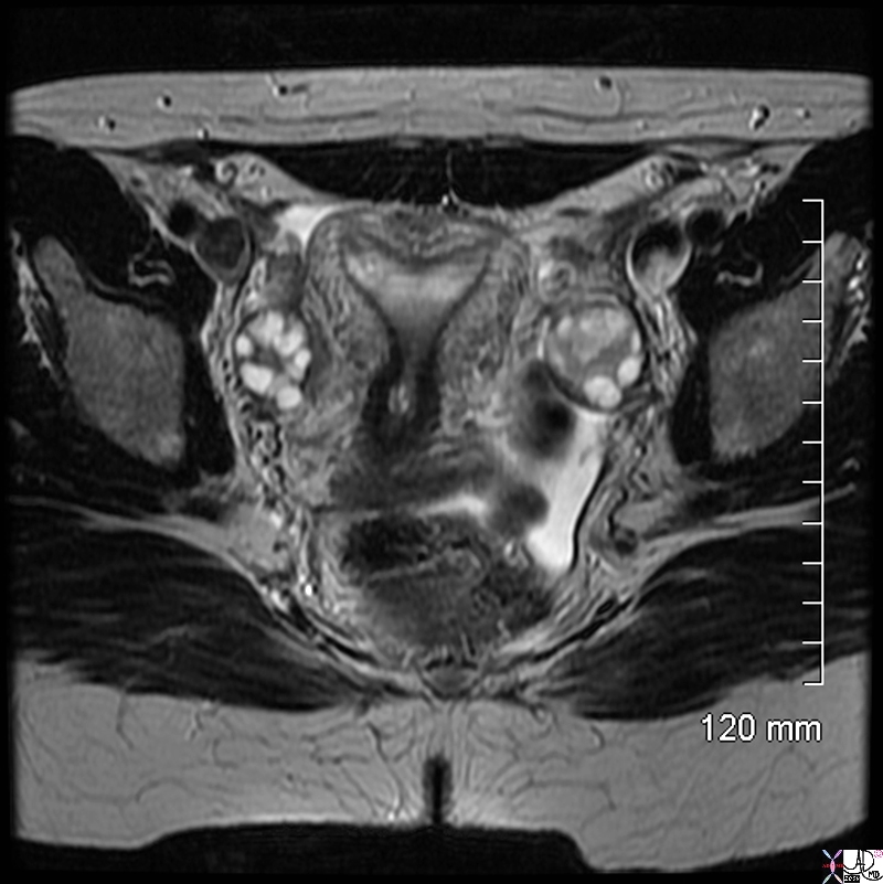
Arcuate Uterus – T2 Weighted MRI |
|
The T2 weighted MRI in axial projection of a 31 year old female shows the endometrial cavity in coronal view with an arcuate shape. This is a normal variant and has no clinical significance. The follicles in the ovaries are also well demonstrated . CODE uterus endometrial cavity ovary ovaries follicles fx arcuate shape dx normal variant
Image Courtesy Ashley Davidoff MD Copyright 2010 99420.8s
|
DOMElement Object
(
    [schemaTypeInfo] =>
    [tagName] => table
    [firstElementChild] => (object value omitted)
    [lastElementChild] => (object value omitted)
    [childElementCount] => 1
    [previousElementSibling] => (object value omitted)
    [nextElementSibling] =>
    [nodeName] => table
    [nodeValue] =>


Arcuate Uterus –  T2 Weighted MRI


The T2 weighted MRI in axial projection of a 31 year old female shows the endometrial cavity in coronal view with an arcuate shape. This is a normal variant and has no clinical significance. The follicles in the ovaries are also well demonstrated . CODE uterus endometrial cavity ovary ovaries follicles fx arcuate shape dx normal variant
Image Courtesy Ashley Davidoff MD Copyright 2010 99420.8s


    [nodeType] => 1
    [parentNode] => (object value omitted)
    [childNodes] => (object value omitted)
    [firstChild] => (object value omitted)
    [lastChild] => (object value omitted)
    [previousSibling] => (object value omitted)
    [nextSibling] => (object value omitted)
    [attributes] => (object value omitted)
    [ownerDocument] => (object value omitted)
    [namespaceURI] =>
    [prefix] =>
    [localName] => table
    [baseURI] =>
    [textContent] =>


Arcuate Uterus –  T2 Weighted MRI


The T2 weighted MRI in axial projection of a 31 year old female shows the endometrial cavity in coronal view with an arcuate shape. This is a normal variant and has no clinical significance. The follicles in the ovaries are also well demonstrated . CODE uterus endometrial cavity ovary ovaries follicles fx arcuate shape dx normal variant
Image Courtesy Ashley Davidoff MD Copyright 2010 99420.8s


)
DOMElement Object
(
    [schemaTypeInfo] =>
    [tagName] => td
    [firstElementChild] => (object value omitted)
    [lastElementChild] => (object value omitted)
    [childElementCount] => 2
    [previousElementSibling] =>
    [nextElementSibling] =>
    [nodeName] => td
    [nodeValue] =>
The T2 weighted MRI in axial projection of a 31 year old female shows the endometrial cavity in coronal view with an arcuate shape. This is a normal variant and has no clinical significance. The follicles in the ovaries are also well demonstrated . CODE uterus endometrial cavity ovary ovaries follicles fx arcuate shape dx normal variant
Image Courtesy Ashley Davidoff MD Copyright 2010 99420.8s

    [nodeType] => 1
    [parentNode] => (object value omitted)
    [childNodes] => (object value omitted)
    [firstChild] => (object value omitted)
    [lastChild] => (object value omitted)
    [previousSibling] => (object value omitted)
    [nextSibling] => (object value omitted)
    [attributes] => (object value omitted)
    [ownerDocument] => (object value omitted)
    [namespaceURI] =>
    [prefix] =>
    [localName] => td
    [baseURI] =>
    [textContent] =>
The T2 weighted MRI in axial projection of a 31 year old female shows the endometrial cavity in coronal view with an arcuate shape. This is a normal variant and has no clinical significance. The follicles in the ovaries are also well demonstrated . CODE uterus endometrial cavity ovary ovaries follicles fx arcuate shape dx normal variant
Image Courtesy Ashley Davidoff MD Copyright 2010 99420.8s

)
DOMElement Object
(
    [schemaTypeInfo] =>
    [tagName] => td
    [firstElementChild] => (object value omitted)
    [lastElementChild] => (object value omitted)
    [childElementCount] => 2
    [previousElementSibling] =>
    [nextElementSibling] =>
    [nodeName] => td
    [nodeValue] =>
Arcuate Uterus –  T2 Weighted MRI
    [nodeType] => 1
    [parentNode] => (object value omitted)
    [childNodes] => (object value omitted)
    [firstChild] => (object value omitted)
    [lastChild] => (object value omitted)
    [previousSibling] => (object value omitted)
    [nextSibling] => (object value omitted)
    [attributes] => (object value omitted)
    [ownerDocument] => (object value omitted)
    [namespaceURI] =>
    [prefix] =>
    [localName] => td
    [baseURI] =>
    [textContent] =>
Arcuate Uterus –  T2 Weighted MRI
)

