

Hi Casey –
I will introduce the concept of how to read a frontal CXR using the Notes Scales and Music approach
Your part will be to apply the concept as to how to diagnose cardiomegaly – global and then each of the chambers as below
There should be no need to go to any of the links – You should be able to demo from this site
This is the basic outline of the lesson
-
- Is the heart enlarged?
- Border forming structures on the frontal
- Elemental shape difference between LVE and RVE
- LVE example is a little subtle but the point to make- need both frontal and lateral sometimes to make the decision of enlargement or not
- Triangular heart examples
- Mitral Stenosis
- Pulmonary Hypertension
- Enlarged LA – Very important since in so many cases it is a marker of CHF So the carinal angle is really an important structure to look for when deciding if there is CHF I find it one of the most useful signs of CHF
- RA enlargement – needs to be quite enlarged to appreciate
- If you have time last case of CHD and frontal CXR is fun
First step – Is the heart enlarged?
- Cardiothoracic ratio as a global method
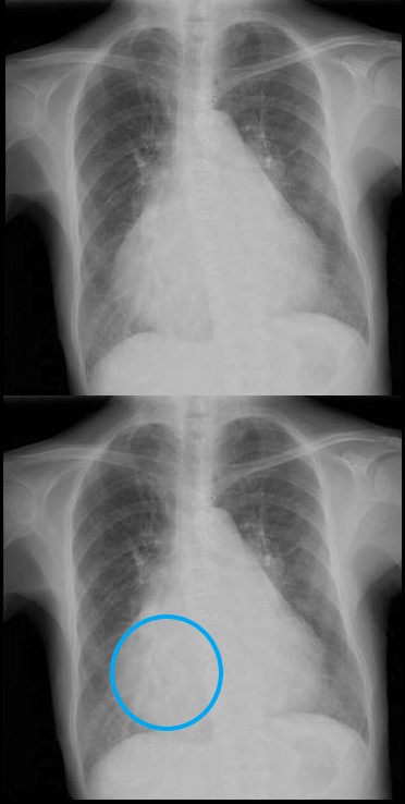
Cardiothoracic Ratio
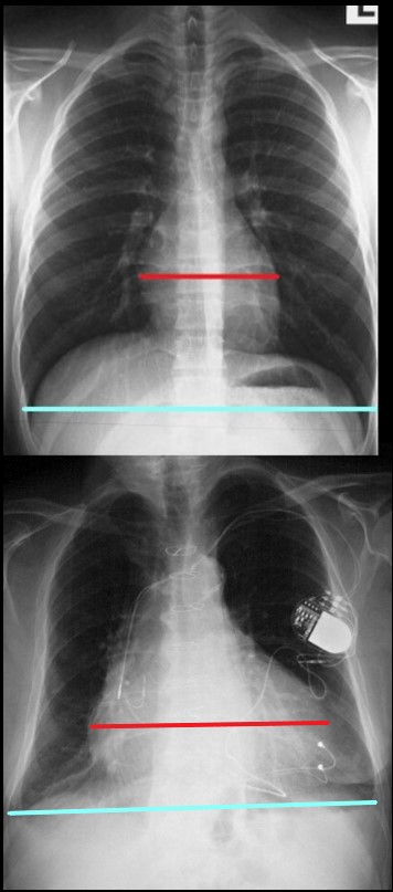
The maximum transverse length of the heart is expressed as a percentage of the maximum length of the internal diameter of the chest. When this ratio – the cardiothoracic ratio (c t r) is greater than 50% cardiomegaly is present. The top image is normal and the bottom reflects cardiomegaly
Ashley Davidoff MD
Border Forming Parts of the Heart
 FRONTAL CXR AND PARTS OF THE HEART
Two Basic Shapes of Cardiomegaly
-
- Oval down and outer of LV
- Triangular RV disease
- Each of the Chambers
- LA, LV, RA, V
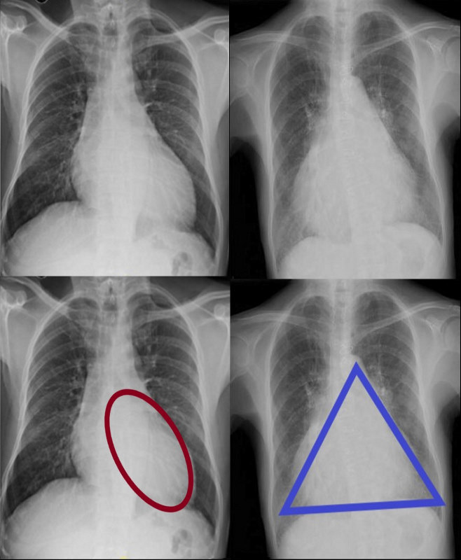
CARDIOMEGALY ? TWO BASIC TYPES -OVOID and TRIANGULAR
The ovoid form which suggests left ventricular dominance and triangular form which suggests right ventricular dominance.
Ashley Davidoff MD
LVE
Subtle Ovoid Form Suggestive on the PA and Confirmed on the Lateral – Using Both Views
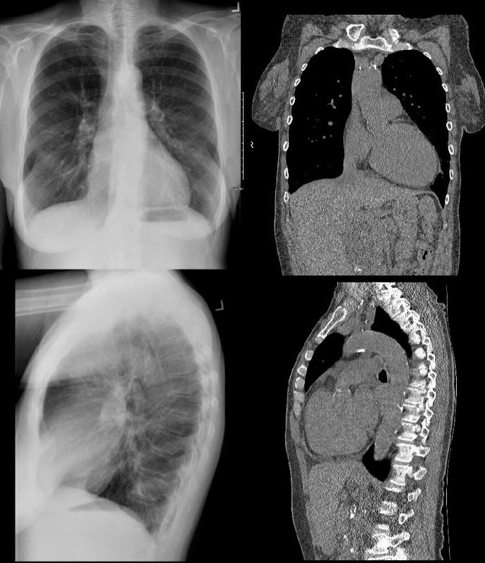
62 year old female with acute chest pain atrial fibrillation, hypotension admitted to ICU. Clinical evaluation was considered to be non-ischemic cardiomyopathy with EF by echo of about 20%. She was hypotensive and, in the ICU, and CXR showed acute CHF with cardiomegaly. The TEE was more in keeping with segmental dyssynergy, Cardiac cath showed occluded RCA bot good collateralization from the LAD. MRI showed subendocardial LGE in the inferior and inferolateral portions of the LV consistent with a prior infarction and EF of 20%
Ashley Davidoff MD
Triangular Heart with RVE
With Mitral Stenosis
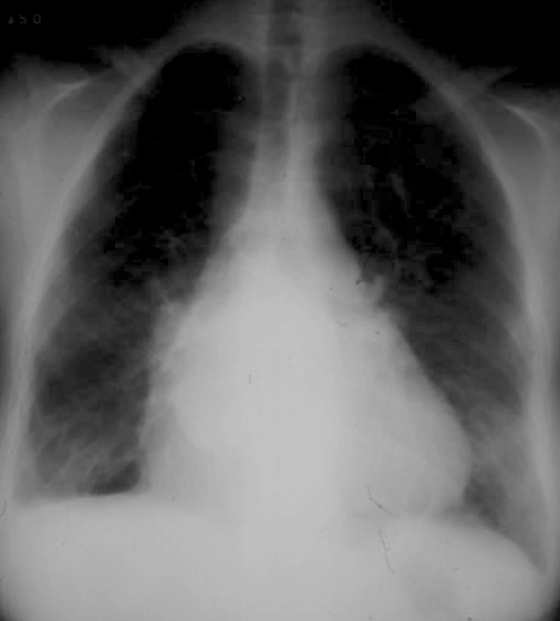
Triangular shaped heart with RVE LAE
Ashley Davidoff MD
With Pulmonary Hypertension

Frontal x-ray with triangular shaped heart due to pulmonary hypertension with enlarged MPA and enlarged descending RPA .
Ashley Davidoff MD
The Enlarged Left Atrium
Widened Carinal Angle
Double Density
Straightened right Heart Border – prominent LA appendage
Triangular Heart

Right Atrial Enlargement
-
- enlarged, globular heart
- narrow pedicle
- gross enlargement of the right atrial shadow, i.e. increased convexity in the lower half of the right cardiac border
- right atrial convexity is more than 50% of the cardiovascular height
- right atrial margin is more than 5.5 cm from the midline
 RIGHT ATRIAL ENLARGEMENT ON FRONTAL X-RAY
The right atrium is the most difficult chamber to assess unless it is very large in which case it will present on the frontal CXR with a very large right paravertebral border. This is a 71 year old female person with rheumatic heart disease with pulmonary hypertension and tricuspid regurgitation hence resulting in a large right atrium (RAE)
Ashley Davidoff MD
If there is time you may want to run through the collage of congenital heart disease cases

The Shapes of the Heart in Health and Disease
From top left ti right and across the rows they are: The normal heart , the ?football? of LV enlargement the ?triangle? or ?proud breast? of RV enlargement, ?snowman? of total anomalous pulmonary venous return, big PA mogul of pulmonary hypertension, ?egg on its side? of D transposition of the great vessels, ?boot shaped? heart seen in both pulmonary atresia and Tetralogy of Fallot, the long smooth combined Ao and PA mogul that has a differential diagnosis of L transposition, absence of the pericardium, and juxtaposition of the atrial appendages, the box shaped large heart of Ebstein?s anomaly, dextrocardia , and the water bottle? heart of a large pericardial effusion.
07197 Images are a combination of images from a personal collection and borrowed from the internet for educational purposes only. Some of the sources are unknown and are used for educational purposes alone 86774b02
TCV Links for extra info